Arq Neuropsiquiatr 2003;61(1):112-114
A CASE OF PRIMARY SPINAL MYOCLONUS
Clinical presentation and possible mechanisms involved
Cynthia Resende Campos
1, João Carlos Papaterra Limongi
1,
Flávia Costa Nunes Machado
1, Mário Wilson Iervolino Brotto
2ABSTRACT - Spinal myoclonus is a rare movement disorder characterized by myoclonic involvement of a group of muscles supplied by a few contiguous segments of the spinal cord. Structural lesions are usually the cause, but in primary spinal myoclonus the etiology remains unknown. We present the case of a 26-year-old woman with cervical spinal myoclonus in which both clinical and electromyographic findings pointed to the segment C1-C3 as the origin of the myoclonus. Laboratorial examinations were normal and no structural lesion was found in magnetic resonance imaging (MRI). Botulinum toxin type A was injected in infrahyoid muscles and cervical paraspinal musculature. The patient remained free of symptoms for almost five months. The pathophysiology of spinal myoclonus remains speculative, but there is evidence that various possible mechanisms can be involved: loss of inhibitory function of local dorsal horn interneurons, abnormal hyperactivity of local anterior horn neurons, aberrant local axons re-excitations and loss of inhibition from suprasegmentar descending pathways.
KEY WORDS: spinal myoclonus, segmental myoclonus, primary myoclonus.
Um caso de mioclonia espinhal primária: apresentação clínica e possíveis mecanismos envolvidos Um caso de mioclonia espinhal primária: apresentação clínica e possíveis mecanismos envolvidosUm caso de mioclonia espinhal primária: apresentação clínica e possíveis mecanismos envolvidos Um caso de mioclonia espinhal primária: apresentação clínica e possíveis mecanismos envolvidosUm caso de mioclonia espinhal primária: apresentação clínica e possíveis mecanismos envolvidos
RESUMO - A mioclonia espinhal é um raro distúrbio do movimento, caracterizado pelo envolvimento mioclônico de um grupo de músculos inervados por segmentos medulares contíguos. Lesões estruturais costumam ser a causa, mas na mioclonia espinhal primária a etiologia não é definida. Apresentamos o caso de uma mulher de 26 anos com mioclonia cervical espinhal em quem os achados clínicos e eletrofisiológicos apontaram o segmento C1-C3 como origem das mioclonias. Os exames laboratoriais foram normais e nenhuma lesão estrutural foi encontrada à ressonância. A toxina botulínica tipo A foi injetada nos músculos infrahioideos e na musculatura paraespinhal cervical. A paciente permaneceu assintomática por cinco meses. A patofisiologia da mioclonia espinhal continua especulativa, mas há evidências de que vários mecanismos possam estar envolvidos: perda da função inibitória de interneurônios da coluna dorsal, hiperatividade anormal de neurônios do corno anterior da medula, re-excitações axonais locais aberrantes e perda do efeito inibitório de vias descendentes suprasegmentares.
PALAVRAS-CHAVE: mioclonia espinhal, mioclonia segmentar, mioclonia primária.
1Department of Neurology, Hospital das Clínicas, University of São Paulo, São Paulo SP, Brazil (USP); 2Electromyography Unit of the
Department of Neurology, Hospital das Clínicas, USP. Received 19 June 2002. Accepted 30 August 2002.
Dra. Cynthia Resende Campos - Rua Capote Valente, 668 / 161 - 05409-002 São Paulo SP - Brasil. E-mail: [email protected]
Myoclonus is defined as a sudden, brief,
shock-like, involuntary movement due to either active
mus-cular contraction (positive myoclonus) or inhibition
of muscle activity (negative myoclonus)
1. Segmental
myoclonus is a rare movement disorder characterized
by myoclonic involvement of a muscle or a group of
muscles supplied by a few contiguous segments of
the brain stem or spinal cord
2. Several causes of spinal
myoclonus have already been described including
spinal tumors, infections, vascular lesions, spinal
anesthesia, AIDS and demyelinating diseases, but in
a few cases the etiology remains unknown
2-5.
We present a case of spinal cord myoclonus in
which no structural lesion was found in magnetic
resonance imaging (MRI) and review some possible
pathophysiological mechanisms.
CASE
Arq Neuropsiquiatr 2003;61(1) 113
movements increased rapidly in the first few days and be-came gradually worse in the last three weeks. General physical examination was normal. On neurological exami-nation, there were involuntary spontaneous synchronous myoclonic jerks of the anterior wall of her neck, subman-dibular region and nape musculature, which resulted in slight extension of her neck. The contractions were rhy-thimic, bilateral and with a rate of approximately 1 Hz. No myoclonus was observed in the tongue. There was no vocalization. These movements could not be controlled by her will or effort. The rate and amplitude of involuntary movements were increased by emotional stress and neck extension and did not ceased during sleep. MRI of the brain and spinal cord, cerebrospinal fluid, routine hema-tological and immunological examinations (including HIV, HTLV I/II, hepatitis B and C, and syphilis), calcium, magne-sium, copper, ceruloplasmin concentrations, and routine electroencephalography were normal.
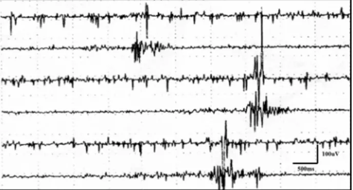
Needle electromyography (EMG) showed rhythmic irre-gular burst discharges of motor units with a rate of 1-3 Hz in the muscles of anterior wall of the neck and cervical paraspinal muscles bilaterally (Fig 1). Trapezius and sterno-cleidomastoid muscles had no abnormal contractions. The bursts of activity were increased by neck extension and almost disappeared with neck flexion.
Sodium valproate 750mg/day was prescribed for two weeks but no improvement was observed. Botulinum toxin type A (Botox ®, Allergan) was injected: 20 units in each side of cervical paraspinal musculature, 20 units in each splenius capitis, 10 units in each geniohyoid muscle and 10 units in the infrahyoid musculature on the left side. Bilateral injection of infrahyoid muscles was avoided in order to prevent disphagia. Two weeks later, there was complete cessation of myoclonus, and electromyographic reduction in both rate and magnitude of the burst dis-charges of myoclonus (Fig 2). Patient was videotaped pre and post toxin injection. She remained free of symptoms for almost five months.
DISCUSSION
Pathophysiologically, myoclonus can be broadly
classified as cortical, subcortical, cortical-subcortical,
segmental, or peripheral
6. In the segmental type,
le-sions placed at different locations along the neuraxis
may be the cause. When the presumed cause is in
the spinal cord, it is called spinal myoclonus. The
responsible site is usually estimated by clinical
observation and electrophysiological examination.
This rare kind of myoclonus involves only the
mus-culature innervated by a few adjacent spinal levels
and is usually rhythmic and slow (<4Hz). The
pre-sence of myoclonus in the mouth floor of our patient
and the lack of involvement of her tongue suggested
that the hypoglossus nuclei themselves were not
in-volved. The affected muscles in the mouth floor
be-longs to a group called infrahyoid muscles. The
in-frahyoid muscles (geniohyoid, thyrohyoid,
sterno-thyroid, sternohyoid and omohyoid) are supplied by
nerve fibers derived from the first cervical nerve (C1)
and from the junction of the second and third cervical
nerves (C2-C3)
7. The EMG showed the involvement
of muscles of the anterior wall of the neck known as
infrahyoid muscles and the simultaneous myoclonic
jerks of cervical paraspinal muscles bilaterally. The
main muscles of this site include semispinalis capitis,
rectus capitis posterior, obliquus capitis superior and
splenius capitis bilaterally, which also depend of C1
and C2 nerves
8. Trapezius and sternocleidomastoid
muscles were free. Both clinical and
electro-myographic findings pointed to the segment C1-C3
as the site of segmental spinal myoclonus.
The pathophysiology of spinal myoclonus remains
speculative. In 1979, Howell et al. suggested that
focal myoclonus could be caused by loss of local
in-hibitory spinal interneuronal function, which allowed
the spontaneous repetitive discharge of local
seg-mental anterior horn cell pools
9. In 1981 Davis et al.
provided histological evidence for this hypothesis in
Fig 2. EMG registered in the same muscles described in Figure 1 (A, C, E – right; B, D, F – left), two weeks after Botulinum toxin A. There was reduction in both rate and magnitude of the burst myoclonic discharges.
114 Arq Neuropsiquiatr 2003;61(1)
a case in which the number of small and medium
sized neurons in the posterior horns of the lumbar
cord was reduced along with relative sparing of large
neurons in the anterior horns
10. On the other hand,
another study showed that physiological suppression
of dorsal horn interneurons failed to occur in
seg-mental myoclonus, indicating that dorsal horn
interneurons could be abnormally hyperactive
11.
Jan-kovic hypothesized that the rhythmical contractions
could be the expression of spontaneous spinal
neu-ronal discharge due to suppression of physiologic
inhibition from suprasegmentar levels but
experi-mental evidence for this hypothesis is lacking
2.
Electrophysiological studies in hemifacial spasm,
which is a form of segmental myoclonus, may
contri-bute to elucidate the possible mechanisms of spinal
myoclonus. Axono-axonal ephatic transmission
among injured fibers due to compression and
demye-lination of the intracranial segment of the facial
nerve
12,13or abnormal central hyperexcitability of
the facial motor nucleus or both
14,17have been
con-sidered as possible mechanisms. Neurophysiological
studies in patients with cryptogenic hemifacial spasm
showed that the self-sustained repetitive firing in facial
nerve axons could result from re-excitations occurring
both at the ephapse site on the axon and at the cellular
level
18or from a permanent antidromic stimulation
from a peripheral ectopic center of excitation
19. In
summary, there is evidence that various possible
me-chanisms can be involved: loss of inhibitory function
of local dorsal horn interneurons, abnormal
hyperac-tivity of local anterior horn neurons, aberrant local
axons re-excitations and loss of inhibition from
supra-segmentar descending pathways.
The worsening of myoclonus in relation to changes
in position has been described
3,20. In our patient, the
worsening of myoclonus during neck extension
suggests that peripheral stimulus may modulate the
abnormal movement, but the symmetrical pattern of
muscle activity points that local spinal generators
could be the source of myoclonus. Our patient
remained asymptomatic for five months after
bo-tulinun toxin injection. This longer duration of benefit
as compared to that observed in focal dystonias have
been described in similar cases and points to a higher
degree of susceptibility of this type of movement to
chemical denervation with botulinum toxin
4.
REFERENCES
1. Caviness JN. Myoclonus. Mayo Clin Proc 1996;71:679-688.
2. Jankovic J, Pardo R. Segmental myoclonus. Arch Neurol 1986;43:1025-1031. 3. Kono I, Ueda Y, Araki K, Nakajuma K, Shibasaki H. Spinal myoclonus
resembling belly dance. Mov Disord 1994;9:325-329.
4. Polo KB, Jabbari B. Effectiveness of botulinum toxin type A against painful limb myoclonus of spinal cord origin. Mov Disord 1994;9:233-235. 5. Fox EJ, Villanueva R, Schutta HS: Myoclonus following spinal
anesthesia. Neurology 1979;29:379-380.
6. Marsden CD, Hallett M, Fahn S. The nosology and pathophysiology of myoclonus. In Marsden CD, Fahn S, (eds.) Movement disorders. London: Butterworths, 1982:196-248.
7. Krieg WJS. Movements of eyes and tongue. In Krieg WJS Functional neuroanatomy. Philadelphia: Blakiston Company, 1942:72-77. 8. Sobotta J. Atlas de anatomia humana. Staubesand J (ed). 19.Ed. Rio de
Janeiro: Guanabara Koogan, 1990.
9. Howell DA, Lees AJ, Toghill PJ. Spinal internuncial neurones in progressive encephalomyelitis with rigidity. J Neurol Neurosurg Psychiatry 1979;42:773-785.
10. Davis SM, Murray NME, Diengdoh JV, Galea-Debono A, Kocen RS. Stimulus sensitive spinal myoclonus. J Neurol Neurosurg Psychiat 1981;44:884-888.
11. Lazzaro V, Restuccia D, Nardone R, et al. Changes in spinal cord excitability in a patient with rhythmic segmental myoclonus. J Neurol Neurosurg Psychiat 1996;61:641-644.
12. Sanders DB. Ephaptic transmission in hemifacial spasm: a single-fiber EMG study. Muscle Nerve 1989;12:690-694.
13. Nielsen VK. Electrophysiology of the facial nerve in hemifacial spasm: ectopic/ephaptic excitation. Muscle Nerve 1985;8:545-555.
14. Kuroki A, Moller AR, Saito S. Recordings from the facial motonucleus in rats with signs of hemifacial spasm. Neurol Res 1994;16:389-392. 15. Moller AR. Hemifacial spasm: ephaptic transmission or hyperexcitability of
the facial motor nucleus? Exp Neurol 1987;98:110-119.
16. Moller AR, Jannetta PJ. Physiological abnormalities in hemifacial spasm studied during microvascular decompression operations. Exp Neurol 1986; 93:584-600.
17. Moller AR, Jannetta PJ. Hemifacial spasm: results of electrophysiologic recording during microvascular decompression operations. Neurology 1985; 35:969-974.
18. Roth G, Magistris MR, Pinelli P, Rilliet B. Cryptogenic hemifacial spasm: a neurophysiological study. Electromyogr Clin Neurophysiol 1990;30:361-370. 19. Poignonec S, Lamas G, Aidan P, Willer JC, Soudant J. Electrophysiological study
of hemifacial spasm. Ann Otolaryngol Chir Cervicofac 1993;110:385-391. 20. Aigner BR, Mulder DW. Myoclonus: clinical significance and an