179 Heart Institute, Rio Grande do Sul/University Heart Foundation
Correspondence address:
Research unit of IC/FUC – Dr. Paulo R. Lunardi Prates
Av. Princesa Isabel, 370 Santana 90620-001 Porto Alegre, RS, Brazil
Phone/Fax: 51-3230-3600 R. 3877 / 3602.
E-mail: [email protected] / [email protected]
Paulo R .L. PRATES, Roberto T. SANT´ANNA, Ivo A. NESRALLA
Rev Bras Cir Cardiovasc 2004; 19(2): 179-182 HOW TO DO IT
Article received in March, 2004 Article accepted in May, 2004
RBCCV 44205-687
Abstract
Robotic surgery is today a reliable method to reduce trauma and related comorbidities in cardiac surgery. In our institution, robotic assistance has been effectively used for thoracoscopic implantation of epimyocardial leads for biventricular pacing. The objective of this article is to describe the technique that we developed to dissect the internal thoracic artery using thoracoscopy assistance with the AESOP robotic system and to report the initial results obtained with its use in nine patients.
Descriptors: Robotics. Cardiac surgical procedures, methods. Surgical procedures, minimally invasive.
Resumo
A cirurgia robótica é hoje um método viável para reduzir o trauma e as morbidades relacionadas em cirurgia cardíaca. Em nossa instituição, o auxílio robótico já foi utilizado de forma bem-sucedida em implantes toracoscópicos de eletrodos epimiocárdicos para marcapassos biventriculares. O objetivo deste artigo é descrever a técnica por nós utilizada para dissecção da artéria torácica interna (ATI), mediante toracoscopia com apoio do robô AESOP e relatar seus resultados iniciais obtidos em nove pacientes.
Descritores: Robótica. Procedimentos cirúrgicos cardíacos, métodos. Procedimentos cirúrgicos minimamente invasivos.
Dissecção endoscópica da artéria torácica interna com auxílio robótico
180
PRATES, PRL ET AL - Endoscopic harvest of internal thoracic artery with robotic assistance
Rev Bras Cir Cardiovasc 2004; 19(2): 179-182
INTRODUCTION
Traditionally, coronary artery bypass graft surgery (CABG) has been performed by sternotomy, an approach that allows access to all the heart structures and great vessels [1,2]. Minimally invasive surgery and total endoscopic surgery aim to diminish the surgical trauma, thereby reducing the hospitalization and hospital costs [1-4].
The ultimate development in terms of minimally invasive CABG, is totally endoscopic CABG (TECABG) [3,4]. Currently, this is successfully performed utilizing telemanipulated robotic systems.
The evolution of TECABG is slow and one of the basic stages for this technique to be successful is endoscopic dissection of the internal thoracic artery (ITA), first performed in Brazil by us and the group of Incor in Sao Paulo [5,6]. The aim of this work is to describe the technique we utilize for the dissection of the ITA using robotic arm-assisted thoracoscopy AESOP. We also report the preliminary results.
METHOD
From April to December 2002, nine patients who were submitted to CABG underwent robot-assisted endoscopic harvesting of the ITA. The ages of the patients varied from 49 to 71 years old (mean 57.5 ± 7.73) and seven patients (78%) were male. All the patients consented to the procedure and the project was approved by the ethics committee of the hospital. The functional class according to the New York Heart Association (NYHA) was class I in seven patients (78%), class II in one patient (12%) and class III in one patient (12%). The ejection fraction (EF) ranged from 48 to 83% (mean 67%). All patients suffered with significant obstructions (98% or more) of the anterior descending coronary artery evidenced by catheterism. Other involved arteries included: the diagonal branch of the anterior descending coronary artery in two patients; circumflex artery in three patients, marginal branch of the circumflex artery in three patients and the right coronary artery in eight.
Operative technique
The surgical procedure is performed under general anesthesia using thiopental, fentanyl, pancuronium, midazolam and isoflurane. The patient is monitored in the surgical room using ECG, pulse oximetry, capnography, measurement of the central venous blood pressure and of the blood pressure in the right radial artery. Orotracheal intubation is achieved using a dual lumen tube for selective ventilation. The patient is placed in the right lateral decubitus position (30 degrees of inclination) using a cushion placed under the left scapula. For a better access to the left lateral wall of the thorax and the axilla, the left upper limb is
positioned on the face of the patient.
After the preparation of the patient and the establishment of selective ventilation to the right lung controlled at a rate of 6 to 8 mL/kg and a PEEP of 3 to 5 cm H2O, the endoscopic harvesting of the of the left ITA is initiated. During the procedure, as far as possible the PAM is kept around 75 mmHg to reduce the risk of bleeding.
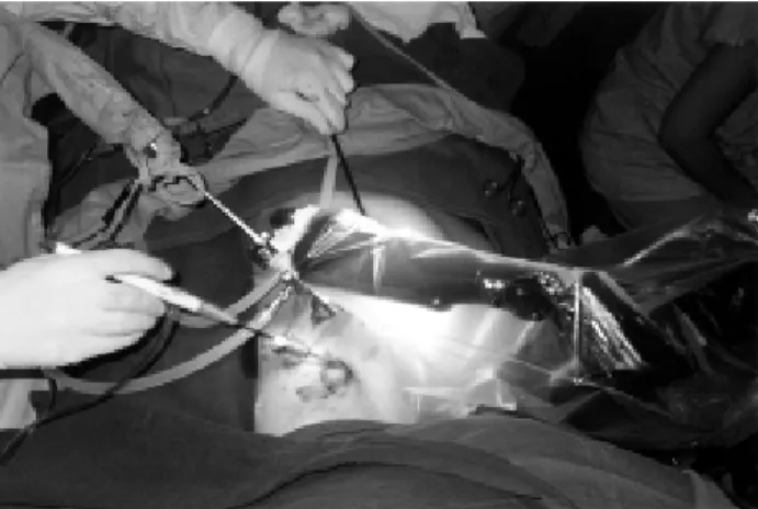
A robotic AESOP arm is utilized, which works as a thoracoscopic-guided camera, which was verbally controlled by the surgeon. It is fixed to the surgical table to the right of the patient (the opposite side to the surgeon), near to the xiphoid appendix (Figure 1). To harvest the ITA, an electrocauterizer (Valleylab) is used, with a dissecting blade of 20 cm supplied with a aspirator to remove the debris and vapor produced during the procedure using a trocar of 3.5 mm inserted in the 2nd and 3rd left intercostal space (LICS), using the same incision, on the medium axillary line. The 21 cm camera with 5 mm of diameter and 0 degrees is inserted in the 4th LICS, also at the same level as the media axillary line using a 5-mm trocar.
Fig. 1 – Positioning of the AESOP robot
A 20-cm long endoscopic tweezers is inserted in the 6th LICS without the help of a trocar, anterior to the axillary line. For better access to the mediastinum, the carbon dioxide flow is maintained by the 5-mm exchange at a pressure of 8 to 10 mmHg in the left hemithorax, increasing the space between the thoracic wall and the mediastinum. The ITA graft is dissected from the first rib until its bifurcation. To dissect the cranial third of the ITA, the 2nd LICS is utilized, to dissect the caudal third the trocar is utilized in the third LICS through the same incision as the electrocauterizer with a 3.5-mm trocar (Figure 2).
181 patients. The CABG is performed in the conventional way,
using median sternotomy and cardiopulmonary bypass (CPB). The other grafts are inserted in the ascending aorta in the usual manner. The postoperative management was the same as used in the traditional CABG.
Fig. 2 – Sites of the incisions for the surgical instruments
septal infarction presented with atheromatous ADA and with a bad distal bed. In the angiographic study performed in this case, the ITA appeared to be integral.
There were no cases of late death. Among the 6 patients regularly consulted in the follow up period, 4 were without symptoms six months after surgery and two presented with functional limitation compatible to a class II angina.
COMMENTS
The constant evolution of surgical treatment apart from having the objective of improving the operative techniques also aims at reducing the comorbidities and improving the esthetic results associated to the procedure. The appearance and evolution of interventionalist procedures created an alternative avoiding the necessity of surgical injury, without anesthesia and with the possibility of a rapid recovery of the patient [3]. However, interventionalist treatment is not indicated for all lesions. CABG continues to be the choice method for complex lesions, principally when the anterior descending artery is involved [1].
TECABG would be an alternative giving advantages to CABG, with a reduced morbidity and an improved esthetical result.
The initial stage to perform this procedure consists of endoscopic harvesting of the ITA. This has previously been described with the utilization of the Harmonic Scalpel (Ethicon Endo-Surgery, Cincinnati, Ohio, USA) and with other more complex robotic systems such as Zeus (Computer motions, Goleta, California, USA) and Da Vinci (Intuitive Surgical, Sunnyvale, California, USA) [4-7].
We believe that using the assistance of the AESOP robot provides a greater stability generated by the video and a better control when compared with conventional video-thoracoscopy. Additionally, it provides better comfort to the surgeon and avoids the fatigue and uncomfortable position of his assistant who normally operates the camera. As, in our series after the endoscopic dissection of the ITA the patients are submitted to sternotomy, the advantages in relation to pain and postoperative morbidity should not be taken into account [8].
Robotic surgery currently is dependent on specific surgical indications, high-cost equipment and greater surgical time than conventional surgeries, in such a way that its benefits tend to be seen over the long term. However, there is a learning curve involved, as we have already described, such that the time necessary to perform the procedure tends to reduce as the surgeon becomes accustomed to the technique. We believe that the initial investment even though it is high, can be reduced as the theoretical benefits reduce time of hospitalization and morbidity.
PRATES, PRL ET AL - Endoscopic harvest of internal thoracic artery with robotic assistance
Rev Bras Cir Cardiovasc 2004; 19(2): 179-182
RESULTS
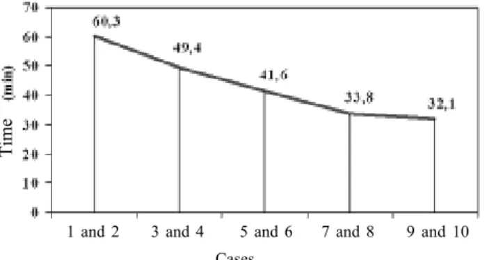
The time necessary for dissection of the left ITA varied from 40 to 90 minutes (mean 66.1 minutes) with the first dissection performed in 90 minutes and the last three in less than 60 minutes (Figure 3). The time gradually diminished with the familiarization with the equipment and learning of the procedure.
There were no hospital deaths. One patient suffered septal infarction in the immediate postoperative period and another suffered a pericardial effusion due to post-pericardiotomy syndrome. The patient who suffered the tweezers camara cauterizer
Fig. 3 – Time necessary for the dissection of the ITA in each case, demonstrating there was a learning curve
T
im
e
182
BIBLIOGRAPHIC REFERENCES
1. Diegeler A, Thiele H, Falk V, Hambrecht R, Spyrantis N, Sick P, et al. Comparison of stenting with minimally invasive bypass surgery for stenosis of the left anterior descending coronary artery. N Engl J Med 2002;347(8):561-6.
2. Dogan S, Aybek T, Andressen E, Byhahn C, Mierdl S, Westphal K, et al. Totally endoscopic coronary artery bypass grafting on cardiopulmonary bypass with robotically enchaced telemanipulation: Report of forty-five cases. J Thorac Cardiovasc Surg 2002;123 (6):1125-31.
3. Falk V, Diegeler A, Walther T, Banusch J, Brucerius J, Raumans J, et al. Total endoscopic computer enhanced coronary artery bypass grafting. Eur J Cardiothorac Surg 2000;17(1):38-45.
4. Jacobs LK, Shayvani V, Sackier JM. Determination of the learning curve of the AESOP robot. Surg Endosc 1997;11(1):54-5.
5. Dallan LAO, Lisboa LAF, Abreu Filho CAC, Platania F, Dallan LAP, Iglesias JC et al. Assistência robótica para dissecção minimamente invasiva da artéria torácica interna na revascularização do miocárdio. Rev Bras Cir Cardiovasc 2003; 18(1):110.
6. Prates PRL, François LMG, Argondizzo MB, Harter MO, Lajus JA, Brasil RV et al. Dissecção endoscópica da artéria torácica interna com auxílio robótico. Rev Bras Cir Cardiovasc 2003; 18(1):110.
7. Bucerius J, Metz S, Walther T, Falk V, Doll N, Noack F, et al. Endoscopic internal thoracic artery dissection leads to significant reduction of pain after minimally invasive direct coronary artery bypass graft surgery. Ann Thorac Surg 2002;73(4):1180-4
8. Sant´Anna JRM, Prates PRL, Kalil RAK, Prates PR, Moretto M, Sant´Anna RT et al. Robotic-Assisted Thoracoscopic Implatation of an Epimyocardial Lead for Biventricular Pacing. Progress in Biomedical Research. 2002;7:32-6.
PRATES, PRL ET AL - Endoscopic harvest of internal thoracic artery with robotic assistance