Cariostatic treatment has been shown to successfully arrest caries. However, it blackens the carious tooth structure. This study evaluated the effects of an experimental cariostatic agent with silver nanoparticles (Ag-Nano) using microhardness (MH) and microbiological tests. The cariostatic agents tested were: Saforide®, Cariestop®, Ancarie® and Ag-Nano. Sixty-six samples from deciduous enamel were submitted to initial (after pH cycling to obtain initial caries-like lesion) and final (after cariostatic application) MH testing and %MH values were calculated. After longitudinal sectioning, internal (I) MH was evaluated. Strains of Streptococcus mutans, Escherichia coli, and Enterococcus faecalis in brain-heart infusion culture were treated with the cariostatic agents. Agar diffusion tests (ADTs) were performed and minimum inhibitory concentrations were determined. The statistical tests used were: Kruskal-Wallis and Dunn (%MD; ADT; MIC) and ANOVA followed by Tukey’s test (I-MH) (p<0.05). The %MH of Saforide® was significantly greater than that of Ag-Nano (p<0.05). Internal MH showed progressive improvement in the enamel remineralization for all cariostatic tested. In ADTs showed greater inhibition of S. mutans, E. faecalis, and
E. coli by Saforide® than by Ancarie® and Ag-Nano. Ag-Nano was able to inhibit 100% microorganism growth at a lower concentration than required for the other agents. It was concluded that Ag-Nano treatment promoted remineralization of deciduous tooth enamel with initial caries-like lesion and bactericidal activity.
I n V i t r o
E v a l u a t i o n o f t h e
R e m i n e r a l i z i n g Po t e n t i a l a n d
Antimicrobial Activity of a Cariostatic
Agent with Silver Nanoparticles
Beatriz Brandão Scarpelli1,2, Marília Franco Punhagui1,2, Márcio Grama Hoeppner3, Ricardo Sergio Couto de Almeida4, Felipe Augusto Juliani4, Ricardo Danil Guiraldo2, Sandrine Bittencourt Berger2
1Department of Paediatric Dentistry, UEL - Universidade Estadual de Londrina, Londrina, PR, Brazil 2Department of Restorative Dentistry, UNOPAR – Universidade Norte do Paraná, Londrina, PR, Brazil 3Department of Restorative Dentistry, UEL - Universidade Estadual de Londrina, Londrina, PR, Brazil 4Department of Microbiology, UEL - Universidade Estadual de Londrina, Londrina, PR, Brazil
Correspondence: Sandrine Bittencourt Berger, Rua Marselha, 183, Jardim Piza, 86041-120 Londrina, PR, Brasil. Tel: +55 43 3371-7820. e-mail: berger.sandrine@gmail.com
Key Words: dental caries, prevention, cariostatic treatment, nanoparticles.
Introduction
The existing standard of care for early childhood caries (ECC) consists of surgical and restorative treatments, with little emphasis on prevention and management of the disease (1). However, the simplicity and accessibility of preventive treatment with silver diamine fluoride (SDF) have stood out since 1969 (2). SDF is considered to be an effective agent for the prevention and control of dental caries, including in deciduous teeth (3,4). An in vitro study (1) indicated that SDF solution is a more effective anticariogenic agent than fluoride varnish in areas with fluoridated water. This product has been used in various concentrations in public and private health services (5) and it is indicated for pediatric patients who do not cooperate with complex restorative procedures and who have high caries risk or activity in deciduous teeth (6,7).
The mechanism of action of SDF has been linked to the formation of silver phosphate on the tooth surface, fluoride uptake in dentin and blocking of dentinal tubules through silver precipitates. The tooth mineral component increases peri- and intertubular dentin resistance to acid demineralization. Cariogenic bacteria (e.g., cariogenic strains of Streptococcus mutans) are inactivated when
enzymatic activity is inhibited and agglutination is induced by dextran (3,5,8,9).
In vitro evaluation of a cariostatic agent peroxidation and thereby interrupting DNA replication
and inhibiting cellular respiration (14,15). An important antimicrobial toxicity parameter of silver nanoparticles is the surface area of the nanomaterial, once higher concentration of silver ions are observed in larger area of silver nanoparticles, leading to greater intracellular bioavailability of silver (15). Once silver nanoparticles are more efficient and/or more convenient to administer, these have lower therapeutic toxicity and can help reduce the costs of health care (16).
The development of new cariostatic agents motivated us to perform this in vitro study in human deciduous enamel, as previous studies have used human (10) or bovine (17) permanent enamel. The study hypothesis was that the tested cariostatic agent with silver nanoparticles would show remineralization potential in deciduous dental enamel and bactericidal activity against S. mutans, Escherichia coli and Enterococcus faecalis.
Material and Methods
This study involved mechanical testing of superficial and internal microhardness (MH) and antimicrobial activity.
Specimens and Block Preparation
A total of 100 intact human deciduous molars that had erupted and exfoliated naturally were used in this study. The teeth were stored in 0.1% thymol solution until the study began. This local ethics committee approved this research (Protocol #913.639).
The teeth were cleaned with periodontal curettes (Hu-Friedy Mfg. Co, Chicago, IL, USA) and debris was removed completely with pumice and a Robinson brush. The roots of all teeth were sectioned transversely 2 mm above the dentinoenamel junction with a double-sided diamond disc (KG Sorensen, Barueri, SP, Brazil). A total of 100 enamel blocks (4x×4×3 mm) were made using a double-sided diamond disc. These blocks were fixed dentin side up in acrylic discs with sticky wax and abraded for 1 min with
silicon carbide (SiC) sandpaper (400 grit; Carburundum Abrasivos, Recife, Brazil) in a polisher (APL4; Arotec, Cotia, SP, Brazil) to plane the dentin surfaces. The blocks were then removed from the discs, inverted and refixed with the enamel surfaces exposed. These surfaces were planed with SiC sandpaper (400, 600 and 1200 grit) and then polished with 1 µm diamond paste and felt discs (Arotec). The blocks were placed in an ultrasonic tank with distilled water (Unique Indústria Comércio Produtos Eletrônicos Ltda, São Paulo, SP, Brazil) for 10 min to remove the waste. Red nail polish was then used to delimit a 7-mm2 area of exposed enamel parallel to the base of the acrylic on each specimen for microhardness testing.
Initial Microhardness Testing and Block Selection
Three impressions of each specimen were taken at a distance of 100 μm, with a static load of 25 g applied for 5 s to the central region of the block using a Knoop-type penetrator (KHN) (HMV-G; Shimadzu, Tokyo, Japan). Initial superficial MH values were obtained and the overall average MH of the 100 enamel blocks was calculated (KHN = 321.89±40.78). Blocks with values 10% above or below this average were excluded from the study. The remaining blocks were divided randomly into six experimental groups (n=11). Analysis of variance (ANOVA) verified the homogeneity of the groups (p=0.463) (Table 1).
Initial Caries-Like Lesion Production and Superficial Microhardness Testing
Blocks in G1-G5 were subjected to pH cycling at 37 °C for 8 days, immersed for 2 h in demineralizing solution (0.05 mol/L acetate buffer [pH 5.0] containing 1.28 mmol/L Ca, 0.74 mmol/L P, 0.03 µg/mL F) and then immersed for 22 h in remineralizing solution (0.1 mol/L Tris buffer [pH 7.0] containing 1.5 mmol/L Ca, 0.9 mmol/L P, 150 mmol/L KCl, 0.05 mg F/mL) (17). The volume of de- and remineralizing solution per area exposed enamel surface was 6.25 and 3.12 mL/mm2 respectively (18). The blocks
Table 1. Division of the experimental groups (n=11) and characterization of the cariostatic agents
Group Treatment Composition Manufacturer
G1 Saforide® 38% Silver diamine fluoride [Ag(NH3)2F] Toyo Seiyaku Kasei CO., LTD
G2 Cariestop® 30% silver nitrate, ammonium hydroxide,
hydrochloric acid and deionized water Biodinâmica Química e Farmacêutica LTDA
G3 Ancarie® 30% silver nitrate, ammonium hydroxide,
hydrochloric acid and purified water Maquira Indústria de Produtos Odontológicos LTDA
G4 Ag-Nano* 0.016% silver nitrate and sodium citrate AAF do Brasil
G5 Enamel with pH-cycling -
-G6 Intact enamel -
B
. B
. Scarpelli et al.
were the subjected to MH testing, as described above, to verify enamel demineralization.
Cariostatic Agent Application and Final Superficial Microhardness Testing
After superficial MH testing of blocks with caries-like lesions, prophylaxis was performed with water, pumice and a Robinson brush on blocks in G1-G4. The enamel blocks were then washed and dried and the cariostatic agents (Table 1) were applied with microbrushes to the enamel surfaces for 3 min, followed by MH testing to evaluate enamel remineralization.
The initial, demineralized, and final MH values in G1-G5 were used to calculate the percentage of superficial remineralization (%MH) using the formula: %MH = (final MH - demineralized MH)/(initial MH - demineralized MH)×100 (17). The negative control samples (group 5) showed decreased %MH because they were subjected to in vitro caries-like lesion production (pH cycling), but no cariostatic agent application. The positive control samples (group 6) showed no change in MH because they were not subjected to pH cycling or cariostatic agent application.
Analysis of Internal Microhardness
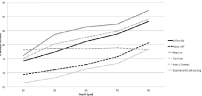
To evaluate the internal effects of the cariostatic agents on dental enamel, blocks in G1-G6 were sectioned along their long axes with a diamond disc in a precision cutter (Isomet 1000; Buehler Ltd., Lake Bluff, IL, USA). The inner surfaces were then abraded with SiC sandpaper (400, 600 and 1200 grit) and polished with felt discs and 1-µm diamond paste. Internal MH was measured at 10-, 30-, 50-, 70- and 90-µm depths with three impressions taken at a distance of 100 µm (19). The percentage of mineral volume (%VM) was determined using the formula: %VM = 4.3(KHN)1/2+11.3 (19).
Microorganism Cultivation
For in vitro evaluation of the inhibitory effects of the cariostatic agents, strains of S. mutans UA 159, E. faecalis ATCC 29212 and E. coli ATCC 25922 were used. The bacteria were maintained on brain-heart infusion (BHI) agar plates (Acumedia Neogen do Brasil, Indaiatuba, SP, Brazil) until cultivation for testing. Bacteria were grown for 12 h in liquid BHI at 37 °C with agitation at 150 rpm. Suspensions of each microorganism were then adjusted in phosphate-buffered saline (PBS) to 0.5 McFarland.
Agar Diffusion Test (ADT)
The bacterial suspensions were spread evenly across the surfaces of plates with solid BHI using sterile cotton swabs. Then, 100 μL each cariostatic agent was added to the SDF base at the adjusted concentration of 300 mg/mL, in agar
plate wells. For the Ag-Nano, 100 µL solution at 0.016 mg/ mL was added to each well. Then, plates with S. mutans were incubated for 24 h at 37 °C with 5% CO2, and plates with E. coli and E. faecalis were incubated without CO2. After incubation, we performed visual analysis, measured the inhibition halos with a caliper and divided these values by the diameter of the well to determine the inhibition zone.
Evaluation of Minimum Inhibitory Concentration (MIC)
The broth microdilution method was performed according to the standards of the National Committee for Clinical Laboratory Standards (M07-A10) (20). The microorganisms were grown in liquid BHI culture medium for 16 h at 37 °C (and 5% CO2 for S. mutans). Suspensions with turbidities of 0.5 McFarland were prepared with PBS, and diluted 1:100 with Mueller-Hinton medium.
Serial dilutions (1:2, 50 µL) of the cariostatic agents were inserted into the wells of a 96-well plate with a U-shaped bottom. Then, 50 µL bacteria suspension was added to each well. Wells without cariostatic agent were used as positive controls, and wells without cariostatic agent or microorganism were used as negative controls. After 24 h incubation at 37 °C, the plates were analyzed visually to determine the MIC of each cariostatic agent able to inhibit 100% bacterial growth.
Statistical Analysis
The data were submitted to the Kolmogorov-Smirnov test for normality. Because %MH, ADT and MIC data showed normality, we used the Kruskal-Wallis test, followed by Dunn’s test (p .05). For internal microhardness data, which were distributed normally, we used ANOVA followed by Tukey’s test (p<0.05).
Results
Internal MH (according to %VM) differed significantly according to treatment and depth (both p<0.001; Fig. 1), with no interaction between these factors (p=0.087).
In vitro evaluation of a cariostatic agent The superficial MH of Saforide® increased significantly
compared with that of Ag-Nano (p<0.05), but Cariestop® and Ancarie® were statistically similar (p>0.05) to Saforide® and Ag-Nano (Table 2).
The ADT results are presented in Table 3; all cariostatic presented inhibition halos. However, the Saforide®- treated specimens showed statistically larger inhibition halos, followed by Cariestop®, Ancarie® and Ag-Nano.
In relation to MIC, all cariostatic tested in this study were able to inhibit the microorganisms. The Ag-Nano to S. mutans and E. coli was necessary a concentration 5 times lower than that of Saforide®, 7.5 times lower than that of Cariestop®, and 36.25 times lower than that of Ancarie®. However, for E. faecalis, the Ag-Nano compared with Saforide® and Cariestop®, was necessary 72.5 times lower than that of Ancarie® (Table 4).
Discussion
SDF is known to effectively prevent and control dental caries (3,6). It is used in different concentrations in deciduous teeth with a high risk of disease or caries activity (7). However, the use of SDF stains caries tissues dark brown and may cause prejudice against those individuals due to their stained teeth (13). Thus, this study evaluated the remineralization and bactericidal potential using an cariostatic agent with silver nanoparticles, without staining the tooth. The results of this study provide support for the hypothesis tested. Ag-Nano showed the ability to remineralize 14.63% of enamel, comparable to the findings of Cardoso et al. (17) for DuraphatTM varnish, which was able to remineralize 12.7% of enamel. Ag-Nano inhibited 100% of microbial activity at a lower concentration than required for commercial formulations of Saforide® and Cariestop® (12-14,21). Thus, the use of SDF can halt the clinical progression of ECC and/or delay definitive care until the child presents sufficient cooperation for conventional treatment (10), without the darkening effect caused by silver precipitations (8,14).
Superficial MH has been used as a reliable indicator of the effectiveness of dental enamel remineralization. Based on this indicator, Saforide® presented the greatest remineralizing effect on the deciduous enamel surface, followed by Cariestop®, Ancarie® and Ag-Nano (Table 2). These results concur with those of Duangthip et al. (9). This study also showed the progressive improvement in internal enamel remineralization (internal microhardness)
(Fig. 1) achieved by Ancarie®, Cariestop® and Saforide®, corroborating the findings of Suzuki et al. (22). SDF prepared with silver nanoparticles effectively increases strength and impedes the progression of caries in dentin in deciduous teeth, with the advantages of no blackening of the dental tissue and no metallic taste (8). In this in vitro study, Ag-Nano solution showed promise for superficial enamel remineralization. These findings demonstrate the effectiveness of SDF solutions, laying the foundation for paradigm changes in pediatric dentistry (8,23).
Despite scientific evidence for the benefits of SDF use on deciduous teeth (1,3-5), its mechanism of action is not fully understood (24). Questions regarding the ideal concentration of silver ions, the appropriate range of clinical application time and the antimicrobial action of SDF persist (9,21,25).
According to Santos et al. (8), nano-silver compounds do not form oxides when contacting oxygen in the medium, not leading to the darkening of the demineralized enamel. Table 2. Percentage of superficial remineralization after cariostatic
agent application
Experimental group %MH
Saforide® 28.55 ± 11.75 a
Cariestop® 24.64 ± 17,67 ab
Ancarie® 16.03 ± 9,89 ab
AG-Nano 14.63 ± 13,38 b
Data are expressed as mean ± standard deviation. Means followed by different letters are significantly different by Dunn’s test (p<0.05).
Table 3. Inhibition zones of cariostatic agents by microorganism
Saforide® Cariestop® Ancarie® Ag-Nano
S. mutans 4.18 ± 0.14 Aa 1.97 ± 0.52 Bb 1.00 ± 0.00 Bc 1.31 ± 0.08 Ac
E. faecalis 4.30 ± 0.16 Aa 3.27 ± 0.42 Ab 2.29 ± 0.40 Ac 1.62 ± 0.19 Ad
E. coli 4.21 ± 0.13 Aa 3.26 ± 0.37 Ab 2.00 ± 0.20 Ac 1.32 ± 0.06 Ac Data (in mm) are expressed as mean (standard deviation). Means followed by different letters (lowercase in line; uppercase in columns) are significantly different by Dunn’s test (p<0.05).
Table 4. Minimum inhibitory concentrationsa of cariostatic agents by microorganism
Microorganism Cariostatic agent (µg/mL)
Saforide® Cariestop® Ancarie® Ag-nano
S. mutans 10.0 15.0 72.5 2.0
E. faecalis 10.0 15.0 145.0 2.0
B
. B
. Scarpelli et al.
The size reduction of the silver nanoparticles involves an increase in the contact surface, which is an important condition for the antimicrobial effects of silver and could prevent black staining in teeth, as occurs after application of SDF. Thus, a powerful antimicrobial activity without esthetic modifications and low production cost may be achieved with the use of silver nanoparticles for the treatment of dental caries (24).
In this study, we performed ADT and determined MICs to qualitatively assess the inhibition potential of SDF and quantitatively evaluate the lowest concentration required to inhibit bacterial growth, respectively, in this study. Although these microbiological tests are simple, they effectively demonstrated the inhibitory activity of SDF on S. mutans, E. faecalis and E. coli. The concentration of the cariostatic agent solution and its microbial effect were positively observed, where the inhibition halos corresponding to Saforide® against S. mutans showed the largest inhibition zone, with 4.18 mm, in agreement with the findings of Montandon and Sperança (21), who have found inhibition halos of 6.70 mm. Regarding the antimicrobial action of experimental solutions of silver nanoparticles in the prevention of dental caries in children, data from Targino et al. (13, 14) are similar to those obtained in the present study. MIC values demonstrate the superiority of Ag-Nano, as it inhibited microbial proliferation 36.25 fold lower than that of Ancarie®, similar to reported by Targino et al. (13) who found values 33.54 fold lower when comparing the antimicrobial activity of chlorhexidine and silver nanoparticles. This may have occurred due to the size of the silver nanoparticles. In the current study was used a low concentration (Table 1) of silver nanoparticles, thus, we believe that the size is more important than the nanoparticles concentration in the antimicrobial activity.
In a clinical evaluation, Santos et al. (8) also showed that cariostatic solutions prepared with silver nanoparticles are promising options for the prevention and treatment of dental caries in children because of their antimicrobial activity.
Fluoride effectively prevents dental caries by inhibiting demineralization and promoting remineralization of the enamel (13). Thus, we must consider the lack of fluoride in the Ag-Nano experimental solution because of the difficulty of stabilizing silver nanoparticles in contact with fluorine. We can speculate that, if fluoride was present in the formulation, there will be an increase on superficial and internal enamel remineralization of the dental enamel. Thus, future studies with the union of fluoride with the silver nanoparticles are necessary. Targino et al. (13) evaluated cytotoxic activity using an experimental solution contains 12,880 μg/mL nano silver fluoride, 399.33 μg/mL silver nanoparticles, 2,334 μg/mL chitosan and 10,147 μg/mL
fluoride, but the effect on enamel surface was not tested. Therefore, based on the results of this study and changes in paradigms related to the prevention and treatment of dental caries, the efficacy of the cariostatic solution based on silver nanoparticles can be confirmed because of its re-mineralizing and antimicrobial action.
Under the conditions of this study, it may be concluded that Ag-Nano re-mineralized deciduous dental enamel and showed bactericidal activity against S. mutans, E. faecalis and E. coli.
Resumo
O tratamento com cariostático tem demostrado sucesso na paralização da cárie. No entanto, causa escurecimento da estrutura dental cariada. Este estudo avaliou os efeitos de um agente cariostático experimental com nanopartículas de prata (Ag-Nano) através de microdureza (MD) e testes microbiológicos. Os cariostáticos testados foram: Saforide®, Cariestop®, Ancarie® e Ag-Nano. Sessenta e seis amostras de esmalte decíduo foram submetidos a MD inicial (após ciclagem de pH para obtenção da lesão de cárie inicial) e final (após aplicação dos cariostáticos), e os valores da porcentagem (%) de MD foram calculados. Após secção longitudinal, a MD interna (I) foi avaliada. Cepas de Streptococcus mutans, Escherichia coli e E. faecalis foram cultivados em ágar infusão de cérebro e coração (BHI) e submetidos aos cariostáticos testados. Além disso, foram realizados teste de difusão em ágar (TDA) e avaliação da concentração mínima inibitória (CIM) dos cariostáticos. Os testes estatísticos usados foram: Kruskall-Wallis e Dunn (%MD; TDA; CIM) e ANOVA seguido de teste de Tukey (MD-I) (p<0.05). A %MD do Saforide® foi significativamente maior do que a de Ag-Nano (p<0,05). A MD interna apresentou melhora progressiva na remineralização do esmalte para todos os cariostático testados. Os resultados do TDA mostraram que S. mutans, E. faecalis e
E. coli sofreram maior inibição pelo Saforide® (p<0,05), em relação ao Ancarie® e Ag-Nano. No entanto, para o teste de CIM o Ag-Nano foi capaz de inibir 100% dos microorganismos, em menor concentração do que os demais cariostaticos. Conclui-se que, o tratamento Ag-Nano foi capaz de promover remineralização do esmalte dental decíduo com lesão de cárie inicial e apresentou atividade bactericida.
Acknowledgements
The authors thank AAF Brazil and Keilla Lenis Vilela for cooperation during the pH cycling and Dione Rodrigues Amgartem, Manager of Innovation and Technical Manager of the SENAI Food Laboratory.
References
1. Fung MHT, Wong MCM, Lo ECM, Chu CH. Arresting early childhood caries with silver diamine fluoride-A literature review. J Oral Health Res 2013;1:1-5.
2. Nishino M, Ono S, Kita Y, Tsuchitani Y. Caries prevention in pits and fissures with diammine silver fluoride solution and fissure sealant. Sealing properties of pits and fissures and adhesive characteristics to enamel. J Osaka Univ Dent Sch 1974;14:1-7.
3. Rosenblatt A, Stamford TC, Niederman R. Silver diamine fluoride: a caries “silver-fluoride bullet”. J Dent Res 2009;88:116-125.
4. Shah S, Bahaskar V, Venkatraghavan K, Choudhary P, Ganesh M, Trivedi K. Silver diamine fluoride: a review and current applications. J Adv Dent Res 2014;5:25-35.
5. Peng JJ, Botelho MG, Matinlinna JP. Silver compounds used in dentistry for caries management: a review. J Dent 2012;40:531-541.
In vitro evaluation of a cariostatic agent 7. Chu CH, Lo EC. Promoting caries arrest in children with silver diamine
fluoride: a review. Oral Health Prev Dent 2008;6:315-321.
8. Santos VE, Jr, Vasconcelos Filho A, Targino AG, Flores MA, Galembeck A, Caldas AF, Jr, et al.. A new “silver-bullet” to treat caries in children-nano silver fluoride: a randomised clinical trial. J Dent 2014;42:945-951. 9. Duangthip D, Chu CH, Lo CM. A randomized clinical trial on arresting
dentine caries in preschool children by topical fluorides-18 month results. J Dent 2015.
10. Klein U, Kanellis MJ, Drake D. Effects of four anticaries agents on lesion depth progression in an in vitro caries model. Pediatr Dent 1999;21:176-180.
11. Sheiham A, James WP. Diet and dental caries: the pivotal role of free sugars reemphasized. Journal Dent Res 2015;94:1341-1347. 12. Holla G, Yeluri R, Munshi AK. Evaluation of minimum inhibitory and
minimum bactericidal concentration of nano-silver base inorganic
anti-microbial agent (Novaron®) against Streptococcus mutans.
Contemp Clin Dent 2012;3:288-293.
13. Targino AG, Flores MA, dos Santos Junior VE, de Godoy Bene Bezerra F, de Luna Freire H, Galembeck A, et al.. An innovative approach to treating dental decay in children. A new anti-caries agent. J Mater Sci Med 2014;25:2041-2047.
14. Targino AGR, Flores MAP, Santos Junior VE, Pessoa HLF, Galembeck A, Rosenblatt A. Antimicrobial activity of silver nanoparticles in treating dental caries RFO - Revista da Faculdade de Odontologia UPF 2013;18:312-315.
15. Durán N, Durán M, Jesus MB, Seabra AB, Fávaro WJ, Nakazato G. Silver nanoparticles: A new view on mechanistic aspects on antimicrobial activity. Nanomedicine 2016;12:789-799.
16. Herman A, Herman AP. Nanoparticles as antimicrobial agents: their toxicity and mechanisms of action. J Nanosci Nanotechnol 2014;14:946–957.
17. Cardoso CA, de Castilho AR, Salomão PM, Costa EN, Magalhaes AC, Buzalaf MA. Effect of xylitol varnishes on remineralization of artificial enamel caries lesions in vitro. J Dent 2014;42:1495-1501.
18. Noronha MS, Romão DA, Cury JC, Tabchoury CPM. Effect of fluoride concentration on reduction of enamel demineralization according to the cariogenic challenge. Braz Dent J 2016;27:393-398.
19. Featherstone JD, ten Cate JM, Shariati M, Arends J. Comparison of artificial carieslike lesions by quantitative microradiography and microhardness profiles. Caries Res 1983;17:385-391.
20. Standards, NNCFL. Methods for dilution antimicrobial susceptibility tests for bacteria that grow aerobically. Approved Standard NCCLS M07-A10 2015;35:01-29.
21. Montandon EM, Sperança PA. Comparative in vitro study of the antimicrobial activity of diammine silver fluoride cariostatic agents. J Bras Odontopediatr Odontol Bebê 2000;3:465-474.
22. Suzuki T, Nishida M, Sobue S, Moriwaki Y. Effects of diamine silver fluoride on tooth enamel. J Osaka Univ Dent Sch 1974;14:61-72. 23. Santos VE Jr., Vasconcelos FM, Ribeiro AG, Rosenblatt A. Paradigm shift
in the effective treatment of caries in schoolchildren at risk. Int Dent J 2012;62:47-51.
24. Hernández-Sierra JF, Ruiz F, Pena DC, Martínez-Gutiérrez F, Martínez
AE, Guillén AJ, et al.. The antimicrobial sensitivity of Streptococcus
mutans to nanoparticles of silver, zinc oxide and gold. Nanomedicine. 2008;4:237-240.
25. Chu CH, Mei L, Seneviratne CJ, Lo ECM. Effects of silver diamine fluoride
on dentine carious lesions induced by Streptococcus mutans and
Actinomyces naeslundii biofilms. Int J Paediatr Dent 2012;22:2-10.