Authors
Tatiana Onofre 1
Julio Flavio Fiore Junior 2 César Ferreira Amorim 1 Suzana Tanni Minamoto 3 Denise de Moraes Paisani 4 Luciana Dias Chiavegato 1
1 Universidade Cidade de
São Paulo, Programa de Fisioterapia, São Paulo - SP, Brazil.
2 McGill University,
Steinberg-Bernstein Centre for Minimally Invasive Surgery and Innovation, Health Centre, Montréal - QC, Canada.
3 Universidade Estadual
Paulista, Escola de Medicina, Departamento de Clínica Médica, Botucatu - SP, Brazil.
4 Universidade de São
Paulo, Escola de Medicina, Departamento de
Fisioterapia, São Paulo - SP, Brazil.
Submitted on: 12/20/2016. Approved on: 05/22/2017.
Correspondence to: Tatiana Onofre. E-mail: tatianaonofre@ hotmail.com
Impact of an early physiotherapy program after kidney
transplant during hospital stay: a randomized controlled trial
Impacto de um programa precoce de fisioterapia após transplante
re-nal durante a internação: um estudo clínico randomizado controlado
Introdução: Distúrbios cardiorrespiratórios e musculoesqueléticos são comuns no período pós-operatório de pacientes de transplante renal, e são frequentemente acompanhados por baixa tolerância a exercícios. Objetivo:
O presente estudo pretendeu avaliar o im-pacto de um programa precoce de fisiotera-pia durante a internação sobre a capacidade funcional e força muscular periférica e respi-ratória após transplante renal. Métodos: Foi realizado um estudo clínico randomizado aberto com pacientes submetidos a trans-plantes renais com doadores vivos. Sessenta e três pacientes foram incluídos (grupo de intervenção - GI: n = 30; grupo de contro-le - GC: n = 33). O GI recebeu o programa precoce de fisioterapia a partir do primeiro dia de pós-operatório até a alta hospitalar e o GC recebeu tratamento padrão. As variáveis de interesse foram medidas no pré-operató-rio e na alta, exceto por força muscular res-piratória e capacidade vital (CV), que foram medidas no primeiro dia de pós-operatório. A capacidade funcional foi avaliada atra-vés do teste da caminhada dos seis minutos (TC6); força muscular periférica e respirató-ria com o uso de um dinamômetro e um ma-novacuômetro, respectivamente; e a CV por meio de um espirômetro. Resultados: Após a cirurgia houve reduções na capacidade fun-cional de caminhar e na força muscular res-piratória sem diferenças entre os grupos (p > 0,05); contudo, a força muscular respiratória foi significativamente mais elevada no GI (p
< 0,001) no momento da alta hospitalar em comparação ao GC. Conclusões: O progra-ma precoce de fisioterapia oferecido duran-te a induran-ternação dos pacienduran-tes submetidos a transplantes renais com doadores vivos pro-duziu uma menor redução da força muscu-lar respiratória e não resultou em benefícios adicionais na capacidade funcional, apesar da relevância clínica desse achado ser incerta.
R
ESUMOPalavras-chave: transplante renal; modali-dades de fisioterapia; força muscular; ca-pacidade vital.
Introduction: Cardiorespiratory and mus-culoskeletal dysfunctions are common in the postoperative period of kidney trans-plant patients and are often accompanied by low exercise tolerance. Objective: The purpose of this study was to evaluate the impact of an early physiotherapy program during hospital stay on functional capaci-ty and peripheral and respiratory muscle strength after kidney transplant. Metho-ds: An open, randomized clinical trial was conducted in patients undergoing living donor kidney transplant. Sixty-three pa-tients were included (intervention group--IG: n = 30; control group-CG: n = 33). IG received an early physiotherapy program from first postoperative day until hospital discharge and CG received standard care. The variables of interest were measured preoperatively and at discharge except for respiratory muscle strength and vital ca-pacity (VC), which were also measured on the first postoperative day. Functional ca-pacity was evaluated through six-minute walk test (6MWT); peripheral and respi-ratory muscle strength using a dynamo-meter and manovacuodynamo-meter, respectively; and VC through spirometer. Results: Af-ter surgery, there was a reduction in func-tional walking capacity and peripheral muscle strength without different between groups (p > 0.05); however, respiratory muscle strength was significantly higher in IG (p < 0.001) at hospital discharge, when comparing with CG. Conclusions:
An early physiotherapy program during hospitalization for patients undergoing living donor kidney transplant caused a lower reduction in respiratory muscle strength and without additional benefits in the functional capacity, when compa-red to a control group, although the clini-cal relevance of this finding is uncertain.
A
BSTRACTKeywords: kidney transplantation; physi-cal therapy modalities; muscle strength; vital capacity.
I
NTRODUCTIONSeveral studies have shown that patients with chronic renal failure have reduced physical exercise capac-ity.1-3 Despite the benefits of kidney transplant for
end-stage renal failure, cardiopulmonary and muscu-loskeletal dysfunctions are common after surgery.4-6
As with any intra-abdominal procedure, patients undergoing kidney transplant have impaired postop-erative pulmonary function due to general anesthesia and diaphragmatic inhibition.7,8 Postoperative muscle
weakness and reduced exercise tolerance9 are also
fre-quently seen after surgery and may have an important impact on a patient’s quality of life.10,11 It is unclear
whether this is a result of disease-related changes in skeletal muscle physiology or due to a reduction in physical activity after the transplantation.12
In recent years, there has been increasing interest in exercise interventions for patients with chronic renal diseases.13 However, there is no specific exercise
pro-gram recommended for kidney transplant recipients. A systematic review suggested, based on trials with low methodological quality, that the benefits of such interventions are nuclear.14 Far too little attention has
been paid to the effects of an exercise program after kidney transplantation,6 especially regarding the best
time to start a program, how long to continue a pro-gram, and the level of intensity the activities should require.
We conducted a randomized clinical trial with the intention of producing evidence about the role of ear-ly exercise intervention for patients undergoing living donor kidney transplant. Thus, the aim of this study was to evaluate the impact of an early physiotherapy program during hospital stay on functional capacity and peripheral and respiratory muscle strength after kidney transplant. We hypothesized that patients who received an early physiotherapy program were more likely to walk longer distances and present greater re-spiratory muscle strength at hospital discharge than those who received only standard care.
M
ETHODSSTUDY DESIGN
This study was a randomized, parallel-group, prag-matic, open trial. The study protocol was registered at The Brazilian Clinical Trials Registry (RBR-65G6XZ) and approved by the local Ethics Committee (protocol 0271/11). All participating patients provided informed
consent. After signing the informed consent document, patients underwent a baseline assessment. After sur-gery patients were randomly assigned into one of two groups: control (CG) and intervention (IG).
The randomization scheme was computer gener-ated and carried out by an investigator who was not involved with the recruitment and treatment of pa-tients. The allocation was concealed by using sequen-tially numbered, sealed, and opaque envelopes. On the first day of treatment, the envelope was opened by the physiotherapist who provided the treatments. Patients were informed that they would receive one of two different physiotherapy approaches. In the post-operative period, all patients were assessed for the same variables from the preoperative period.
PARTICIPANTS
Adult patients (> 18 years old) admitted for living donor kidney transplantation in a tertiary hospital (Hospital do Rim e Hipertensão, São Paulo, Brazil) were included in this study. Patients were excluded using the following criteria: longer than 24 hours spent in mechanical ventilation and the intensive care unit, reoperation, intraoperative death, or any con-traindications to performing the proposed measure-ments and/or treatment.
INTERVENTIONS
All groups received preoperative information about the importance of coughing and early mobilization by a physiotherapist. Patients in the CG received the standard care at our institution, with daily visits from a physiotherapist to encourage unsupervised mobili-zation, which included deambulation in the corridor and three sets of 10 repetitions of deep breaths.
Patients in the IG received daily supervised phys-iotherapy sessions with duration of approximately 30 minutes from postoperative Day 1 (1st PO) until
hos-pital discharge. The specific physiotherapy protocol was as follows: 1st PO Day: patients (1) performed
three sets of 10 repetitions of breathing exercises as-sociated with elevation of the upper limbs in a seated position, (2) walked in a 30-meter corridor (four laps) assisted by a physiotherapist who encouraged an in-crease in intensity and speed according to the patient’s tolerance and (3) performed five repetitions of step exercises using a 25 cm step.
2nd PO Day: the same as 1st PO Day with the
sets of 10 repetitions of shoulder diagonals and elbow flexions) with the training load determined by toler-ance (on average, women and men used 2 Kg and 4 Kg weights, respectively), and (2) two repetitions of stair climbing exercises (going up and down a 12-step flight of stairs, 25 cm each step). From 3rd PO Day
until discharge: the same as 2nd PO Day with an
in-crease of the stair climbing (one repetition per day) and walking exercises (one lap per day). During the sessions, all patients performed the exercises with rest intervals of at least two minutes between the sets.
OUTCOMES
The outcome measure of primary interest was func-tional walking capacity (6MWT) measured preop-eratively and at discharge. The secondary variables were: respiratory muscle strength (maximum inspira-tory pressure (MIP) and maximum expirainspira-tory pres-sure (MEP) and vital capacity, which were meapres-sured preoperatively, on the first postoperative day, and at discharge; and peripheral muscle strength of upper and lower limbs, which were measured preoperative-ly and at discharge. The functional walking capacity was assessed by the 6MWT and performed according to the procedures described by the American Thoracic Society (ATS, 2002).15
Peripheral muscle strength was assessed by mea-suring the maximum force of voluntary isometric con-traction using a dynamometer sensor “strain gauge” (TRF_200, EMG System of Brazil) with a sensitivity of 2 mV/V and a maximum capacity to measure up to 200 kgf.16
To record the signals from the dynamometer, we used the signal acquisition system model EMG200 (EMG System of Brazil), composed of an A/D con-verter with a16-bit resolution and a sampling fre-quency programmed at 200 Hz per channel, a low pass filter of 100 Hz and gain amplification of 600 times, thus, rejecting the common module > 100 dB. The data were stored in files using the same software, V1.2 EMGLab, by the same manufacturer.17
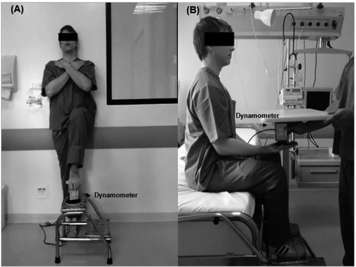
For the lower limbs (LL) in a straight position, we assessed the chain of the knee extensor muscles in the limb contralateral to the patient’s surgical incision (Figure 1a). The muscle group analyzed for upper limb (UL) strength was the chain of the elbow flexor contralateral to the arteriovenous fistula, which is
the dominant member in the absence of the fistula (Figure 1b). Normal predicted values of UL and LL strength were calculated using the values proposed by Bohannon.16
Respiratory muscle strength was assessed using a manovacuometer (GlobalMed) connected to a nozzle with an orifice diameter of 2 mm to prevent the facial muscles contributing by generating pressure. The pa-tients remained seated during the assessment. Three measurements were made, each starting from func-tional residual capacity for MIP and total lung capac-ity for MEP. The highest of the three measurements were recorded.
The values were expressed as absolute values and percentages, predicted according to Neder et al.18 The
vital capacity was measured using a Wright’s spirom-eter (Ferraris Mark 8) connected to a mouthpiece. A nose clip was used to prevent air escaping through the nose. Patients were asked to perform a maximal deep inspiration and then exhale completely.19 At
all-time points (preoperative, the first postoperative day, and discharge from hospital) measurements were per-formed in the following order: VC, MIP, and MEP.
STATISTICALANALYSIS
Sample size was calculated based on the functional parameters for the 6MWT, waiting a minimum dif-ference of 75 ± 100 meters20 between pre- and
post-intervention with a power of 80%. An alpha of 5% was determined and a sample of 28 patients per group were used. The interest variables were submitted to Kolmogorov-Sminov (K-S) normality test, and un-less specified, the data were presented as mean and standard deviation. Was used the chi-square test to analyze the differences between groups of categorical variables.
To test the difference hypothesis of intra and inter-group variables (MIP, MEP, CV), we used a ANOVA repeated measures considering the factor group (CG e IG) and time (preoperative, 1st PO, discharge). The
R
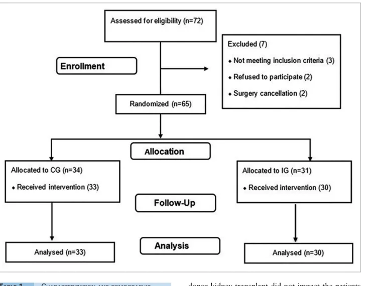
ESULTSSeventy-two patients were recruited and 63 were included in the study, divided into a CG (n = 33) and IG (n = 30). Seven patients were excluded for the reasons presented in Figure 2. The preoperative and surgical characteristics of the patients are shown in Table 1. There was no dif-ference in age, dialysis time, length of stay and clinical characteristics between the IG and the CG (p > 0.05).
Although there was a significant reduction (p < 0.001) in both groups compared to the 6MWT dis-tance at discharge, no difference was found in rela-tion to Δ6MWT between IG and CG (p = 0:29; Table 2). In the preoperative period, both groups presented similar predicted values (p > 0.05) for upper and lower limb strength (UL = CG: 97.8 ± 44.1% vs. IG: 116.1 ± 44.2%, and LL = CG: 102.1 ± 31.1% vs. IG: 122.5 ± 56.9%), without significant difference at hospital discharge (Table 2).
As expected, our results showed a significant reduc-tion in the 1st PO respiratory muscle strength in both the
CG (MIP = 9.0%, MEP = 34.9%) and the IG (MIP = 11.6%, MEP = 26.2%). While in hospital discharge, these values have not taken to the preoperative period, the IG showed a lower reduction of MIP (0.4% vs. 3.1%) and MEP (11.3% vs. 21.8%) relative to the CG (Table 3).
Both groups also showed reduction of VC on the first postoperative day (CG: 27.2% and IG: 20.5%; p < 0.001), with increase at hospital discharge, but no difference compared to preoperative values. However, IG showed values near the baseline (Table 3). The re-peated measures ANOVA showed significant differ-ence (p < 0.001) between the groups only for MIP and MEP. Both groups showed a significant reduction in serum creatinine at discharge, 2.1 (1.3) and 2.7 (1.1) mg/dL respectively for CG and IG but no statistical differences between groups.
D
ISCUSSIONTABLE 1 CHARACTERIZATIONANDDEMOGRAPHIC
FACTORSOFCONTROLANDINTERVENTION
GROUPS
CG (n = 33) IG (n = 30) p-value
Male, n (%) 23 (69.7) 17 (56.7) 0.28* Age (years),
mean (SD) 35.6 (10.4) 37.0 (9.2) 0.56** Hypertension,
n (%) 32.0 (97.0) 26.0 (87.0) 0.34* Diabetes, n (%) 3.0 (1.0) 0.0 (0.0) 0.42* Serum Cr (mg/
dL), mean (SD) 9.6 (1.9) 9.1 (1.7) 0.84** Hemoglobin
(mg/dL), mean (SD)
12.4 (2.7) 11.9 (2.2) 0.79**
Dialysis (months), mean (SD)
18.7 (17.8) 13.8 (13.1) 0.72**
Length of stay (days), mean (SD)
7.1 (3.5) 6.7 (2.2) 0.64**
CG, control group; IG, intervention group; SD, standard deviation; Cr, creatinine. *Chi-Square test **Independent samples t-test.
donor kidney transplant did not impact the patients functional walking capacity and peripheral muscle strength when compared to standard care. However, patients in the IG had better respiratory muscle strength when comparing with CG. To the best of our knowledge, only one study evaluated the effect of exercise training in the immediate postoperative pe-riod of kidney transplant patients, however, focused on biochemical markers of graft function.21
We hypothesized that the intervention used in the IG would be at least 20% more effective than simple orien-tations in improving 6MWT distance. However, this hy-pothesis has not been confirmed. In a recent systematic review, Heiwe and Jacobson14 showed evidence that an
exercise program after transplantation has potential ben-efits for physical activity and walking capacity. However, in our study, we observed that functional walking capaci-ty was similar between the group performing a supervised exercise program and the group receiving standard care.
including peripheral muscles14,22 our study
popula-tion was younger and presented a lower durapopula-tion of the disease and dialysis period compared to previous studies. Our patients had normal predicted values for UL and LL17 right before surgery, which
corrob-orates this hypothesis. On the other hand, Petersen et al.23 demonstrated abnormal skeletal muscle
func-tion and an increase in muscle fatigability in hemodi-alysis patients with no other difference after kidney transplantation.
In our study, an exercise protocol commencing im-mediately after renal transplantation did not increase the distance walked in the 6MWT or peripheral mus-cle strength. It is well known that the capacity to per-form physical activity, such as walking, may be lim-ited by the muscular, cardiac or pulmonary systems.23
However, our patients have good clinical conditions without significant reductions in peripheral muscle strength and functional walking capacity in the pre-operative period, which might explain our findings.
Corroborating our findings, Heiwe and Jacobson14
showed that there is no evidence that an exercise pro-tocol after transplantation promotes improvement in peripheral muscle strength. In relation to the influ-ence of cardiac limitations on walking distance, it was not our aim to evaluate cardiac parameters. However, cardiovascular disease remains the major cause of mortality in kidney recipients24-26 and the majority
of our study participants had arterial hypertension. Moreover, the duration of the intervention session and the number of sessions could be insufficient to promote measurable muscle strength differences with the outcome measures utilized. However, there is no consensus about these concerns and our exercise pro-gram was based on our institution’s physiotherapy routine.
We did observe a reduction in the distance walked at discharge compared to the preoperative period in both groups and this could be related to the effects induced by the surgery. Based on previous studies, the
TABLE 2 MUSCULARSTRENGTHANDFUNCTIONALCAPACITYOFPATIENTSBEFOREANDAFTERTRANSPLANTATIONOF
CONTROLGROUPANDINTERVENTIONGROUP
CG (n = 33) IG (n = 30)
Preoperative Discharge p-value Preoperative Discharge p-value
UL strength (N) 23.7 (8.7) 21.4 (9.9) 0.13* 25.2 (8.3) 22.2 (5.8) 0.06* LL strength (N) 52.5 (18.0) 51.3 (16.7) 0.67* 53.2 (16.0) 50.8 (13.0) 0.44* 6MWD (m) 584.9 (99.2) 502.4 (100.9) < 0.001* 598.7 (72.2) 537.6 (83.7) < 0.001* ∆6MWT (m) -82.5 (78.9) -61.1 (81.8) 0.29**
%predicted 6MWD 86.9 (12.6) 75.0 (15.6) < 0.001* 88.7 (11.9) 79.5 (11.1) < 0.001* ∆%predicted 6MWT -11.9 (11.3) -9.2 (12.0) 0.36**
Data are expressed as mean (standard deviation). CG, control group; IG, intervention group; UL, upper limbs; LL, lower limbs; N, newtons; 6MWD, six-minute walking distance; m, meters. *Paired t-test between preoperative and discharge. **Independent samples t-test between CG and IG.
TABLE 3 RESPIRATORYMUSCLESTRENGTHANDVITALCAPACITYOFPATIENTSBEFOREANDAFTERTRANSPLANTATIONOF
CONTROLGROUPANDINTERVENTIONGROUP
CG (n = 33) IG (n = 30) p-values*
Preoperative 1st PO Discharge Preoperative 1st PO Discharge Time Group Interaction
MIP (cmH2O)
78.6 (27.4)
69.6 (27.8)
75.2 (27.2)
89.2 (26.5)
76.6 (27.8)
88.2
(23.1) 0.03
a,c < 0.001 0.38
%predicted MIP
67.1 (21.2)
58.1 (22.6)
64.0 (20.4)
80.4 (23.0)
68.8 (21.7)
80.0
(20.7) < 0.001
a,c < 0.01 0.56
MEP (cmH2O)
102.3 (19.1)
61.7 (23.3)
75.2 (26.3)
107.2 (17.4)
77.0 (22.2)
102.0
(21.3) < 0.001
a,b,c < 0.001 < 0.01
%predicted MEP
84.4 (19.3)
49.5 (20.8)
62.6 (23.9)
94.7 (23.3)
68.5 (26.7)
83.4
(24.1) < 0.001
a,b,c < 0.001 < 0.01
VC (L/min) 3.3 (1.1)
2.4 (1.0)
3.0 (1.0)
3.4 (1.1)
2.7 (1.0)
3.4
(1.0) < 0.001
a,c 0.35 0.19
majority of kidney transplant recipients do not prac-tice any type of exercise up to 12 months before and after transplant.24,27 The fact that our results show
that patients from both groups had a lower functional capacity at discharge could be a predictor variable to maintain physical inactivity after transplantation.
However, we believe that the training group will become more physically active after discharge com-pared to the control group, since they were submitted to systematic and supervised physiotherapy immedi-ately after surgery. Recently, Greenwood et al.,26 in a
12-week pilot randomized controlled trial, concluded that both aerobic training and resistance training in-terventions appear to be feasible and clinically ben-eficial in kidney transplant recipients. Based on this, we can imagine that an average of about seven days of training, as in our study, is not sufficient to achieve statistically significant differences. However, the aim of our study was to evaluate the effect of exercise pro-gram just during hospitalization.
Several studies have shown that compared to up-per abdominal surgery lower abdominal surgery is less likely to promote a reduction in pulmonary vol-umes and respiratory muscle strength during the post-operative period.28 Our data are supported by results
obtained from the Literature,29,30 however, we also
observed that the exercise protocol induced a lower reduction in respiratory muscle strength and VC com-pared to the control group.
Grams et al.,31 in a recent meta-analysis assessing
the effects of breathing exercises on the recovery of pulmonary function, showed that a significant im-provement of maximal respiratory pressure can oc-cur in patients who perform breathing exercises af-ter upper abdominal surgery. Although the findings of Grams et al.31 relate to upper abdominal surgery,
it is well known that patients who undergo abdomi-nal surgery, whether upper or lower, usually develop a restrictive lung pattern. Therefore, we believe that breathing exercises probably increase diaphragm mo-bility and improve respiratory muscle synergism.32
In addition, the concept of the breathing exercise has changed over the years and only recently includes active upper and lower limb exercises, supervised walking, and the use of steps. These latter changes in body position, as well as breathing exercises, optimize basal area ventilation.33 In the present study, the IG
performed upper and lower limb exercises combined
with breathing exercises and used training loads of 2 kg and 4 kg for female and male participants, re-spectively. However, there is no consensus regarding the weight of the training load, the number of series, and the frequency of the exercise protocol, each of which could have been insufficient and could have in-fluenced our results.
Our study has a few limitations. First, we did not evaluate clinical outcomes such presence of postoper-ative complications (pneumonia, etc.) or evaluation of the level of pain in the surgical incision. However, this topic was not one of the aims of our study. Second, these results cannot be generalized and extrapolated for other populations of kidney recipients (older pa-tients, deceased donors recipients) because our study was carried out in non-elderly patients with a more recent diagnosis with few co-morbidities and only in living donor recipients with few time on dialysis.
In addition, we could not be evaluated the impact of graft function in a sample only with live donor recipi-ents (very low probability of delayed graft function for instance). Finally, short intervention time may not have time enough for providing a positive outcome and we could not evaluate how much the unsupervised group exactly exercised, but as a matter of ethics, we could nev-er stop them to do exnev-ercise. Probably our results show that an early postoperative physiotherapy protocol after kidney transplantation does not promote an additional benefit when it comes to functional capacity and periph-eral muscle strength after renal transplantation because a short intervention time maybe not have time enough for providing a positive changes.
C
ONCLUSIONSA
CKNOWLEDGEMENTSWe are grateful for MECOR - ATS (Methods in Epidemiologic, Clinical and Operations Research) Program Director A. Sonia Buist M.D, MECOR Latin-American Program Director Ana Maria Menezes M.D.; and the MECOR Faculty, especially William Vollmer by all contribution in the manuscript.
R
EFERENCES1. Heiwe S, Clyne N, Dahlgren MA. Living with chronic renal failure: patients’ experiences of their physical and functional capacity. Physiother Res Int 2003;8:167-77. DOI: http://dx.doi. org/10.1002/pri.287
2. Faria Rde S, Fernandes N, Lovisi JC, Reboredo Mde M, Marta MS, Pinheiro Bdo V, et al. Pulmonary function and exercise tol-erance are related to disease severity in pre-dialytic patients with chronic kidney disease: a cross-sectional study. BMC Nephrol 2013;14:184. DOI: http://dx.doi.org/10.1186/1471-2369-14-184 3. Teplan V, Mahrova A, Piťha J, Racek J, Gürlich R, Teplan
V Jr, et al. Early exercise training after renal transplantation and asymmetric dimethylarginine: the effect of obesity. Kid-ney Blood Press Res 2014;39:289-98. DOI: http://dx.doi. org/10.1159/000355806
4. Surgit O, Ersoz G, Gursel Y, Ersoz S. Effects of exercise train-ing on specific immune parameters in transplant recipients. Transplant Proc 2001;33:3298. PMID: 11750411 DOI: http:// dx.doi.org/10.1016/S0041-1345(01)02400-9
5. Richard R, Verdier JC, Doutreleau S, Piquard F, Gény B, Rieu M. Exercise limitation in trained heart and kidney transplant re-cipients: central and peripheral limitations. J Heart Lung Trans-plant 2005;24:1774-80. DOI: http://dx.doi.org/10.1016/j. healun.2005.03.017
6. Didsbury M, McGee RG, Tong A, Craig JC, Chapman JR, Chadban S, et al. Exercise training in solid organ transplant recipients: a systematic review and meta-analysis. Transplan-tation 2013;95(5):679-87. DOI: http://dx.doi.org/10.1097/ TP.0b013e31827a3d3e
7. Hedenstierna G, Edmark L. The effects of anesthesia and muscle pa-ralysis on the respiratory system. Intensive Care Med 2005;31:1327-35. DOI: http://dx.doi.org/10.1007/s00134-005-2761-7
8. Qaseem A, Snow V, Fitterman N, Hornbake ER, Lawrence VA, Smetana GW, et al.; Clinical Efficacy Assessment Subcom-mittee of the American College of Physicians. Risk assessment for and strategies to reduce perioperative pulmonary compli-cations for patients undergoing noncardiothoracic surgery: a guideline from the American College of Physicians. Ann Intern Med 2006;144:575-80. PMID: 16618955 DOI: http://dx.doi. org/10.7326/0003-4819-144-8-200604180-00008
9. van den Ham EC, Kooman JP, Schols AM, Nieman FH, Does JD, Franssen FM, et al. Similarities in skeletal muscle strength and exercise capacity between renal transplant and hemodi-alysis patients. Am J Transplant 2005;5:1957-65. DOI: http:// dx.doi.org/10.1111/j.1600-6143.2005.00944.x
10. Kanat F, Golcuk A, Teke T, Golcuk M. Risk factors for postop-erative pulmonary complications in upper abdominal surgery. ANZ J Surg 2007;77:135-41. PMID: 17305986 DOI: http:// dx.doi.org/10.1111/j.1445-2197.2006.03993.x
11. Kovelis D, Pitta F, Probst VS, Peres CPA, Delfino VDA, Mo-celin AJ, et al. Função pulmonar e força muscular respiratória em pacientes com doença renal crônica submetidos à he-modiálise. J Bras Pneumol 2008;34:907-12. DOI: http://dx.doi. org/10.1590/S1806-37132008001100004
12. Adams GR, Vaziri ND. Skeletal muscle dysfunction in chronic renal failure: effects of exercise. Am J Physiol Renal Physiol 2006;290:F753-61. PMID: 16527920
13. Greenwood SA, Lindup H, Taylor K, Koufaki P, Rush R, Macdougall IC, et al. Evaluation of a pragmatic exercise reha-bilitation programme in chronic kidney disease. Nephrol Dial Transplant 2012;27:iii126-34. DOI: http://dx.doi.org/10.1093/ ndt/gfs272
14. Heiwe S, Jacobson SH. Exercise training for adults with chronic kidney disease. Cochrane Database Syst Rev 2011:CD003236. PMID: 21975737
15. ATS Committee on Proficiency Standards for Clinical Pulmo-nary Function Laboratories. ATS statement: guidelines for the six-minute walk test. Am J Respir Crit Care Med 2002;166:111-7. PMID: 12091180
16. Bohannon RW. Reference values for extremity muscle strength obtained by hand-held dynamometry from adults aged 20 to 79 years. Arch Phys Med Rehabil 1997;78:26-32. PMID: 9014953 DOI: http://dx.doi.org/10.1016/S0003-9993(97)90005-8 17. Rougier PR. Relative contribution of the pressure variations
under the feet and body weight distribution over both legs in the control of upright stance. J Biomech 2007;40:2477-82. PMID: 17196210 DOI: http://dx.doi.org/10.1016/j.jbio-mech.2006.11.003
18. Neder JA, Andreoni S, Lerario MC, Nery LE. Reference val-ues for lung function tests. II. Maximal respiratory pressures and voluntary ventilation. Braz J Med Biol Res 1999;32:719-27. PMID: 10412550 DOI: http://dx.doi.org/10.1590/S0100-879X1999000600007
19. Chiavegato L, Medina-Pestana J, Tedesco-Silva H, Paisani D, Fiore J Jr, Faresin S. Surgical approach does not affect peri-operative respiratory morbidity in living donor nephrectomy: comparison between anterior subcostal incision and flank in-cision. Transplant Proc 2010;42:1472-5. DOI: http://dx.doi. org/10.1016/j.transproceed.2009.12.064
20. Tomczak CR, Warburton DE, Riess KJ, Jendzjowsky NG, Esch BT, Liang Y, et al. Pulmonary oxygen uptake and heart rate kinetics during the six-minute walk test in transplant recipients. Transplantation 2008;85:29-35. PMID: 18192908 DOI: http:// dx.doi.org/10.1097/01.tp.0000296056.00863.f0
21. Juskowa J, Lewandowska M, Bartłomiejczyk I, Foroncewicz
B, Korabiewska I, Niewczas M, et al. Physical rehabilitation and risk of atherosclerosis after successful kidney transplanta-tion. Transplant Proc 2006;38:157-60. PMID: 16504691 DOI: http://dx.doi.org/10.1016/j.transproceed.2005.12.077 22. Carli F, Charlebois P, Stein B, Feldman L, Zavorsky G, Kim DJ,
et al. Randomized clinical trial of prehabilitation in colorectal surgery. Br J Surg 2010;97:1187-97. PMID: 20602503 DOI: http://dx.doi.org/10.1002/bjs.7102
23. Petersen AC, Leikis MJ, McMahon LP, Kent AB, Murphy KT, Gong X, et al. Impaired exercise performance and muscle Na(+),K(+)-pump activity in renal transplantation and haemo-dialysis patients. Nephrol Dial Transplant 2012;27:2036-43. DOI: http://dx.doi.org/10.1093/ndt/gfr586
24. Salzman SH. The 6-min walk test: clinical and research role, technique, coding, and reimbursement. Chest 2009;135:1345-52. DOI: http://dx.doi.org/10.1378/chest.07-1682
25. Rosas SE, Reese PP, Huan Y, Doria C, Cochetti PT, Doyle A. Pretransplant physical activity predicts all-cause mortality in kidney transplant recipients. Am J Nephrol 2012;35:17-23. DOI: http://dx.doi.org/10.1159/000334732
28. Gordon EJ, Prohaska TR, Gallant MP, Sehgal AR, Strogatz D, et al. Longitudinal analysis of physical activity, fluid intake, and graft function among kidney transplant recipients. Transpl Int 2009;22:990-8. DOI: http://dx.doi.org/10.1111/j.1432-2277.2009.00917.x
29. Dureuil B, Cantineau JP, Desmonts JM. Effects of upper or lower abdominal surgery on diaphragmatic function. Br J An-aesth 1987;59:1230-5. PMID: 2960367 DOI: http://dx.doi. org/10.1093/bja/59.10.1230
30. Lunardi AC, Paisani Dde M, Tanaka C, Carvalho CR. Im-pact of laparoscopic surgery on thoracoabdominal mechan-ics and inspiratory muscular activity. Respir Physiol Neuro-biol 2013;186:40-4. PMID: 23313854 DOI: http://dx.doi. org/10.1016/j.resp.2012.12.012
31. Grams ST, Ono LM, Noronha MA, Schivinski CI, Paulin E. Breathing exercises in upper abdominal surgery: a systematic review and meta-analysis. Rev Bras Fisioter 2012;16:345-53. DOI: http://dx.doi.org/10.1590/S1413-35552012005000052 32. Barbalho-Moulim MC, Miguel GP, Forti EM, Campos Fdo A,
Costa D. Effects of preoperative inspiratory muscle training in obese women undergoing open bariatric surgery: respira-tory muscle strength, lung volumes, and diaphragmatic excur-sion. Clinics (São Paulo) 2011;66:1721-7. DOI: http://dx.doi. org/10.1590/S1807-59322011001000009