Ivo H. Saraiva
Dissertation presented to obtain the Ph.D degree in Biochemistry
Instituto de Tecnologia Química e Biológica | Universidade Nova de Lisboa
Oeiras, December, 2012
Structural and functional characterization of
the gene products responsible for
phototrophic iron oxidation by purple bacteria
From left to right: Professor Miguel Teixeira, Professor Carlos Geraldes, Dr. Marta Bruix, Ivo Saraiva,
Professor Paula Tamagnini, Dr. Catarina Paquete and Dr. Ricardo Louro.
December 3rd 2012.
President of the Juri:
Professor Miguel Teixeira.
Main opponents:
Dr. Marta Bruix
Dr. Paula Tamagnini
Thesis committee:
Dr. Ricardo Louro
Professor Dianne Newman
Professor Carlos Geraldes
Acknowledgements
i
Acknowledgements
I would like to thank
Doctor Ricardo O. Louro for the friendship, supervision, the guidance, the advices, for all that he taught me and for accepting me in his lab, where I found a fun and relaxed work environment always prone to scientific debate.
Professor Dianne K. Newman for kindly receiving me in her home and in her lab, where I had the privilege of working with a team of scientific and human excellence, and where I found the perfect work environment that always made
me feel at home. I thank in particular Itzel Ramos-Solis and Lars Dietrich for the support in the lab and Lina Bird for her collaboration on the study of PioC.
Professor Carlos Salgueiro with whom I enjoyed a successful collaboration and who has kindly provided necessary support in the study of PioC.
Doctor Catarina Paquete for the support and useful discussions on the kinetics of FoxE.
Doctor Carlos Frazão and Luís Pereira for the X-ray crystallography studies of FoxE.
Doctor Manolis Matzapetakis for the friendship and for support and key
Acknowledgements
ii
Professor Miguel Teixeira for obtaining the EPR spectra of FoxE and for subsequent useful discussion on the data.
Professor Carlos Geraldes for accompanying my work during these years and for the continuous support.
My lab colleagues who´s merry nature always made it hard to do some actual work in the lab. I thank in particular Bruno Fonseca for the friendship and Eduardo Calçada for the friendship and for the contributions in the study of
PioC.
Agradeço em especial à minha irmã e aos meus pais que iniciaram a formação necessária a escrever esta tese e agradeço à Mafalda que a concluiu. A eles dedico esta tese.
Abstract
iii
Abstract
Iron is an essential element in life. It is used in a variety of different processes
in the energetic metabolism of different organisms. Among these bioenergetic processes is photoferrotrophy, characterized by the utilization of Fe(II) as the sole electron source for photosynthesis. The metabolic activity of photoferrotrophs is proposed to have had a relevant role in the formation of
ancient geological structures consisting of Fe(III) minerals, such as the Banded Iron Formations.
Photoferrotrophy was only recently discovered (in the 1990s) and lacks a detailed molecular description. The objective of this work was to characterize photoferrotrophy at a molecular level, in order to understand how
photoferrotrophs transfer electrons from Fe(II) to the photosynthetic reaction center, while avoiding Fe(III) precipitation inside cells.
The first target of this work was the bacterium Rhodobacter ferrooxidans SW2.
In this organism, the three-gene fox operon was proposed to be responsible
for Fe(II) oxidation. The first gene, foxE, encodes a diheme c-type cytochrome, which is predicted to oxidize Fe(II), performing the first step in photoferrotrophy. Using NMR, EPR and UV-visible spectroscopies, together with electrochemistry techniques, the reduction potentials of the hemes of FoxE were determined, providing a thermodynamic characterization.
Stopped-flow experiments, monitored by UV-visible spectroscopy, allowed for the study of the kinetics of Fe(II) oxidation by FoxE. Finally, X-ray crystallography was used to determine the structure of this protein. All together, the data show that, in vitro, FoxE forms a homo-trimer in solution that is thermodynamically
and kinetically capable of oxidizing Fe(II). This reaction is pH-dependent in a
Abstract
iv
The second focus of this work was on the bacterium Rhodopseudomonas palustris TIE-1. In this organism, the three-gene pio operon was shown to be responsible for photoferrotrophy. The third gene of this operon, pioC, encodes
a High Potential Iron-Sulfur Protein (HiPIP), predicted to transfer electrons to the cyclic photosynthesis apparatus. Cyclic voltammetry was used to
determine the reduction potential of PioC. NMR and EPR spectroscopic characterizations were performed and NMR was used to determine the solution structure of this HiPIP. The reduction potential is in agreement with the predicted function of PioC, and the solution structure contains conserved
features observed in all HiPIP structures determined so far.
Resumo
v
Resumo
O ferro é um elemento essencial à vida. É usado de várias maneiras diferentes
no metabolismo energético de diferentes organismos. A fotoferrotrofia é uma destas estratégias bioenergéticas e caracteriza-se pela utilização de Fe(II) como fonte exclusiva de electrões para a fotossíntese. Foi proposto que a actividade de organismos fotoferrotróficos teve um papel relevante na formação de
estruturas geológicas constituídas por minerais de Fe(III), como as Formações de Ferro em Banda.
Tendo a fotoferrotrofia sido recentemente descoberta, na década de 1990, uma descrição molecular detalhada ainda não foi efectuada. O objectivo deste trabalho é obter esta descrição de modo a compreender como os organismos
fotoferrotróficos transferem electrões do Fe(II) até ao centro reactivo fotossintético e, ao mesmo tempo, evitam a precipitação de Fe(III) nas células. O primeiro alvo deste trabalho foi a bactéria Rhodobacter ferrooxidans SW2.
Nesta bactéria o operão com três genes fox foi proposto ser responsável pela
oxidação fotossintética de Fe(II). O primeiro gene, foxE, codifica um citocromo di-hémico tipo c, que se prevê oxidar Fe(II), sendo responsável pelo primeiro passo no metabolismo fotoferrotrófico. Conjugando espectroscopias de RMN, RPE e UV-visível com técnicas de electroquímica, os potenciais de redução dos hemos do FoxE foram determinados, para uma caracterização termodinâmica.
A conjugação das técnicas de fluxo-interrompido e espectroscopia de UV-visível permitiu estudar a cinética de oxidação de Fe(II) pelo FoxE e cristalografia de raios-X foi usada na determinação da estrutura desta proteína. Os dados adquiridos indicam que, in vitro, o FoxE forma um homo-trímero em
solução que é termodinâmica e cineticamente apto para oxidar Fe(II). Esta
Resumo
vi
solubilidade do Fe(III). Esta correlação pode ser relevante na prevenção da precipitação de Fe(III) nas células.
O segundo alvo deste trabalho foi a bactéria Rhodopseudomonas palustris
TIE-1. Neste organismo, foi demonstrado que o operão com três genes pio é
responsável pela fotoferrotrofia. O terceiro gene deste operão, pioC, codifica
uma Proteína Ferro-Enxofre de Alto Potencial (HiPIP), que se prevê que transfere electrões para o sistema de fotossíntese cíclica. O método de Voltametria Cíclica foi usado para determinar o potential de redução do PioC. Foi feita uma caracterização espectrocópica por RMN e RPE e a estrutura do
PioC em solução foi determinada por RMN. O potencial de redução está de acordo com a função prevista e a estrutura mantém todas as características conservadas nas estruturas de HiPIPs determinadas até agora.
Os dados obtidos levaram à proposta de mecanismos de oxidação de Fe(II) pelas bactérias estudadas. Estes envolvem uma cadeia de transferência
Table of contents
vii
Table of contents
Acknowledgements ... i
Abstract ... iii
Resumo ... v
Table of contents ... vii
List of figures ... ix
List of tables ... xi
1 Introduction ... 3
1.1 Iron redox cycle ... 4
1.1.1 Microbial iron reduction ... 7
1.1.2 Microbial iron oxidation ... 8
1.1.2.1 Aerobic iron oxidation ... 8
1.1.2.2 Anaerobic iron oxidation ... 11
1.2 Photoferrotrophy: shining light on iron oxidation ... 12
1.2.1 Photoferrotrophs ... 13
1.2.1.1 Rhodobacter ferrooxidans SW2 ... 13
1.2.1.2 Rhodomicrobium vannielii BS-1 ... 14
1.2.1.3 Chlorobium ferrooxidans KoFox ... 14
1.2.1.4 Rhodovulum iodosum N1 and Rhodovulum robiginosum N2... 14
1.2.1.5 Thiodictyon strain F4 ... 15
1.2.1.6 Rhodopseudomonas palustris TIE-1 ... 15
1.2.2 Photoferrotrophic genes... 16
1.3 Multi-heme c-type cytochromes ... 20
1.3.1 Functional mechanism ... 21
1.4 High potential iron-sulfur proteins (HiPIP) ... 23
1.4.1 Biological function ... 23
Table of contents
viii
1.5 Biotechnological applications ... 24
1.5.1 Hydrogen production ... 24
1.5.2 Bioremediation ... 25
1.5.3 Microbial fuel cells ... 26
2 The FoxE iron oxidoreductase of Rhodobacter ferrooxidans SW2 ... 31
2.1 Introduction ... 31
2.2 Experimental procedures ... 32
2.3 Results ... 37
2.4 Discussion ... 45
3 PioC: a photoferrotrophic HiPIP... 57
3.1 Introduction ... 57
3.2 Experimental procedures ... 57
3.3 Results ... 60
3.4 Discussion ... 66
4 Concluding remarks ... 75
4.1 Photoferrotrophy in Rhodobacter ferrooxidans SW2 ... 75
4.2 Photoferrotrophy in Rhodopseudomonas palustris TIE-1 ... 77
4.3 Comments on biotechnological applications ... 81
Appendix A: Expression and production of c-type cytochromes ... 85
Appendix B: Paramagnetic NMR spectroscopy ... 87
Appendix C: Bioelectrochemistry ... 90
Appendix D: Matlab routine ... 92
Appendix E: PioC NMR signals assignment... 95
Appendix F: CCPNMR CFS definition ... 104
Table of contents
ix
List of figures
Figure 1.1: redox cycling of iron at different pH and O2 concentrations. ... 6
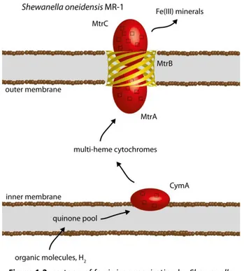
Figure 1.2: cartoon of ferric iron respiration by Shewanella oneidensis MR-1. ... 7
Figure 1.3: cartoon of ferrous iron oxidation by Acidithiobacillus ferrooxidans 23270T. 9 Figure 1.4: cartoon of ferrous iron oxidation by Sideroxidans lithotrophicus ES-1. ... 11
Figure 1.5: scanning electron microscopy micrograph of SW2. ... 13
Figure 1.6: transmission electron microscopy micrograph of TIE-1. ... 15
Figure 1.7: genes involved in photoferrotrophy in Rhodopseudomonas palustris TIE-1 and Rhodobacter ferrooxidans SW2. ... 16
Figure 1.8: predicted location of the products of the pio operon based on sequence analysis. ... 17
Figure 1.9: predicted location of the products of the fox operon based on sequence analysis. ... 18
Figure 1.10: schematic representation of an MFC. ... 28
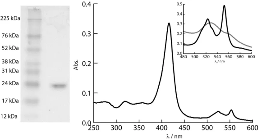
Figure 2.1: SDS-PAGE and UV-visible spectrum of FoxE. ... 37
Figure 2.2: X-ray crystal structure model of FoxE. ... 39
Figure 2.3: X-band EPR spectrum of fully oxidized FoxE. ... 40
Figure 2.4: 1D 1H-NMR spectra of FoxE poised at different degrees of oxidation. ... 40
Figure 2.5: 1D 1H-NMR spectra of FoxE fully re-oxidized with K3Fe(CN)6. ... 42
Figure 2.6: redox titrations of FoxE at pH 6 and pH 7 at 25°C.... 43
Figure 2.7: kinetic traces of FoxE reduction with Fe(II)-EDTA at pH 6 and pH 7 at 25°C. ... 44
Figure 2.8: electron flow diagram in the periplasm of SW2. ... 47
Figure 2.9: schematic representation of reduction of each FoxE monomer. ... 49
Figure 3.1: X-Band EPR spectrum of oxidized purified PioC. ... 61
Figure 3.2: 1H-NMR spectrum of PioC at 25 °C.... 62
Figure 3.3: cyclic voltammogram of PioC. ... 63
Figure 3.4: superposition of the backbone of the 20 best solutions of the CYANA calculation of the structure of PioC. ... 64
Table of contents
x
Figure 3.6: distance restraints used in the calculation of the structural model of PioC. 65
Figure 3.7: organization of the membrane structures of R. palustris. ... 66
Figure 3.8: superposition of HiPIP structures deposited in PBD. ... 68
Figure 3.9: superposition of the backbone of the 20 best solutions of the CYANA calculation of the structure of PioC with the additional restraints of Table 3.2. .. 69
Figure 3.10: sequence alignment of HiPIPs with structures deposited in PDB. ... 69
Figure 3.11: rmsd values of the models present in Figure 3.9. ... 70
Figure 3.12: electron flow diagram in the periplasm of TIE-1. ... 72
Figure 4.1: proposed mechanism of photoferrotrophy in SW2. ... 76
Figure 4.2: proposed mechanism of photoferrotrophy in TIE-1. ... 78
Table of contents
xi
List of tables
Table 2.1: Speciation and reduction potential of FeEDTA. ... 35
Table 2.2: reduction potentials of the two hemes of FoxE and kinetic parameters for the reduction of FoxE determined at pH 6 and pH7 at 25°C. ... 44
Table 3.1: statistics of the CYANA calculations with NOEs and with NOE plus the restraints in Table 3.2 ... 71
Table 3.2: restraints determined from the conserved structural features of HiPIPs. ... 71
1
Introduction
3
1
Introduction
Since life began on Earth, it has changed and shaped the planet’s geochemistry. In every niche where thermodynamics is favorable and kinetics allows, evolution has given life strategies to thrive. The understanding of the geochemical evolution of the planet is tightly linked to the evolution of life and of its metabolic strategies. Evidence shows that major geochemical events, like
the Great Oxidation Event (1) or the origin of Banded Iron Formations (BIF) (2), were triggered by specific microbial metabolisms. In order to answer the questions on how and when these events happened, it is necessary to know the biochemistry of the microorganisms associated with them. This is a goal that can only be achieved by studying the present microbial communities and
their metabolic strategies that might answer these questions. Ultimately, the correlation of evolution of life and of the geochemistry of the planet should allow the association of particular geochemical events with the appearance of particular metabolic pathways, their respective genes and proteins, and allow
understanding how specific metabolic strategies shape the environment (3). Of the many strategies living organisms use to obtain energy, this thesis focuses on photoferrotrophy, i.e. the utilization of ferrous iron oxidation as an electron source for photosynthesis (4, 5). This photosynthetic iron oxidation is described by the following reaction:
4Fe2+ + CO2 + 11H2O + h → [CH2O] + 4Fe(OH)3 + 8H+
This particular metabolic strategy is a good illustration of how life and Earth have co-evolved since the origin of Life on the planet. First, photoferrotrophy is
Introduction
4
probable cause of BIFs, stratified geological structures that compose the largest iron ore deposits in the world (2). This work focuses on the biochemical and functional characterization of proteins involved in photoferrotrophy, namely the products of genes recently discovered to be involved in this metabolism in the Gram-negative bacteria Rhodopseudomonas palustris TIE-1
and Rhodobacter ferrooxidans SW2. The detailed biochemical characterization of this metabolism will help understand its role in the biogeochemistry of iron (Figure 1.1).
1.1 Iron redox cycle
Iron is the most abundant element on Earth and the fourth most abundant on
the Earth’s crust. It belongs to the fourth period and eighth group in the
periodic table and has a [Ar] 3d6 4s2 electron configuration. The most common oxidation states in the biosphere are +2 and +3 with electronic configurations d6 (ferrous) and d5 (ferric), respectively. Depending on environmental conditions (ligand field, pH, redox conditions) iron can be readily convertible
between these two states.
The reduction potential of the Fe(II)/Fe(III) pair is conditioned by the complexing agents in solution. Non-complexed soluble ferric and ferrous iron ions only exist at low pH with a reduction potential of 770 mV. At
circumneutral pH, iron forms complexes with other species in solution, which results in a decrease of the reduction potential to values between 100 mV and 400 mV, depending on the complexing agents. These can be phosphates, carbonates or organic acids present in the environment. The nature of these
Introduction
5 With O2 having a reduction potential of 870 mV at pH 7 and of 1.12 V at pH 2 the oxidation of iron by O2 (autooxidation) is always thermodynamically favorable. The rate of this reaction is given by the following equation (7):
[ ]
[ ][ ]
[ ] [ ][ ] 1-1
According to this equation the rate of autooxidation decreases two orders of magnitude for each unit of pH decrease, indicating that this reaction is slow in
acidic environments.
Given iron’s availability and chemical properties, it is not surprising that Life,
throughout evolution, has incorporated this metal in its many bioenergetic strategies. Microorganisms are able to use the forward and backward
Introduction
6
Figure 1.1: redox cycling of iron at different pH and O2 concentrations.
Introduction
7 1.1.1 Microbial iron reduction
By the time respiratory metabolisms evolved on Earth, the variability of
electron acceptors was limited (9, 10). One available electron acceptor was Fe(III) that resulted from abiotic photochemical oxidation of abundant Fe(II).
This, together with the widespread of iron respiration in today’s
microorganisms, either in Bacteria or in Archea, suggests that Fe(III) respiration was one of the first respiratory metabolisms to evolve (11). Fe(III) is reduced
mostly in anaerobic conditions either by strict anaerobes, like members of the genus Geobacter, or by facultative anaerobes, like members of the genus Shewanella. Both of which are Gram-negative bacteria. At circumneutral pH,
the available Fe(III) can be in dissolved chelated form or in solid ferric minerals. The mentioned genera are able to use solid ferric minerals as terminal electron acceptors (12, 13), for which there is the need to transfer electrons to the
Introduction
8
extracellular space. Depending on the species, electrons from oxidation of different organic compounds (acetate, lactate, palmitate, aromatic compounds) or H2 in the cytoplasm are transferred through the periplasm, to terminal iron reductases in the outer membrane. This transfer is performed by a complex chain of inner membrane, periplasmatic and outer membrane
multi-heme cytochromes (Figure 1.2) (14, 15). The reduction of ferrous minerals may lead to their solubilization and mobilization of iron.
1.1.2 Microbial iron oxidation
1.1.2.1 Aerobic iron oxidation
Low pH slows down autooxidation (equation 1-1) allowing the competition of
microorganisms for Fe(II). Many acidophiles oxidize iron using O2 as terminal electron acceptor (16, 17). The most studied organism performing this metabolism is the bacterium Acidithiobacillus ferrooxidans (18), a chemolithoautotroph belonging to the -proteobacteria. This microorganism is
associated with acidified environments rich in iron and sulfur ores (19). Different strains of A. ferrooxidans seem to have different mechanisms of iron
oxidation (20). For the type strain, A. ferrooxidans ATCC 23270T, the proteins involved are coded by the rus operon (Figure 1.3) (21–23). Iron is oxidized
extracellularly by the outer membrane c-type cytochrome Cyc2. The electrons
are then transferred to the periplasmatic blue copper protein rusticyanin,
where the path bifurcates. The electrons can follow an electrochemical downhill path through the cytochrome c4 Cyc1 to the inner membrane aa3 -type terminal oxidase that couples the reduction of O2 to the translocation of H+ from the cytoplasm to the periplasm. This path helps keep the proton
Introduction
9
bc1 complex, the quinone pool and finally to the NADH dehydrogenase NADH1 ending in the reduction of NAD+. This uphill path is driven forward by using the transmembrane proton gradient. A. ferrooxidans uses iron as source of energy
and of reducing power.
Other identified acidophiles oxidizing iron (24) belong to the genera
Acidithiobacillus (25), Thiobacillus (26) and Acidiferrobacter (27), which are all -proteobacteria, and to the genus Ferrovum (24), which belongs to the -proteobacteria.
Recently, there has been increasing interest in the research of aerobic iron oxidizers that grow at circumneutral pH (17). In the presence of O2, Fe(II) is
rapidly autooxidized (equation 1-1). As such, microorganisms can only compete with autooxidation of iron in micro-aerobic Fe(II) rich conditions. These are found in habitats where Fe(II) rich anoxic water comes in contact with O2, like
Figure 1.3: cartoon of ferrous iron oxidation by
Introduction
10
fresh water iron seeps (28–32), marine hydrothermal vents (33–35) or the rhizosphere of plants that grow in soils saturated with anoxic water (36–38). Because of these specific environmental conditions, the laboratory study of aerobic iron oxidizers at circumneutral pH requires techniques that mimic oxic-anoxic interfaces (39). So far, all characterized strains belong to the
proteobacteria. The most common bacteria found in the described fresh water
habitats belong to the -proteobacteria genera Gallionella, Leptothrix or
Syderoxydans. The bacterium Gallionella ferruginea is the most studied. It was first described in 1836 (40). The cells from this bacterium form stalks that are proposed to position the cell in the right Fe(II)/O2 gradient and to provide a
nucleation site for Fe(III) precipitation, thus preventing cell encrustation (17). Recently, the genome of the bacterium Syderoxydans lithotrophicus ES-1 was sequenced. Analysis of the genome and search for homologous of genes from iron oxidizers and reducers that are known to be involved in iron metabolism led to the proposal of an iron oxidation electron chain coded by a three gene
operon (41). The first two genes, mtoA and mtoB, code for a periplasmatic deca-heme cytochrome and its associated outer-membrane -barrel, respectively. These cytochromes are known to be able to transfer electrons between the periplasm and the extracellular space through the pore of the
associated -barrel (42). The third gene cymA, codes for a homologue of the
inner-membrane associated tetra-heme cytochrome CymA, from the iron reducer Shewanella oneidensis MR-1. In the proposed iron oxidation mechanism MtoB allows access of MtoA to the extracellular space to receive electrons from ferrous iron, thus preventing ferric iron precipitation inside the
Introduction
11 In marine environments, the best known neutrophilic aerobic iron oxidizer is the bacterium Mariprofundus ferrooxydans. This species and closely related clones from deep-sea environmental samples form the new proposed class
-proteobacteria (43). Based on the whole genome analysis of the type strain
Mariprofundus ferrooxydans PV-1T and analysis of protein expression patterns,
a group of genes was suggested to be involved in iron oxidation (44). Among these are a molydopterin and 4Fe-4S cluster binding protein, a c-type cytochrome and two terminal oxidases, type ccb3 and type bd. Experimental evidence supporting this hypothesis is still lacking.
1.1.2.2 Anaerobic iron oxidation
The reduction potentials of complexed iron species at pH 7 are below 400 mV. These values allow the utilization of electron acceptors with reduction potentials lower than oxygen (870 mV). The majority of anaerobic iron
Introduction
12
oxidizers can be divided into photosynthetic (with the potential of the reaction center near 450 mV) and nitrate-reducing (with the potential of the NO3-/NO2 -at 430 mV). The utiliz-ation of nitr-ate as final electron acceptor in iron oxid-ation was first detected in 1996 (45). Microorganisms performing this metabolism are phylogenetically spread in Bacteria and Archea (8). Several strains have
been isolated from fresh-water, brackish-water or marine environments (45–
49). Sustained growth of iron-oxidizing nitrate reducers is proposed to only occur in the presence of additional organic substrates, like acetate (50). This mixotrophic metabolism, even though present only in a small fraction of
nitrate reducers, is widespread among this group of organisms and contributes significantly to iron oxidation in their habitats (50).
Photosynthetic bacteria that use oxidation of iron as electron source for photosynthesis form a second group of organisms that oxidize iron in anaerobic circumneutral environment. This metabolism is the focus of this
thesis and is described in more detail.
1.2 Photoferrotrophy: shining light on iron oxidation
The oxidation of ferrous iron by anoxygenic photosynthetic bacteria was first proposed a few decades ago (51), as an hypothesis for the origin of Banded Iron Formations (BIF). These geological structures form the largest geological
iron deposits in the planet but their origin is still debated. The activity of oxygenic photosynthetic cyanobacteria was proposed first to be the main cause that led to this iron oxidation and deposition. In the beginning of the 1990s photoferrotrophy was detected and photoferrotrophs were first isolated
Introduction
13 Several photoferrotrophs have been isolated in the last twenty years. However, the identification of the molecular mechanisms of this iron oxidation was still stalled, probably due to the lack of genetically tractable organisms. This obstacle has recently been overcome twice (52, 53), opening the window for the biochemical study of photoferrotrophy.
1.2.1 Photoferrotrophs
Until today seven photoferrotrophs from different sources have been cultured and characterized. They are all Gram-negative bacteria that appear to have different strategies to oxidize iron.
1.2.1.1 Rhodobacter ferrooxidans SW2
Rhodobacter ferrooxidans SW2 (SW2) is among the first isolated organisms
capable of using electrons from iron oxidation to feed photosynthesis (5), and it is one of the most studied. It was isolated from fresh water sediments, belongs to the -proteobacteria and is a purple non-sulfur bacterium. It is able
to grow photoheterotrophically on a variety of substrates (low molecular weight carboxylates, saccharides and amino acids). In the dark it can grow on oxygen.
The SW2 operon foxEYZ was identified as possibly responsible for
Introduction
14
photoferrotrophy, as it was shown to enhance phototrophic iron oxidation in the related bacterium Rhodobactercapsulatus SB1003 (53).
1.2.1.2 Rhodomicrobium vannielii BS-1
BS-1 was also isolated from fresh water sediments and is an -proteobacterium (54). Iron oxidation by BS-1 is enhanced by the addition of organic co-substrates like acetate, butyrate, malate, succinate and pyruvate.
This organism, upon oxidizing Fe2+, becomes encrusted in Fe3+ minerals that inhibit further growth. The authors state that phototrophic iron oxidation has little physiological importance in BS-1 metabolism.
1.2.1.3 Chlorobium ferrooxidans KoFox
KoFox was also isolated from fresh water sediments, but, unlike the previous organisms, it is a green sulfur bacterium belonging to the Chlorobia (55). Iron
oxidation is accelerated by additional substrates either organic (pyruvate, fumarate, acetate and cysteine) or inorganic (H2, S2O3-). KoFox grows in co-culture with the bacterium Geospirillum strain KoFum. KoFum does not oxidize
iron but it becomes incrusted in ferric iron precipitates from the activity of KoFox, suggesting that photoferrotrophs have specific properties to avoid
incrustation.
1.2.1.4 Rhodovulum iodosum N1and Rhodovulum robiginosum N2
These two bacteria are the only marine photoferrotrophs isolated so far (56). They are also -proteobacteria and have a versatile metabolism being able to
grow photoheterotrophically with a variety of small organic molecules and also
Introduction
15 1.2.1.5 Thiodictyon strain F4
F4 belongs to the -proteobacteria and was isolated from a fresh water marsh.
It is a purple sulfur bacterium (57). This organism has shown the highest iron
oxidation rates so far (58).
1.2.1.6 Rhodopseudomonas palustris TIE-1
TIE-1 is also a purple non-sulfur bacterium from the -proteobacteria, but is
phylogenetically apart from strains SW2, N1 and N2 (Figure 1.6) (59). It was isolated from fresh water sediments. This bacterium has a very versatile metabolism being able to grow photoauto-, photohetero-, chemohetero- and chemoautotrophycally. This organism has the particularity of being genetically
tractable. This allowed the identification of the pioABC operon, as required for photoferrotrophy (52).
Introduction
16
1.2.2 Photoferrotrophic genes
Despite the biological and geological importance of photoferrotrophy, genes
responsible for this metabolism were identified in two organisms only recently. In Rhodopseudomonas palustris TIE-1, which is genetically tractable, a three
gene operon was identified by analysis of protein expression patterns in cultures grown with or without Fe2+ as electron source (52) (Figure 1.7). In Fe2+ oxidizing conditions a c-type cytochrome was detected to be differently
expressed. The identification of this protein by mass spectroscopy and subsequent sequence analysis led to the identification of the pio operon. The
first gene, pioA, codes for a 540 amino acid protein with ten canonical c-type
heme binding sites (CXXCH). It has a Sec system peptide signal for co-translational translocation to the periplasm, as is required for the maturation of c-type cytochromes (60). By studying the effect on iron oxidation of
individual deletions of the pio genes, PioA was predicted to be the iron
oxidase. In BLAST runs of PioA the first half of the sequence hits only proteins
from other strains of the R. palustris species and has no predicted function. The second half of the PioA sequence, which contains the ten heme binding sites, falls into the deca-heme c-type cytochrome family. One member of this
family, MtrA, is involved in ferric iron reduction by Shewanella oneidensis
MR-Figure 1.7: genes involved in photoferrotrophy in
Rhodopseudomonas palustris TIE-1 and Rhodobacter ferrooxidans SW2.
Introduction
17 1. This suggests a common evolutionary path between iron oxidation and iron reduction (61). The second gene of the pio operon, pioB, codes for an 811 amino acid protein also with a Sec system peptide signal. A BLAST run of PioB
places it in a deca-heme associated outer-membrane -barrel family (Figure 1.8). To this family belongs the beta-barrel MtrB, which is coded in the same
operon of MtrA in Shewanella oneidensis MR-1. It was shown that these
-barrels allow the transfer of electrons from their associated periplasmatic deca-heme cytochromes, through the outer membrane, to electron acceptors on the outside of the cells (42). This interaction allows the reduction of extracellular solid minerals. In the case of MtrAB, this association ultimately
allows the reduction of ferric minerals with electrons gathered from different donors in the cytoplasm. Again, these data suggest a common evolutionary path for iron oxidation and reduction. The third gene, pioC, codes for a High
Figure 1.8: predicted location of the products of the pio
operon based on sequence analysis.
PioA has the two domains represented, with the deca-heme cytochrome domain interacting with PioB in the outer membrane as it happens with this family of proteins. All the
Introduction
18
Potential Iron-Sulfur protein (HiPIP). These proteins are commonly seen in photosynthetic electron transfer pathways. PioC has a Tat system peptide signal for post-folding translocation to the periplasm, as is common in iron-sulfur proteins. This protein has similarity with several other annotated HiPIPs, including a HiPIP from the acidophilic iron oxidizer Acidithiobacillus ferrooxidans, which shows a precedence of the involvement of HiPIPs in iron oxidation.
Because SW2 is not genetically tractable, a different strategy was used to identify the genes involved in photoferrotrophy (53). A cosmid library of SW2
genome was expressed in Rhodobacter capsulatus SB1003. This bacterium is related to SW2 but is not capable of growing photoautotrophically exclusively on Fe2+. The cosmids that led to enhanced iron oxidation by SB1003 allowed the identification of the fox operon as the probable responsible for
photoferrotrophy in SW2 (Figure 1.7 and Figure 1.9). The first coded gene,
foxE, is a di-heme c-type cytochrome with 291 amino acids. Expressing this
Figure 1.9: predicted location of the products of the fox
Introduction
19 genealone in SB1003 enhances iron oxidation. Therefore, a c-type cytochrome was again predicted to be the iron oxidase. A BLAST run of FoxE has no significant hits, indicating a new member of the diverse cytochrome family. The second gene foxY, codes for a 362 amino acid protein with a Sec system
peptide signal. A BLAST run suggests that this protein binds a pyrroloquinoline
quinone (PQQ) redox cofactor. As such, FoxY may be involved in electron transfer between FoxE and the other components of the cyclic photosynthetic electron transfer chain. The last gene, foxZ, codes for a 301 amino acid protein
with a Sec system signal. Comparison with the databases suggests that this
protein is a cytoplasmatic membrane protein with unknown transport function.
In order to try to predict genes that may be involved in photoferrotrophy in the other organisms described here, the protein sequences of the pio and fox
operons were blasted against the available genomes. Only the genomes of the
bacteria Chlorobium ferrooxidans KoFox and of the type strain of the
Rhodomicrobium vannielii species, which is also capable of iron oxidation, are available. KoFox has no significant homologues of the Fox or Pio proteins.
Rhodomicrobium vannielii however, has three consecutive genes (Rvan_3213,
Rvan_3214 and Rvan_3215) that have significant homology to the pioABC
operon and encode a deca-heme cytochrome, its associated outer membrane -barrel and a hypothetical periplasmatic HiPIP, respectively. The products of
these three genes might be able to oxidize iron and transfer electrons to the reaction center as predicted for TIE-1. However, the iron oxidation activity in
Introduction
20
1.3 Multi-heme c-type cytochromes
The proteins predicted to be iron oxidizers in TIE-1 and SW2, PioA and FoxE
respectively, are multi-heme c-type cytochromes. Cytochromes are electron transfer proteins containing a heme cofactor. This cofactor is composed of an iron atom coordinated by a porphyrin ring. Cytochromes can be classified as a, b, c or d according to the substituents of the porphyrin ring (62). c-type
cytochromes are the only in which the porphyrin ring is covalently bound to
the polypeptide chain. These connections are thioether bonds between substituents 3 and 8 and the cysteine residues of the binding motif CXXCH, in which the histidine is the proximal ligand to the iron. The distal ligand is found
in an unpredictable region of the polypeptide and, in most cases, is another histidine or a methionine, or a less common ligand like lysine, tyrosine, cysteine, asparagine or the N-terminal. Several factors determine the reduction potential of a heme. Depending on protein fold, nature of the axial ligands or the geometry of the cofactor, c-type hemes have a reduction
potential between -400 and 450 mV (63).
The number of hemes present in a c-type cytochrome varies considerably. The protein with the highest number of hemes per peptide to have its structure solved has sixteen c-type hemes (64) and the available genomic sequences
show that cytochromes can have up to 58 heme binding motifs (65). Some of
these numbers are more common like two, three and four hemes. Frequently, the folding of these smaller cytochromes is repeated in tandem in larger cytochromes, probably due to gene duplication and fusion.
Multi heme c-type cytochromes are present in almost all groups of Bacteria
Introduction
21
Shewanella provide the ability to use many different electrons acceptors (66–
69).
The covalent binding of several hemes to their apo-protein is a complex process that requires several enzymes in sequential steps. The enzymatic machinery used by the organisms studied in this thesis is the product of the
ccm operon (60). This operon codes up to nine membrane and periplasmatic proteins that, in a stereo specific way, attach the hemes to their binding sites after the polypeptide is translocated to the periplasm by the Sec pathway. As a consequence, no cytoplasmatic c-type cytochromes have been found.
In order to fully understand the functional mechanisms of multi heme cytochromes, a detailed characterization of the role of each individual heme is required. This means determining the heme intrinsic reduction potentials and electron transfer rate constants and understanding how the several cofactors interact. This detailed characterization has been performed for several
cytochromes (70–83).
1.3.1 Functional mechanism
With each heme having 2 possible redox states, a multi-heme cytochrome having n hemes has 2n possible ways of electron distribution, each way
defining a redox microstate. These microstates can be thermodynamically described by considering the individual reduction potentials of its reduced
hemes. However, due to the close packing of the redox centers, there are electrostatic interactions between hemes. This results in anti-cooperativity in heme reduction due to electron repulsion, and these interactions need to be considered in the functional characterization of a multi-heme cytochrome (84).
Introduction
22
therefore cooperative. The heme is not only a redox co-factor but also has two propionate groups that are important in the redox-Bohr effect (76). Taken together the n hemes and m acid-base centers, there are 2n+m possible
microstates. Each of these can be thermodynamically defined by a set of n
reduction potentials, m pKa values, corresponding heme-heme interactions and redox-Bohr effects (85). Through the characterization of several multi-heme cytochromes, these parameters were shown to be defined in order to originate only a few thermodynamically favorable microstates (84). That is in the activity of a multi-heme cytochrome, instead of 2n+m possible microstates, only a few
are significantly populated in physiologically significant conditions (76). This gives specificity to the cytochrome by allowing only the population of microstates that have a physiological and functional meaning. This is illustrated by the c7 tri-heme cytochrome family. These cytochromes having similar structures have different functional roles, which are dictated by the different
sets of favorable microstates (71, 72, 86). Another example is the cytochrome
c3 family. Due to the redox-Bohr effect, only specific microstates are populated that result in energy transduction by converting weakly acidic protons into more acidic protons through coupling with electron transfer (87).
Another important consequence of the close packing of the hemes in
multi-heme cytochromes is that it allows for a fast intramolecular electron transfer. This means that, upon the entry or exit of an electron, there is rapid equilibrium between the possible different microstates (88). Therefore, whatever heme receives or donates an electron, only the thermodynamically favorable microstates remain significantly populated. The functional
consequences of this rapid equilibrium are illustrated by the small tetra-heme cytochromes (STC) from the bacteria Shewanella oneidensis and Shewanella frigidimarina (82). In these cytochromes electrons enter the protein only
Introduction
23
frigidimarina. However, the rapid equilibrium in the distribution of electrons in the cytochrome keeps the microstates, in which these hemes are reduced, scarcely populated. This allows the proteins to be ready to receive more electrons until full reduction.
1.4 High potential iron-sulfur proteins (HiPIP)
The third gene present in the pio operon is a HiPIP. HiPIPs together with ferredoxins form a group of proteins that contain a 4Fe-4S cluster arranged as a cubane and coordinated by four cysteine residues. Under physiological conditions this cluster in HiPIPs changes between +2 and +3 oxidation states (89) and its reduction potential ranges from 50 to 500 mV (90). In Ferredoxins
it changes between oxidation states +1 and +2 and has an average reduction potential of -400 mV (89). What defines the accessible oxidation states of these two protein families is still debated, but exposure to the solvent and hydrogen-bond network are important aspects (91). The 4Fe-4S clusters are assembled in the cytoplasm of bacteria and the holoprotein is then
translocated folded by the Tat translocation system to the periplasm where it functions.
1.4.1 Biological function
HiPIPs are single electron transfer proteins. Having a high reduction potential HiPIPs are primarily found in purple photosynthetic bacteria where they
function as electron mediators between the cytochrome bc1 and the reaction center tetra-heme cytochrome in cyclic photosynthesis (92, 93). A few exceptions are the case of Rhodothermus marinus (94, 95) or of Acidithiobacillus ferrooxidans (96, 97) where the HiPIPs are involved in
Introduction
24
1.4.2 HiPIP structure
HiPIPs are small proteins without highly conserved sequences. According to the
Protein data Bank the structure of HiPIPs of eight different organisms has been solved, either by X-ray diffraction or NMR spectroscopy (Allochromatium vinosum, Thermochromatium tepidum, Halorhodospira halophila, Rhodocyclus
tenuis, Marichromatium purpuratum, Rhodoferax fermentans,
Ectothiorhodospira shaposhnikovii and Rhodothermus marinus). All these are photosynthetic Proteobacteria with the exception of Rhodothermus marinus, which belongs to the Bacteroidetes group. Even though the sequences of HiPIPs are not highly conserved, all the structures have a common folding
around the cluster. The 4Fe-4S cluster is secluded from the solvent and surrounded by aromatic residues that contribute to its stabilization (98, 99). Another common feature is an alfa-helix near the N-terminal. Away from the cluster, HiPIPs present loops of variable length originated by non-conserved insertions.
1.5 Biotechnological applications
Besides the described biological and geological relevance of the study of photoferrotrophy, studying this metabolism will ultimately lead to biotechnological relevant information. The organisms under study have versatile metabolisms with potential to be used in different biotechnological
applications.
1.5.1 Hydrogen production
Given the current world population growth and the evidence for climate change, finding new sustainable and clean energy sources is a priority. Hydrogen is a viable alternative fuel as it causes no CO2 emissions and has a
Introduction
25 from fossil fuels, and as demand for H2 increases (100) sustainable production mechanisms are required. Several sustainable approaches are currently researched (101), including biological photosynthetic production (102).
In phototrophs H2 is produced by hydrogenases and nitrogenases, which are O2 sensitive enzymes. In oxygenic phototrophs this creates the requirement for
approaches that separate photosynthesis and H2 production either spatially or temporally (103, 104). Anoxygenic phototrophs, like the ones under study here, have the advantage of not having to deal with oxygen. In these organisms, H2 is produced mostly by nitrogenases. These enzymes catalyze the
reduction of N2 and have H2 as an obligate product, being able to produce only H2 in the absence of N2. In the genomes of the organisms under study two different nitrogenases are coded: Fe-Fe and Mo-Fe nitrogenases. Several different strains of R. palustris have been used in H2 production (105–107), as well as different species of the genus Rhodobacter (108–110). This makes TIE-1
and SW2 good candidates for H2 production, in particular TIE-1 as it is genetically tractable.
1.5.2 Bioremediation
Bioremediation is the utilization of an organism’s metabolism for the removal or treatment of pollutants. The bacteria under study in this work have a
versatile metabolic capacity, being able to feed on a variety of organic and inorganic substrates. In their genomes they code for several enzymes involved in the degradation of aromatic pollutants (111, 112) and also several cytochromes P450 (113) and S-transferases (114), enzymes involved in
Introduction
26
The iron precipitation process also has potential applications. The precipitation of ferric iron promotes co-precipitation of other molecules and ions in solution. This has been suggested as strategy to clean contaminated waters (24).
1.5.3 Microbial fuel cells
Microbial fuel cells (MFC) are a rising technological field with clean energy
production potential (115) combined with the capacity to perform bioremediation. An MFC is a bioelectrochemical device that uses the metabolism of bacteria to produce electrical current (Figure 1.10). Bacteria gather energy by transferring electrons from low potential substrates to high potential ones. In a MFC an electrical circuit is placed in this electron flow in
order to harness electrical energy. The bacteria in a MFC feed on different substrates, preferably organic or inorganic wastes, and use an anode as an electron acceptor. The electrons then flow through the electrical circuit to a cathode and are ultimately transferred to a terminal electron acceptor.
Many advances have been made in this technology, and several issues are
research targets for optimization. Although MFCs have a theoretical potential that depends on the standard potentials of the substrates and final electron acceptors being utilized, these values are never reached. These potential losses are due to ohmic losses and overpotentials, which depend on the design of the
cell and on the choice of final electron acceptor and microbial culture at the anode. Different cultures, either axenic or mixed, can be used at the anode (116). Depending on the working conditions of the cell, different species thrive at the anode. Important factors in this adaptation are inoculum source, the fed
substrates or the anode potential (117).
Introduction
27 Some species use small organic electron shuttles (118–120). These molecules are reduced by the cells, then diffuse to the anode to deliver the electrons and are then reused. Another strategy is the production of electron conducting nanowires that connect the different cells in a biofilm and the electrode (121, 122). A third mechanism is the direct electron transfer from the cells to the
electrode through outer-membrane bound electron transfer proteins, a mechanism that requires a close interaction between the cells and the electrode (42, 123). A better understanding of these electron transfer mechanisms will allow an optimization of this step in the electron flow, thus
limiting potential losses.
Another aspect is the choice of terminal electron accepter at the cathode. The most used acceptor is O2. Oxygen has several advantages: it has a high reduction potential, it is easily available and it is free. It has the disadvantages of having slow reduction kinetics, creating the need for expensive catalysts,
like platinum, and of being able to diffuse into the anode chamber and inhibit bacterial activity. Another commonly used acceptor is ferricyanide. This molecule has low overpotential and has allowed a higher power density then oxygen (115). However, it needs to be regularly replaced, as it is insufficiently reoxidized by oxygen. It is also possible to reduce protons at the cathode
producing H2 (124). So far the potentials in MFC do not allow the reaction without an increase in potential through an external source, but production of H2 is a promising option in MFCs (125, 126). Another approach is the utilization of a second bacterial culture that receives the electrons at the cathode (127). Biocathodes where Mn (128) or Fe (129) are oxidized have been studied. In this
last approach the acidophilic iron oxidizer Acidithiobacillus ferrooxidans was utilized with the electrons ultimately transferred to O2.
Introduction
28
photosynthetic these organisms would have the advantage of using CO2 as final electron acceptor, thus creating an MFC that should be CO2 neutral. The understanding of molecular mechanisms of photoferrotrophy should allow determining if this metabolic capacity can be used in this biotechnological application and to determine how a prototype could be optimized.
Figure 1.10: schematic representation of an MFC.
Bacteria at the anode chamber feed on organic or inorganic wastes and transfer electrons to the anode through: a) electron shuttles (ES), b) nanowires or c) directly through outer membrane cytochromes. The protons produced flow through the selectively permeable membrane (dashed line) to the cathode chamber and the electrons flow through an electrical circuit to the cathode. The electrons are then transferred to the final electron acceptor. This can be d) abiotic or e)
2
The FoxE iron oxidoreductase
of Rhodobacter ferrooxidans
SW2
The work presented in this chapter was published in:
Ivo H. Saraiva, Dianne K. Newman, Ricardo O. Louro. 2012. Functional
characterization of the FoxE iron oxidoreductase from the photoferrotroph Rhodobacter ferrooxidans SW2. J Biol Chem.
doi:10.1074/jbc.M112.360636
Ivo H. Saraiva designed and performed all the experiments and
participated in the writing of the manuscript
Luís Pereira, Ivo H. Saraiva, Ricardo Coelho, Dianne K. Newman, Ricardo O.
Louro, Carlos Frazão. 2012. Crystallization and preliminary crystallographic studies on FoxE from Rhodobacter ferrooxidans SW2, a Fe(II)
oxidoreductase involved in photoferrotrophy. Acta Crystalogr F. In press.
Ivo H. Saraiva performed the purification of FoxE.
The FoxE iron oxidoreductase of Rhodobacter ferrooxidans SW2
31
2
The FoxE iron oxidoreductase of
Rhodobacter
ferrooxidans
SW2
2.1 Introduction
Despite the importance of photoferrotrophy for understanding the co-evolution of life and Earth, little is known about its molecular mechanisms.
Rhodobacter ferrooxidans SW2 has been a subject of studies aiming to
understand biologically induced mineralization because ferric (hydr)oxides
precipitate away from the cell and no mineral is found inside the cells or encrusting the cell surface (130–132). It is also known that SW2 utilizes only soluble Fe(II) compounds as the electron source for photoferrotrophy and does not actively dissolve Fe(II) minerals (132), suggesting that iron oxidation does not occur at the cell surface. Recently, the operon foxEYZ from SW2 was found
to stimulate light-dependent Fe(II) oxidation by the bacterium Rhodobacter capsulatus SB1003 (53). This operon is composed of three genes: foxE, that encodes a 29 kDa protein containing two canonical heme c binding sites, foxY,
that encodes a predicted pyrroloquinoline quinone binding protein, and foxZ,
that encodes a predicted inner membrane transport protein. FoxE was
predicted to be the iron oxidoreductase and to perform the main step in photoferrotrophy because its expression alone is enough to significantly enhance light-dependent Fe(II) oxidation activity on SB1003 (53). This c-type
cytochrome has no significant sequence homology with other predicted
The FoxE iron oxidoreductase of Rhodobacter ferrooxidans SW2
32
for FoxE that is in agreement with the requirement of soluble Fe(II) for the photoferrotrophic metabolism of SW2 (132). However, this cellular location implies the occurrence of mechanisms to avoid iron precipitation in the periplasm. Detailed characterization of the iron oxidoreductase FoxE is crucial for the understanding of this photosynthetic metabolism.
To investigate the first step of photoferrotrophy in SW2, detailed functional and structural characterization of FoxE was performed.
2.2 Experimental procedures
Cloning
The foxE gene was amplified by PCR from the plasmid pfoxE (53) and cloned
without its periplasmic signal sequence (predicted with SignalP) into the
NcoI/EcoRI sites of the pET20b(+) plasmid, which contains the periplasmic signal sequence pelB from E. coli. The fragment pelB:foxE was amplified by PCR and cloned into the BglII/EcoRI restriction site of plasmid pUX19 (133). The resulting plasmid was used to transform E. coli JCB7123 cells containing the pEC86 plasmid (134), which codes for the c-type cytochrome maturation (ccm)
operon that allows E. coli to produce cytochromes in aerobic conditions. All constructs were confirmed by DNA sequencing.
Expression and growth
Cells were grown in Lysogeny Broth (LB) supplemented with 50 g.mL-1 kanamycin and 34 g.mL-1 chloramphenicol to maintain selective pressure for
the pUX19 and pEC86 plasmids respectively. Frozen stocks of E. coli JCB7123 containing both plasmids were used to inoculate 200 mL of LB. The culture was grown for 24 h at 30° C and stirred at 140 rpm. The resulting cell culture was used for a 1% inoculum of four 5 L conical flasks with 3.5 L of LB each and the
The FoxE iron oxidoreductase of Rhodobacter ferrooxidans SW2
33 mM EDTA, 100 mg.L-1 lysozyme, pH 8) and incubated in ice for 30 min with gentle stirring. The resulting spheroplasts were pelleted and the supernatant, containing the periplasm, was cleared by ultracentrifugation at 138000 g for 30 min and then frozen at -80° C.
Purification
The periplasmic fractions of four growth batches were pooled, concentrated in an ultrafiltration cell with a 30 kDa cut-off membrane and dialyzed twice against 20 mM TrisHCl pH 7.6 for 12 h. The resulting fraction was loaded into a DEAE chromatography column equilibrated with 20 mM TrisHCl pH 7.6. A step
gradient with 20 mM TrisHCl, 1 M NaCl, pH 7.6 was used to elute the bound proteins and FoxE was eluted at 150 mM NaCl. The presence of the cytochrome was determined by UV-visible spectroscopy and SDS-PAGE stained to detect hemes (135). Fractions containing FoxE were pooled, concentrated and washed with 5 mM potassium phosphate buffer at pH 7 in an
ultrafiltration cell with a 30 kDa cut-off membrane. The resulting fraction was loaded into a hydroxylapatite chromatography column equilibrated with 5 mM potassium phosphate buffer at pH 7. Bound proteins were eluted with a step gradient and FoxE was eluted at 50 mM potassium phosphate buffer at pH 7. The pure protein yield was 1.5 mg per liter of culture medium. Both
chromatography steps were performed in a GE Äktaprime Plus purification system.
The binding of two heme groups to the FoxE polypeptide was confirmed by mass spectrometry (MALDI-TOF) analysis.
Dynamic light scattering
Dynamic light scattering (DLS) experiments were performed in a Malvern Instruments Zetasizer Nano at 25 °C. FoxE samples were prepared in 50 mM potassium phosphate buffer at pH 7. The molecular weight of the FoxE particle
The FoxE iron oxidoreductase of Rhodobacter ferrooxidans SW2
34
UV-visible spectroscopy
UV-visible spectra were recorded in a Shimadzu UV-1700 spectrophotometer at room temperature (~25°C).
EPR spectroscopy
EPR spectroscopy was performed in a Bruker EMX spectrometer equipped with
an ESR-900 continuous-flow helium cryostat. The spectrum was recorded
under the following conditions: microwave frequency, 9.391 GHz; microwave power, 2 mW; modulation frequency, 100 kHz; modulation amplitude, 1 mT; temperature, 13 K. FoxE was prepared in 50 mM potassium phosphate pH 7
buffer and oxidized with potassium hexachloroiridate(IV).
NMR spectroscopy
1H-NMR spectroscopy was performed in a 500 MHz Bruker Avance II+
spectrometer at 25 °C. FoxE samples in 50 mM potassium phosphate buffer pH 7 were lyophilized twice and solubilized in D2O before spectra acquisition. The
residual water signal was suppressed by pre-saturation. The sample was reduced with small amounts of sodium dithionite or sodium ascorbate and oxidized with potassium ferricyanide. A 2D NOESY spectrum of a 4.9 mM FoxE sample was obtained with 8192x144 points, 512 scans, a 45kHz sweep width and a mixing time of 25 ms. Longer mixing times caused the loss of cross peaks.
A superWEFT 180° pulse was used to suppress the water signal and reduce the intensity of sharp diamagnetic signals (136).
Redox titration
Redox titrations were performed at 25 °C inside an anaerobic glove box (Coy Laboratory Products, Inc.) in an OTTLE cell using a potentiostat (CH
The FoxE iron oxidoreductase of Rhodobacter ferrooxidans SW2
35 (SHE) by adding 241 mV to the imposed potentials. The FoxE concentration was 20 M prepared in 50 mM phosphate buffer with 300 mM KCl. A mixture at 40 M of each of the following mediators was used: potassium ferricyanide,
n,n-dimethyl-p-phenyldiamine, p-benzoquinone, 1,2-naphtoquinone-4-sulphonic acid, 1,2-naphtoquinone, trimethyl hydroquinone, phenazine methosulphate and 1,4-naphtoquinone. Different mediator concentrations
were used to check for their possible interference with FoxE. No effects in the reduction potentials were detected. Data were collected in reductive and oxidative directions and were reproducible within 10 mV.
Stopped-flow
Stopped-flow experiments were performed in a KinetAsyst SF-61DX2 Hitech Scientific apparatus placed inside an anaerobic glove box (MBraun 150-GI). FoxE at 1 M (after mixing) in 50 mM phosphate buffer with 300 mM KCl was reduced with Fe(II)-EDTA at different concentrations (150, 100 and 50 M after
mixing). Reduction of FoxE was followed at the Soret band (417 nm) and the reduction fractions were calculated by normalization using the fully reduced
and fully oxidized absorbance values. Fe(II)-EDTA was prepared by mixing FeSO4 and NaEDTA in a 1:1.5 ratio inside the anaerobic glove box. Speciation in solution was calculated using the PHREEQC Interactive 2.18.5314 software with the minteq.v4.dat database (137). Fe(II)-EDTA was always the dominant
species in solution (Table 2.1).
Table 2.1: Speciation and reduction potential of FeEDTA.
Values were determined with the minteq.v4.dat database for the experimental conditions used.
% of Fe(EDTA)2- Em of FeEDTA
pH 6 99% 130 mV
The FoxE iron oxidoreductase of Rhodobacter ferrooxidans SW2
36
Kinetic analysis
The kinetics of reduction of FoxE was analyzed considering a collisional model, in which the association and dissociation of the electron donor and the protein are in fast equlibrium. Therefore:
[ ( ) ][ ] 2-1
Experiments were performed in pseudo-first order conditions. A linear dependence of the pseudo-first order rate constants with Fe(II)-EDTA concentration allowed the determination of second order rate constants.
According to Marcus theory the rate constants depend on the driving force, on the reorganization energy and on structural factors. To separate the structural factors from the contribution of the driving force, the reduction rate constants (Ki) of each heme i were described by the following equation:
[ ( ) ] 2-2
where ki is the intrinsic rate constant that accounts for the structural factors of the ith heme, E0i is the potential of reduction of the i
th heme by the electron
donor , F is the Faraday constant, R is the gas constant, T is the temperature
and is the reorganization energy, taken as 1 eV (88). The reorganization energy and the structural factors were considered constant during reduction. The kinetics of the reduction of FoxE were analyzed considering the two hemes
independent and therefore described by the sum of two exponentials. The model was implemented in Matlab and fit to the experimental data using the Nelder-Mead simplex algorithm (138). Standard errors of the fit parameters were obtained from the diagonal elements of the covariance matrix (139).
X-ray crystal structure
The FoxE iron oxidoreductase of Rhodobacter ferrooxidans SW2
37
2.3 Results
The purity of FoxE was assessed by SDS-PAGE (Figure 2.1). The UV-spectrum of
FoxE as purified has maxima at 553, 523, 416, 319 and 274 nm. The cytochrome was considered pure with a ratio Abs416/274~5. The presence of the
peaks at 553 and 524 nm, the and bands respectively, indicate partial reduction of the cytochrome during purification, even though all procedures
were performed in aerobic conditions. Full reduction with sodium ascorbate originates peaks at 553 nm for the band, 523 nm for the band and 417 nm
for the Soret band. In the fully oxidized state, obtained by adding potassium
ferricyanide, the Soret band shifts to 411 nm and the and bands become broad, typical of the low spin hexacoordinated hemes.
The degree of homogeneity of the sample was determined by DLS. The results
indicate that the sample is monodispersive with a diameter of 8.3 nm. Assuming a globular shape, this translates into an approximate molecular weight of 99.4±10.1 kDa. This is approximately three times the molecular
Figure 2.1: SDS-PAGE and UV-visible spectrum of FoxE.
The gel shows a single band from FoxE. The spectrum is from FoxE as purified and the inset
The FoxE iron oxidoreductase of Rhodobacter ferrooxidans SW2
38
weight deduced from the annotated sequence plus two hemes (29 kDa). The preliminary structural model of FoxE was determined by X-ray crystallography. For the crystallization conditions used FoxE forms a homotrimer complex (Figure 2.2A). Each monomer has two domains, where each domain binds a c-type heme (Figure 2.2B). The hemes were numbered
according to the order by which their respective binding site appears in the
amino acid sequence. The secondary structure is composed of -helices and loops. Monomer contacts are performed mostly through the Heme I domain
plus two N-terminal -helices that form an arm embracing the neighboring monomer. Even though the two domains have little sequence homology, they
present the same structural folding (Figure 2.2C). The Heme II domain is sequentially inserted in the Heme I domain, and the two hemes have similar coordination, with the distal ligands of Heme I and II being M230 and M116, respectively. The hemes have a similar binding cavity, with the exception of the
two N-terminal -helices that form part of the binding cavity of the Heme I
from the neighboring monomer. The distance between redox cofactors is an important factor controlling electron transfer. The intra monomeric heme-heme distance is 18 Å, with the shortest inter monomeric heme-heme distance being 25 Å between Hemes I. Additionally, the Heme I domain shows one intra monomeric disulfide bond between C80 and C258, at 16 Å from Heme I and 32
The FoxE iron oxidoreductase of Rhodobacter ferrooxidans SW2
39
Figure 2.2: X-ray crystal structure model of FoxE.
A) Stereo view of the FoxE trimer showing the three monomers in different colors, with the hemes in stick representation. B) Stereo view of the FoxE monomer with the Heme I domain in