1413-8670/© 2012 Elsevier Editora Ltda. All rights reserved.
www.elsevier.com/locate/bjid
The Brazilian Journal of
INFECTIOUS DISEASES
Original Article
Prevalence and risk factors for cervical intraepithelial
neoplasia among HIV-infected women
Nara Chartuni Pereira Teixeira
a, Angela Cristina Labanca Araújo
a,
Christine Miranda Correa
b, Claudia Teixeira da Costa Lodi
c, Maria Inês Miranda Lima
a,
Nara de Oliveira Carvalho
d, Dora Mendez del Castillo
d, Victor Hugo Melo
e*
aDepartment of Health Services, Belo Horizonte, MG, Brazil bDepartment of Health Services, Três Rios, MG, Brazil
cFaculdade de Ciências Médicas de Minas Gerais, Belo Horizonte, MG, Brazil
dCenter for Research and Diagnosis (NUPAD), School of Medicine, Universidade Federal de Minas Gerais, Belo Horizonte, MG, Brazil eSchool of Medicine, Universidade Federal de Minas Gerais, Belo Horizonte, MG, Brazil
A R T I C L E I N F O
Article history:
Received 7 October 2011 Accepted 11 December 2011
Keywords: HIV
DNA probes, HPV
Cervical intraepithelial neoplasia Immunosuppression
*Corresponding author at: School of Medicine, Universidade Federal de Minas Gerais, Av. Alfredo Balena 190/2nd floor, Belo Horizonte, MG , 30130-100, Brazil
E-mail address: victormelo@terra.com.br (Victor Hugo Melo) A B S T R A C T
Objectives: To evaluate the prevalence and the risk factors for cervical intraepithelial neoplasia (CIN) among HIV-infected women.
Methods: Cross-sectional study of 494 HIV-infected women in Brazil, between 1998 and 2008. Gynecologic exam was performed, and samples were collected for cervical cytology and for HPV DNA detection. Cervical biopsy was carried out when indicated. HPV infection, CD4 T-lymphocyte count and HIV viral load were compared with cervical histopathology. Univariate and multivariate statistical analyses were performed to evaluate the statistical association of several risk factors.
Results: CIN prevalence detected by histopathology was 23.4% (6% of CIN2/3 and 17.4% cases of CIN1). Multivariate analysis confirmed an independent association of CIN with CD4 T-lymphocyte count below 200 cells/mm3 (OR 5.0, 95% CI 2.5-10.1), with a positive detection
of HPV DNA (OR 2.0, 95% CI 1.2-3.5), and with age ≤ 34 years old (OR 1.5, 95% CI 1.0-2.4). HIV viral load and antiretroviral use were not independent risk factors for CIN.
Conclusions: Severity of immunosupression, presence of HPV infection and younger age are strong predictors of CIN among HIV-infected women.
© 2012 Elsevier Editora Ltda. All rights reserved.
Introduction
HIV infection has become a burden on the female population. In developing countries during the past decade, the HIV/AIDS pandemic has overloaded the health care system
with an enormous impact on women, particularly those of reproductive age.1 In Brazil, although men still account for the
(NaCl, 0.09%); samples were sent to the laboratory within 24 hours. HPV DNA method of extraction was previously described.12 The globin gene was amplified in all samples,
in order to control for DNA quality. The samples in which the globin gene was not amplified were excluded from the study. HPV detection by PCR was carried out in a nested-PCR system, using the primers MY09/11 and GP5+/6+. DNA was amplified with specific primers for HPV types 6, 11, 16, 18, 31, 33 and 35, in independent reactions. Sequencing reaction of nested-PCR product was used when HPV types were not identified by the former primers. The nested-PCR product was purified following the precipitation protocol by alcoholic purification adapted from the Automated DNA Sequencing-Chemistry Guide (Applied Biosystems). Sequences of 30 nucleotides were aligned using the Bioedit program (version 7.0), with HPV reference sequences obtained from the ICTVdB database (http://www.ictvdb.rothamsted.ac.uk). A complementary analysis of sequences was obtained from the NCBI (http://www.ncbi.nlm.nih.gov/blast), enabling viral genotype identification.
Cytological samples were diagnosed according to the 1991 Bethesda system. After cervical specimens were collected, a colposcopic exam was performed. If indicated, lesions were further evaluated by biopsy or by loop electrosurgical excision procedure (LEEP). Definitive surgical treatment was provided as necessary. Colposcopic evaluation followed the International Federation for Cervical Pathology and Colposcopy (IFCPC) classification. CIN grades were defined according to Richard’s classification as CIN1, CIN2 and CIN3.13
Additional data included CD4 T-lymphocyte count and HIV viral load quantification collected within six months before or after biopsy. Data about antiretroviral use were abstracted from medical charts or patient information. Antiretroviral therapy was prescribed by the current health care provider.
The outcome variable was histopathologic result of cervical biopsies, categorized as CIN or normal. Patients, who did not undergo biopsy, because they had both normal cytology and colposcopy, were categorized as with normal results. Those who presented cervicitis at biopsy were also considered normal.
Predictor variables included CD4 T-lymphocyte count, either as a continuous variable or categorized as < 200 cells/mm³; HIV viral load, either as a continuous variable or whether viral load was detected or not; and the presence of HPV infection. Other covariates evaluated were age, lifetime sexual partners, smoking history (yes/no), condom use (yes/no), parity, age at first intercourse and reported route of HIV acquisition.
For univariate statistical analyses, X² test was used to compare categorical variables between CIN and normal groups. For continuous variables, the Mann-Whitney test was used. The p-value was considered statistically significant below 0.05. Multiple logistic regression models were used to assess the effects of predictor variables of CIN, adjusted for confounding variables. Adjusted odds ratios (OR) were computed with 95% confidence intervals (CI). Variables were included in the multivariate model if they were associated with CIN with a p-value below 0.20 in the univariate analysis. We tested the interaction among variables before the modeling step, using the X² test. Several models Human papillomavirus (HPV) infection, another viral
outbreak of epidemic proportions, is also a sexually transmitted disease, and has been etiologically linked to both pre-invasive lesions and invasive cervical carcinoma.3
Each year, approximately 490,000 women are newly diagnosed and 274,000 women die from invasive cancer of the uterine cervix induced by oncogenic types of HPV.4 The overwhelming
majority of women affected by this completely preventable disease live in resource-constrained nations, where access to screening services is limited or non-existent.5
Several studies have shown that HPV infection is significantly more common among HIV-positive women compared to those that are not infected.6 HIV leads to an
increased risk of cervical intraepithelial neoplasia (CIN) and cervical cancer.7 Up to 20% of these co-infected patients
develop HPV-induced premalignant lesions of the uterine cervix within three years of HIV diagnosis.8 Progression of an
untreated HPV-induced dysplastic lesion can lead to invasive cervical cancer, an AIDS-defining illness.9
Decreased CD4 T-lymphocyte count and increased HIV-RNA levels are risk factors for CIN.10 In addition, it has also been
shown that with decreasing numbers of CD4 T-lymphocytes, there is an increase in both frequency and severity of cervical dysplasia in HIV-infected women.11
There are few studies in the literature that use histopathologic diagnosis, rather than cytological results, as the endpoint to confirm cervical intraepithelial lesions. The aim of the present study was to evaluate the prevalence and risk factors for CIN, as confirmed by cervical biopsy, among HIV-infected women.
Methods
This is a cross-sectional multicenter study involving HIV-infected women enrolled in health units from five different cities in Minas Gerais, Brazil, between 1998 and 2008: Belo Horizonte (Hospital das Clínicas, Federal University of Minas Gerais), Betim, Barbacena, Divinópolis and Conselheiro Lafaiete. Inclusion criteria were: HIV infection by two positive HIV tests: ELISA and western blot; ≥ 18 years old; willing to take part in the study (signing the approved informed consent form after the explanation of the study objectives and the clinical procedures). Exclusion criteria were: difficulties in obtaining information (barrier of language, disorientation); unanalyzable samples; pregnant women; history of hysterectomy; and refusal to participate. The Research Ethics Committee at the Universidade Federal de Minas Gerais approved this study.
A standardized questionnaire was used to interview the women to collect information about sociodemographic and clinical data. Each of them underwent a complete gynecologic examination, including HPV DNA cervical screening and Pap smear sampling from the ectocervix and endocervix using a plastic Ayres’s spatula and cytobrush.
Characteristics Patients (n = 494)
(%)
Route of HIV transmission
Sexual 440 89.1
Blood 9 1.8
Missing 45 9.1
Marital status
Single/divorced 220 44.5
Widow 68 13.7
Stable union 204 41.2
Missing 2 0.4
Condom use
Yes 217 43.9
No 171 34.6
Sexually inactive 83 16.8
Missing 23 4.7
Smoking
Yes 143 28.9
No 280 56.6
Former smoking 65 13.1
Missing 6 1.2
Injection drug use
Yes 7 1.4
No 469 94.9
Former use 8 1.6
Missing 10 2.0
Table 1 - Socio-demographic characteristics of HIV-infected women enrolled in the study
Characteristics Patients
(n = 494)
(%)
CDC classification
AIDS 215 43.5
No AIDS 264 53.4
Missing 15 3.8
Antiretroviral use
Yes 308 62.3
No 163 32.9
Missing 23 4.6
Cytological resultsa
Cervical cancer 1 0.2
HSIL 8 1.6
LSIL 49 9.8
ASCUS/AGUSb 13 2.6
Negative 423 85.8
Histopathologic resultsc
Cervical cancer 1 0.2
CIN2/3 30 6.0
CIN1 86 17.3
Negative 71 14.3
Not done 306 62.2
HPV presence
Yes 342 69.2
No 135 27.3
Inadequate material 17 3.4
High-risk HPV presence
Yes 221 44.5
No 256 51.8
Inadequate material 17 3.4
aIncluded one case of invasive cervical cancer; bBethesda classification,
1991; cIncluded one case of invasive cervical cancer; CDC, Centers for Disease Control and Prevention; AIDS, acquired immune deficiency syndrome; HSIL, high-grade squamous intraepithelial lesion; LSIL, low-grade squamous intraepithelial lesion; ASCUS, atypical squamous cells of undetermined significance; AGUS, atypical glandular cells of undetermined significance; CIN, cervical intraepithelial neoplasia; HPV, human papillomavirus.
Table 2 - Clinical and laboratory characteristics of HIV-infected women included in the study
were evaluated to determine the most parsimonious multivariate model for analysis and the variables that were included. All analyses were conducted using SPSS software (Statistical Package for the Social Sciences), version 12.0.
Results
Between August 1998 and April 2008 a total of 510 HIV-infected women were enrolled. The analysis was limited to 494 women because 16 patients were excluded. These women presented Pap smear diagnosis of squamous intraepithelial lesions (SIL) with a normal colposcopy, and no biopsy was performed.
Median age was 34 (ranged between 18 and 71); median age at first sexual intercourse was 17 (ranged between 10 and 46); median number of sexual lifetime partners was 3; and median parity was 2 (ranged from 0 to 14).
As it can be seen on Table 1, the majority of women (89.1%) acquired HIV-infection through heterosexual intercourse; 44.5% of these patients were single or divorced, and 41.2% were married. Condoms were used by 43.9% of the group, and 16.8% denied current sexual intercourse. Only 28.9% of patients smoked, and 1.4% of patients admitted the use of illicit injected drugs.
Continuous variables CIN (median)
Normal (median)
p-valuea
Age (years) 33 35 0.004
CD4 T-lymphocyte count (cells/mm³)
336 429 0.000
HIV viral load (copies/mL) 5550 895 0.000
a Mann-Whitney test; CIN, cervical intraepithelial neoplasia. Table 3 - Univariate analysis of continuous variables among HIV-infected women with or without cervical intraepithelial neoplasia (CIN)
Univariate analysis revealed no significant association between CIN and any of the behavioral and biologic factors investigated, such as: reported route of HIV acquisition; number of lifetime sexual partners; age at first intercourse; marital status; condom use; parity, and smoking or use of illicit injected drugs.
Table 3 shows the univariate analysis of continuous variables, comparing women with and without CIN. Median
age was between 33 and 35, respectively (p = 0.004). Median viral load and CD4 T-lymphocyte count of patients with or without CIN were also highly significantly different in the groups (p = 0.000).
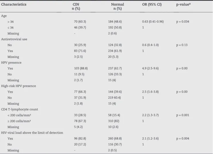
As it can be seen on Table 4, women older than 34 years presented lower frequency of CIN (p = 0.034). CIN distribution was similar, regardless of antiretroviral therapy: 25.9% among patients with CIN and 32.8% of women without CIN (p = 0.13). In contrast, other variables were associated with CIN development, such as HPV infection (p = 0.00) or high-risk HPV genotype (p = 0.00); CD4 T-lymphocyte count < 200 cells/mm³ (p = 0.00); and detectable HIV viral load (p = 0.004).
The variable “use of antiretrovirals” was excluded from the multivariate model, since we found an interaction between both CD4 T-lymphocyte count and detectable HIV viral load, and also because adherence to drugs was not measured. Five variables were included in the initial model: marital status (p = 0.067); HPV presence (p = 0.000); age under 34 years (p = 0.004); CD4 T-lymphocyte count bellow 200 cells/mm³ (p = 0.001); and detectable HIV viral load
Characteristics CIN
n (%)
Normal n (%)
OR (95% CI) p-valuea
Age
> 34 70 (60.3) 184 (48.6) 0.63 (0.41-0.96) p = 0.034
≤ 34 46 (39.7) 192 (50.8) 1
Missing - 2 (0.6)
Antiretroviral use
No 30 (25.9) 124 (32.8) 0.6 (0.4-1.0) p = 0.13
Yes 83 (71.6) 234 (61.9) 1
Missing 3 (2.5) 20 (5.3)
HPV presence
Yes 103 (88.8) 237 (62.7) 4.9 (2.5-9.6) p = 0.00
No 11 (9.5) 126 (33.3) 1
Missing 2 (1.7) 15 (4)
High-risk HPV presence
Yes 77 (66.3) 144 (39.6) 2.5 (1.6-3.8) p = 0.00
No 37 (31.9) 219 60.4) 1
Missing 2 (1.8) 15 (4)
CD4 T-lymphocyte count
< 200 cells/mm³ 33 (28.5) 58 (15.4) 2.2 (1.3-3.7) p = 0.001
≥ 200 cells/mm³ 78 (67.3) 310 (82) 1
Missing 5 (4.2) 10 (2.6)
HIV-viral load above the limit of detection
Yes 96 (82.8) 260 (68.8) 2.1 (1.2-3.6) p = 0.004
No 20 (17.2) 116 (30.7) 1
Missing - 2 (0.5)
(p = 0.004). However, as shown on Table 5, only three factors were independently associated with a higher prevalence of CIN in the final multivariate model: age under 34 years at study entry; detection of HPV; and a CD4 T-lymphocyte count below 200 cells/mm³. The Hosmer-Lemeshow goodness of fit value was 0.344, indicating that it was a good model.
Discussion
Previous study has shown that HIV-infected women present higher prevalence of CIN and cervical cancer if compared to uninfected women.14 Levi et al. in a cross-sectional study of
265 HIV-infected women from São Paulo (Brazil), aiming to evaluate HPV (detected by Hybrid Capture II) and CIN (detected by cytology) prevalence, found abnormal cervical smears in 19.0% of these women, in which 7.0% had high-grade lesions (corresponding to CIN II and CIN III). They have not found any case of cervical invasive carcinoma.15 In the present study,
we found a CIN prevalence of 23.6%, with 6% of CIN2/3 and only one case of cervical carcinoma. In addition, two large based multi-center cohort studies of HIV-infected women conducted in the United States – the Women’s Interagency HIV Study (WIHS) and the HIV Epidemiology Research Study (HERS) – have demonstrated a similar prevalence of CIN (17% and 18%) based on cytology, and a lower prevalence of high-grade squamous intraepithelial lesions and cancer: less than 3% in both cohorts.16,17 These studies were limited,
though, because these women were taking part in a screening program for cervical cancer; the treatment that followed could have eliminated previous lesions before cancer developed. In countries where women have poor access to medical care, rates of CIN2/3 or more severe lesions could be higher.18
HIV-infected women have a higher prevalence of HPV infection overall and high-risk HPV infection in particular.19
We found an overall HPV prevalence of 69.2% among 494 HIV-infected women. Other Brazilian studies have found similar presence of HPV infection: 64.5% (n = 265)15 and 60%
(n = 140).20 Studies performed in other countries have shown
similar HPV prevalence among HIV-infected women, ranging between 67.8% and 74%.21,22 The link between HPV infection
and CIN corroborates the results of previous studies and supports the causal pathway of HPV in cervical cancer. HPV infection is significantly associated with CIN progression.23
We found that women infected with any type of cervical HPV were at major risk of presenting CIN (OR 5.0, 95% CI 2.5–10.1) if compared to women without HPV.
The association between CIN prevalence and HIV-related immunodeficiency has been previously reported. One study showed significant correlation between low levels of CD4 T-lymphocytes, high HIV viral load and risk of CIN.24
A Brazilian study demonstrated that immunosuppressed women had a higher risk of lesion recurrence compared to women with a CD4 T-lymphocyte count > 200 cells/mm3.25 In our
study, there was significant association between HIV-related immunodeficiency measured by CD4 T-lymphocyte count, and the presence of CIN. This association remained statistically relevant even after controlling for HPV detection. This finding may be interpreted as an effect of immunodeficiency on the carcinogenic effect of HPV.
Some authors have found an irrelevant difference between HIV viral load in plasma and the presence of CIN.26
Although HIV viral load in our study was not predictive of CIN, the association of CIN with higher HIV viral load warrants further investigation.
There was no significant association of CIN with antiretroviral therapy in our study. Related data were obtained through medical prescriptions or through information provided by the patient concerning antiretroviral therapy. There is therefore no way to guarantee that these women were truly compliant with antiretrovirals. The effect of antiretroviral therapy on cervical cancer and other malignancies is not fully understood yet.27
Age data and CIN development are not well established. We have found that being under 34 is a risk factor for CIN. Koffi et al. 28
found that the average age of onset of CIN was lower among HIV-infected individuals, regardless of the grade of the lesion.
We know that observational retrospective studies such as this present limitations in what concerns the lack of information about long-term changes in immune status or outcome. Another limitation of this study relates to the unknown duration of HPV/HIV co-infection in these women. However, several studies have shown that the severity of CIN in HIV-infected women can be measured using cervical cytology as an endpoint, without histopathologic confirmation. It is worth pinpointing that the present study based the CIN diagnosis on histopathologic results, and cervical biopsy was guided by colposcopy. It should be pointed out the large sample size of this investigation, providing considerable statistical power for robust conclusions about the potential risk factors for developing CIN. Furthermore, our findings may be an important incentive for all HIV-infected women to be engaged in a gynecological care program. Cervical cancer screening directed to HIV-infected women who have experienced a low nadir of CD4 T-lymphocyte count need to be modified to encourage closer monitoring.
In conclusion, the prevalence of CIN was high among HIV-infected women, with low rates of high-grade lesions. We observed that immunosuppression, younger age and HPV infection were predictive of cervical intraepithelial neoplasia. As these women were enrolled for long-term care of their HIV infection, future opportunities for repeating gynecological evaluations will provide longitudinal data for other conclusions concerning risk factors of CIN. It is necessary to further investigate these risk factors to identify HIV-infected women with higher risk of CIN and establish appropriate strategies for management, including cervical cancer screening.
Variables Multivariate analysis
OR (95% CI) p-value
Age ≤ 34 years 1.5 (1.0-2.4) 0.049
HPV detection 5.0 (2.5-10.1) 0.000
CD4 T-lymphocyte count below 200 cells/mm³
Acknowledgements
We would like to thank the Fundação para o Amparo da Pesquisa em Minas Gerais (FAPEMIG) for institutional support.
Conflict of interest
All authors declare to have no conflict of interest.
R E F E R E N C E S
1. UNAIDS (2010) Report of the Global HIV/AIDS Epidemic. http://www.unaids.org/globalreport/Global_report.htm. Acessed 18 August 2011.
2. Ministério da Saúde (2010) http://www.aids.gov.br/publicacao/ boletim-epidemiologico-2010. Accessed 31 August 2011. 3. Lizano M, Berumen J, Carranca A. HPV-related
carcinogenesis: Basic Concepts, Viral Types and Variants. Arch Med Res. 2009;40(6):428-34.
4. Insinga RP, Dasbach EJ, Elbasha EA. Epidemiologic natural history and clinical management of Human Papillomavirus (HPV) Disease: a critical and systematic review of the literature in the development of an HPV dynamic transmission model. BMC Infect Dis. 2009;29(7):9-119. 5. Dain PK, Holmes KK, Hughes JP, Koutsky LA. Determinants
of cervical cancer rates in developing countries. Int J Cancer. 2002;100(2):199-205.
6. Averbach SH, Gravitt PE, Nowak RG, et al. The association between cervical human papillomavirus infection and HIV acquisition among women in Zimbabwe. AIDS. 2010; 24(7):1035-42.
7. Jamieson DJ, Duerr A, Burk R, et al. Characterization of genital HPV infection in women who have or who are at risk of having HIV infection. Am J Obstet Gynecol. 2002;186(1):21-7.
8. Massad LS, Ahdieh L, Benning L, et al. Evolution of cervical abnormalities among women with HIV-1: evidence from surveillance cytology in the Women’s Interagency HIV Study. J Acquir Immune Defic Syndr. 2001;27(5):432-42.
9. Chatuvedi AK, Madeleine MM, Biggar RJ, Engels EA. Risk of human papillomavirus –associated cancers among persons with AIDS. J Natl Cancer Inst. 2009;101(16):1120-30.
10. Harris TG, Burk RD, Palefsky JM, et al. Incidence of cervical squamous intraepithelial lesions associated with HIV serostatus, CD4 cell counts and human papillomavirus virus test results. JAMA. 2005;293(12):1471-6.
11. Hawes SE, Critchlow CW, Sow PS, et al. Incident high-grade squamous intraepithelial lesions in Senegalese women with and without human immunodeficiency virus type 1 (HIV-1) and HIV-2. J Natl Cancer Inst. 2006;98(2):100-109.
12. Carvalho NO, del Castillo DM, Perone C, et al. Comparison of HPV genotyping by type-specific PCR and sequencing. MIOC. 2010;105(1):73-78.
13. Richard RM. A modified terminology for cervical intraepithelial neoplasia. Obstet Gynecol. 1973;75:131-33.
14. Massad LS, Seaberg EC, Wright RL, et al. Squamous cervical lesions in women with human immunodeficiency virus: long-term follow-up. Obstet Gynecol. 2008;111(6):1388-93. 15. Levi JD, Fink MCS, Canto CLM, et al. Human Papillomavirus
prevalence, viral load and cervical intraepithelial neoplasia in HIV-infected women. Braz J Infect Dis. 2002;6(3):129-34. 16. Massad LS, Riester KA, Anastos KM, et al. Prevalence and
predictors of squamous cell abnormalities in Papanicolaou smears from women infected with HIV-1. J Acquir Immune Defic Syndr. 1999;21(1):33-41.
17. Duerr A, Kieke B, Warren D, et al. Human papillomavirus associated cervical cytology abnormalities among women with or at risk of infection with HIV. Am J Obstet Gynecol. 2001;184(4):584-90.
18. Agaba PA, Thacher TD, Ekwempu CC, Idoko JA. Cervical dysplasia in Nigerian women infected with HIV. Int J Gynecol Obstet. 2009;107(2):99-102.
19. Peedicayil A, Thiyagarajan K, Gnanamony M, et al. Prevalence and risk factors for human papillomavirus and cervical intraepithelial neoplasia among HIV-positive women at a tertiary level hospital in India. J Low Genit Tract Dis. 2009;13(3):159-64.
20. Melgaço F, Rosa M, Augusto E, et al. Human papillomavirus genotypes distribution in cervical samples from women living with human immunodeficiency virus. Arch Gynecol Obstet. 2011;283:809-17.
21. Bollen LJ, Chuachoowong R, Kilmarx PH, et al. Human papillomavirus (HPV) detection among human
immunodeficiency virus–infected pregnant Thai women: implications for future HPV immunization. Sex Transm Dis. 2006;33:259-64.
22. Mbulawa ZZ, Marais DJ, Johnson LF, et al. Influence of human immunodeficiency virus and CD4 count on the prevalence of human papillomavirus in heterosexual couples. J Gen Virol. 2010;91:3023-31.
23. Clifford GA, Gonçalves MA, Franceschia S. Human papillomavirus types among women infected with HIV: a meta-analysis. AIDS. 2006;20(18):2337-44.
24. Heard I, Tassie JM, Schmitz V, Mandelbrot L, Kazatchkine MD, Orth G. Increased risk of cervical disease among human immunodeficiency virus infected women with severe immunosuppression and high human papillomavirus load. Obstet Gynecol. 2000;96 :403-9.
25. Lima MA, Tafuri A, Araújo AC, Lima LM, Melo VH. Cervical intraepithelial neoplasia recurrence after conization in HIV-positive and HIV-negative women. Int J Gynecol Obstet. 2009;104(2):100-04.
26. Taylor G, Wolff T, Khanna N, Furth P, Langenberg P. Genital dysplasia in women infected with human immunodeficiency virus. J Am Board Fam Pract. 2004;17(2):108-12.
27. Minkoff H, Zhong Y, Burk RD, et al. Influence of adherent and effective antiretroviral therapy use on human papillomavirus infection and squamous intraepithelial lesions in human immunodeficiency virus-positive women. J Infect Dis. 2010;201(5):681-90.