Serum markers thrombophilia in pregnant women with Systemic Lupus
Erythematosus
Vanessa Marcon de Oliveira 1
Ernesto Antonio Figueiró-Filho 2
Cristiane Munaretto Ferreira 3
Erica Freire de Vasconcelos Pereira 4
1,3,4Faculdade de Ciências Farmacêuticas, Alimentos e Nutrição. Universidade Federal de Mato Grosso do Sul. Caixa Postal 549. Cidade Universitária.
Campo Grande, MS, Brasil. CEP: 79.0709-000. E-mail: [email protected]
2Departamento de Ginecologia e Obstetricia. Universidade Federal de Mato Grosso do Sul. Campo Grande, MS, Brazil.
Abstract
Objectives: to determine the frequency of serum markers for hereditary and acquired thrombophilia and their association with pregnancy in women with Systemic Lupus Erythematosus (SLE).
Methods: a case-control study was conducted among 25 pregnant women with SLE (study group) and 32 pregnant women without known disease and with at least one previous preg-nancy (control group). The presence of antiphospholipid antibodies and hereditary throm-bophilia were examined in both groups. We used the χ2 Test with Yates correction or Fisher’s Exact Test to verify the associations and calculate the relative risk.
Results: thrombophilia was present in 72.0% of pregnant women with SLE and in 6.0% of patients in the control group. A significant association was found between the presence of SLE and serum markers for hereditary thrombophilia / antiphospholipid antibodies (p<0.05). The relative risks for antiphospholipid antibodies were 13.20 (ICR95%= 1.81 - 96.46) in pregnant women with SLE, 7.26 (CI95%= 1.77 - 29.86) for the presence of serum markers of hereditary thrombophilia and 7.92 (CI95%= 2.62 - 3.94) for the presence of hereditary thrombophilia and/or antiphospholipid antibodies.
Conclusions: the identification of markers for hereditary and/or acquired thrombophilia in pregnant women with lupus may be clinically useful to determine which patients have a higher risk of obstetric complications.
Introduction
Systemic lupus erythematosus (SLE) is a chronic autoimmune disease induced by multiple factors, characterized by the production of autoantibodies, which lead to the damage of multiple organs and changes in immunological patterns.1It is a serious
disease with significant mortality and morbidity, and approximately 90% of affected patients are women of childbearing age.2
The disease has a variety of signs and symptoms that are often unspecific, thereby making its diag-nosis difficult. The diagdiag-nosis of SLE is made when at least four of the 17 criteria defined by the American College of Rheumatology for SLE are present (Table 1).3
As the manifestations of a normal pregnancy may sometimes be confused with lupus activity, it is important to understand the changes that occur during pregnancy.2 During normal pregnancy,
changes occur in clotting factors, which result in protein S deficiency, increased fibrinogen levels, and changes in factor II and factor VII. These changes increase the risk of thromboembolism by five-fold.4
Thrombocytopenia is usually mild and does not cause bleeding problems.4
Thrombophilias are mainly related to the pres-ence of antiphospholipid antibodies that are present in one-third of patients with SLE (currently a diag-nostic criteria according to the American College of Rheumatology), and their presence is associated with poor pregnancy outcomes.5These antibodies
Table 1
Clinical and Immunologic Criteria for SLE.
continue Criteria are cumulative and need not be present concurrently. Adapted from Petri et al.3
1) Acute cutaneous lupus
a) including lupus malar rash (do not count if malar discoid) i) bullous lupus
ii) toxic epidermal necrolysis variant of SLE iii) maculopapular lupus rash
iv) photosensitive lupus rash
(1) in the absence of dermatomyositis b) or subacute cutaneous lupus
i) nonindurated psoriaform and/or annular polycyclic lesions that resolve without scarring, although occasionally with postinflammatory dyspigmentation or telangiectasias)
2) Chronic cutaneous lupus a) including classical discoid rash
i) localized (above the neck)
ii) generalized (above and below the neck) b) hypertrophic (verrucous) lupus
c) lupus panniculitis (profundus) d) mucosal lupus
e) lupus erythematosus tumidus f) chillblains lupus
g) discoid lupus/lichen planus overlap
3) Oral ulcers: palate a) buccal b) tongue c) or nasal ulcers
d) in the absence of other causes, such as vasculitis, Behcets, infection (herpes), inflammatory bowel disease, reactive arthritis, and acidic foods
Table 1 concluded
Clinical and Immunologic Criteria for SLE.
Criteria are cumulative and need not be present concurrently. Adapted from Petri et al.3
4) Nonscarring alopecia (diffuse thinning or hair fragility with visible broken hairs)
a) in the absence of other causes such as alopecia areata, drugs, iron deficiency and androgenic alopecia
5) Synovitis involving two or more joints, characterized by swelling or effusion OR tenderness in 2 or more joints and thirty minutes or more of morning stiffness.
6) Serositis
a) typical pleurisy for more than 1 day i) or pleural effusions
ii) or pleural rub
b) typical pericardial pain (pain with recumbency improved by sitting forward) for more than 1 day i) or pericardial effusion
ii) or pericardial rub iii) or pericarditis by EKG
(1) in the absence of other causes, such as infection, uremia, and Dressler's pericarditis 7) Renal
a) Urine protein/creatinine (or 24 hr urine protein) representing 500 mg of protein/24 hr or b) Red blood cell casts
8) Neurologic a) seizures b) psychosis
c) mononeuritis multiplex
i)in the absence of other known causes such as primary vasculitis d) myelitis
e) peripheral or cranial neuropathy
i) in the absence of other known causes such as primary vasculitis, infection, and diabetes mellitus f) acute confusional state
i) in the absence of other causes, including toxic-metabolic, uremia, drugs 9) Hemolytic anemia
10) Leukopenia (< 4,000/mm3at least once)
i)in the absence of other known causes such as Felty's, drugs, and portal hypertension b) Lymphopenia (< 1,000/mm3at least once)
ii)in the absence of other known causes such as corticosteroids, drugs and infection 11)Thrombocytopenia (<100,000/mm3) at least once
a)in the absence of other known causes such as drugs, portal hypertension, and TTP
Immunological Criteria
1) ANA above laboratory reference range
2) Anti-dsDNA above laboratory reference range, except ELISA: twice above laboratory reference range 3) Anti-Sm
4) Antiphospholipid antibody: any of the following a) lupus anticoagulant
b) false-positive RPR
c) medium or high titer anticardiolipin (IgA, IgG or IgM d) anti-β2glycoprotein I (IgA, IgG or IgM)
5) Low complement a) low C3 b) low C4 c) low CH50
promote arterial and venous thrombosis, thrombocy-topenia and pregnancy loss.6,7In despite of these
information, previous reports regarding hereditary thrombophilia serum markers are addressed in nonpregnant women with SLE.8-10
Large numbers of complications can occur in pregnant women with SLE. Possible adverse events include reactivation of the disease, thrombosis and miscarriage in women with antiphospholipid anti-bodies, neonatal lupus, hypertension and toxicity of drugs used to treat SLE.11The high disease activity
during pregnancy results in an increased frequency of preterm births and a decreased likelihood of live-birth, with almost 25% of pregnancies resulting in fetal death.12It is recommended that the
immuno-suppressive drugs needed to control SLE continue to be administered, particularly if the patient has major organ involvement, such as lupus nephritis.12
Corticosteroids are relatively safe for use during pregnancy, therefore it may contribute in increasing maternal hypertension and gestational diabetes. Prednisone is the choice drug option in cases of active SLE disease upon the maternal organism, for it does not cross the placental barrier. By the other hand, dexamethasone should enter fetal circulation, so that it becomes the best choice in treatment of congenital heart block.2
The objective of the present study was to deter-mine the frequency of serum markers for hereditary and acquired thrombophilia and antiphospholipid antibodies and to examine their association with pregnancy in women with SLE.
Methods
We carried out a prospective, observational, cross-sectional controlled study in pregnant women from the Service of Gynecology and Obstetrics at the Hospital University Maria Aparecida Pedrossian (HUMAP) from June 2007 to June 2012. These were two distinct groups of patients, who were classified according to the following criteria:
Control Group (CG) - consisted of pregnant women without known disease who had at least one previous pregnancy, whose previous pregnancies were all low-risk and resulted in term deliveries and who had no history of miscarriage. Patients with a prior history of serum markers of hereditary throm-bophilias and antiphospholipid antibodies were excluded from this group.
Study Group (SG) – consisted of pregnant women with a diagnosis of Systemic Lupus Erythematosus (SLE) prior to pregnancy based on clinical and laboratory criteria3(Table 1) who also
might not have been diagnosed with hereditary thrombophilia or antiphospholipid antibodies prior to the current pregnancy.
Pregnant women whose SLE was in the active phase, who were less than 18 years of age and those whose diagnosis of SLE was made in the current pregnancy were excluded from the study.
The selection of patients was conducted prospec-tively and at random according to the order in which cases meeting the inclusion criteria of the outpatient high-risk pregnancy (SG) group and the low-risk (CG) group presented during the study period.
The calculation of the simple random sampling minimum number of pregnant women with a prior history of SLE to be included in SG took the following factors into account: the total population of primary care patients at the Clinic of High-Risk Pregnancy HUMAP (1,000 pregnant patients/year); the prevalence of antiphospholipid antibodies in this group of patients, whose values are well established in the literature and are approximately 40%,13the
prevalence of thrombophilia in the general popula-tion, which is approximately 5%,14and test power of
85% and a sampling error of 5% using a two-tailed test. With these parameters, the minimum number of pregnant women required in both groups was 24.
All patients were screened for the presence of antiphospholipid antibodies by serum IgM and IgG anticardiolipin, lupus anticoagulant and anti-β2 glycoprotein I (anti-β2GPI). In despite of the
litera-ture recommendation of repeating the dosage within 12 weeks interval, it was not performed due to specific characteristics of our routine service.12
methylenetetrahy-drofolate reductase (MTHFRC677T) polymorphism are not screened as routine in our service.
The anticardiolipin antibodies IgM and IgG were processed using enzyme immunoassay (ELISA) following the methodology proposed in international standardization.15 The results were considered
reac-tive for IgM and IgG when they had values higher than 11 UMPL and 11 UGPL, respectively.
The presence of lupus anticoagulant was conducted in three stages, and the first test was performed with the screening techniques aPTT (acti-vated partial thromboplastin time) and dRWT (time of the dilute Russell viper venom). When the obtained values were greater than 1.26 and/or 1.14 in the aPTT tests and dRWT, respectively, they were held at the second step, where the aPTT test proceeded using a mixture of 50% plasma and 50% of the patient’s normal plasma. If no correction was needed, the test proceeded to the third stage. This consisted of dRWT confirmatory testing, which tested positive for lupus anticoagulant when the concentration was greater than 1.21.15
The search for anti-β2GPI antibodies was
performed by ELISA, where values above 15 U/mL were considered to be reactive.15
The dosage of protein C was measured using the chromogenic method,16,17the result of which is the
finding of a normal serum value greater than or equal to 65%. Values less than 65% are considered to indicate protein C deficiency.16,17
The dosage of protein S was measured using the chromogenic method,16,17the result of which is the
finding of a normal serum value greater than or equal to 55%. Values below 30% are considered to indicate protein S deficiency.16,17
The dosing of antithrombin was measured using the chromogenic method,16,17the result of which is
the finding of a normal serum value of greater than or equal to 60%. Values below 60% are considered to indicate antithrombin deficiency.16,17
Homocysteine was measured using high-pres-sure liquid chromatography (HPLC) with isocratic elution and fluorimetric detection,18whose reference
value is 4 to 12 μmol/L. Hyperhomocysteinemia is evidenced by values greater than 12 μmol/L.18
The research Q506 mutation in the factor V (factor V Leiden) was measured using polymerase chain reaction (PCR) using to the restriction enzyme Mnli,16 and is considered to be a positive mutation (homozygous or heterozygous ).
Carriers of hereditary thrombophilia were all pregnant women who had test results that indicated protein C deficiency, protein S deficiency, anti-thrombin deficiency, hyperhomocysteinemia or the
presence of Factor V Leiden mutation (heterozygous or homozygous affected). Carriers of antiphospho-lipid antibodies were all patients whose results were positive for anticardiolipin IgM/IgG, lupus anticoa-gulant and/or anti-β2GPI, according to the
metho-dology explained.
We emphasize that serum markers for acquired thrombophilias were not repeated after 12 weeks, because the present study did not aim to perform the diagnosis of APS.
The parametric variables (age and parity) are expressed as the mean ± standard deviation and were compared using the Student t test. The nonpara-metric variables (presence/absence of hereditary thrombophilia and antiphospholipid antibodies) were evaluated in double-entry contingency tables using the χ2test with Yates correction or Fisher’s Exact
Test. Significant associations were those with p value <0.05. Relative risks were calculated and are presented with 95% confidence intervals (CI).
The patients included in the study signed an informed consent form (ICF). The study and its ICF were approved by the Ethics Committee on Human Research of the Federal University of Mato Grosso do Sul - UFMS, Protocol 884, as of May 18, 2007.
Results
Of the 57 pregnant women studied, 25 (43.9%) were in the study group and 32 (56.1%) were in the control group. The mean age in the SG was 29.4 years, and the average number of pregnancies was 2.1±1.4. In the CG, the mean age was 30.3 ±4.4 years, and the mean number of pregnancies was 1.2±0.4. No difference between the groups was found for maternal age or parity.
Positive screening for hereditary thrombophilia and/or antiphospholipid antibodies was found in 72.0% (18/25) of pregnant women in the SG. Among the CG patients, the presence of thrombophilia and/or antiphospholipid antibodies was found in 9.4% (3/32) of the sample (Table 2).
Screening for antiphospholipid antibodies in pregnant women with lupus demonstrated positive results in ten women (40%), out of these, six (24%) patients were identified with isolated antibodies and 4 (16%) patients had antibodies associated with other thrombophilia serum markers. In the pregnant women from the CG the presence of antiphospho-lipid antibodies was detected in only one patient (3.1%) (Table 2).
. B ra s. S a ú d e M a te rn . I n fa n t. , R e ci fe , 1 7 ( 4 ): 8 3 3 -8 4 2 o u t. / d e z ., 2 0 1 7 a l . Table 2
Association between the presence of SLE and the occurrence of serum markers of thrombophilias in pregnant women.
*Fisher’s Exact Test; 1pregnant women with SLE; 2pregnant women with at least one successful pregnancy.
Study group 1
Control group 2
Total RR (IC95%)
P
Serum markers of antiphospholipid antibodies*
Serum markers of hereditary thrombophilia
Serum markers of hereditary thrombophilia and/or antiphospholipid
antibodies
Yes No Total Yes No Total Yes No Total
n % n % n % n % n % n % n % n % n %
10 1 11 40.0 3.1 19.3 15 31 46 60.0 96.9 80.7 25 32 57 100.0 100.0 100.0
13.20 (1.81 - 96.46) 0.001 11 2 13 44.0 6.3 22.8 14 30 44 56.0 93.7 77.2 25 32 57 100.0 100.0 100.0
7.26 (1.77 - 29.86) 0.001 18 3 21 72.0 9.4 36.8 7 29 36 28.0 90.6 63.2 25 32 57 100.0 100.0 100.0
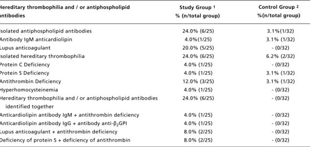
Table 3
Frequency of serum markers for thrombophilias in pregnant women with and without SLE.
1pregnant women with SLE; 2pregnant women with at least one successful pregnancy.
Isolated antiphospholipid antibodies Antibody IgM anticardiolipin Lupus anticoagulant
Isolated hereditary thrombophilia Protein C Deficiency
Protein S Deficiency Antithrombin Deficiency Hyperhomocysteinemia
Hereditary thrombophilia and / or antiphospholipid antibodies identified together
Anticardiolipin antibody IgM + antithrombin deficiency Anticardiolipin antibody IgG + antibody anti-β2GPI
Lupus anticoagulant + antithrombin deficiency Deficiency of protein S + deficiency of antithrombin Hereditary thrombophilia and / or antiphospholipid antibodies
Study Group 1 % (n/total group)
Control Group 2 %(n/total group)
24.0% (6/25) 4.0%(1/25) 20.0% (5/25) 24.0% (6/25) 4.0% (1/25) 4.0% (1/25) 12.0% (3/25)
4.0% (1/25) 24.0% (6/25)
4.0% (1/25) 4.0% (1/25) 8.0% (2/25) 8.0% (2/25)
3.1%(1/32) 3.1% (1/32) - (0/32) 6.2% (2/32)
- (0/32) 3.1% (1/32) 3.1% (1/32) - (0/32) - (0/32)
- (0/32) - (0/32) - (0/32) - (0/32)
and/or antiphospholipid antibodies (Table 2). The relative risk for the presence of antiphospho-lipid antibodies in pregnant women with SLE was 13.20 (CI95%= 1.81 - 96.46). The presence of serum markers for hereditary thrombophilia had a relative risk of 7.26 (CI95%= 1.77 - 29.86). The relative risk was 7.92 (CI95%= 2.62 - 23.94) when considering the occurrence of serum markers for hereditary thrombophilia and/or antiphospholipid antibodies.
Isolated hereditary thrombophilia was present in 24.0% of the pregnant women diagnosed with SLE (6/25) and 6.1% (2/32) of pregnant women in control group. Two or more hereditary thrombophilias and / or the presence of antiphospholipid antibodies together were found in 24% (6/25) of patients EG (Table 3). Among pregnant women in the SG, the most frequent isolated hereditary thrombophilias were as follows: antithrombin deficiency (12%), protein C and S deficiencies, and hyperhomocys-teinemia (4%). In screening for the presence of antiphospholipid antibodies, the presence of lupic anticoagulant alone was demonstrated in 20% of the sample group (5/25) (Table 3).
Discussion
The prevalence of thrombophilic factors in pregnant women with SLE is not well established in the lite-rature. Thus, the investigation of the prevalence of these factors in this group of patients helps to clarify
its importance both in these clinical profiles and in preventing obstetric complications. The presence of antiphospholipid antibodies in patients with SLE has been described,11,13,19but markers for hereditary
thrombophilia are not well characterized, and varying methodologies make it difficult to compare data.8-10
The prevalence of antiphospholipid antibodies in SLE patients is approximately 40%, and between 12% and 44% of patients are positive for anticar-diolipin, 15% to 34% are positive for lupus antico-agulant and 10% to 19% are positive for the anti-β2 -glycoprotein I (β2-GPI anti).13These data agree with
the results of the present study, where 40% (10/25) of patients had markers for antiphospholipid anti-bodies. Anticardiolipin antibody was found in 12% (3/25) of patients, lupus anticoagulant was found in 24% (6/25) and anti-β2GPI was found in 4% (1/25), which is the only antibody whose value was below that described in the literature.
Thrombotic events occur more frequently in lupus patients with antiphospholipid antibodies than in patients without SLE or other autoimmune disease who have these antibodies.20A study conducted in
2009 showed similar results and found a higher frequency of thrombosis and pregnancy loss in patients with SLE, which were associated with the presence of markers for thrombophilia in patients with acquired primary APS.21
antibodies was found to be 3.1 times higher in women experiencing pregnancy loss, especially after 20 weeks gestation. This was an independent risk factor for additional losses in a pregnancy cohort study of 166 pregnant women in the Hopkins Lupus Center.22
Maternal lupus activity and the presence of antiphospholipid antibodies were associated with concomitant major causes of obstetric complica-tions,12,22It is estimated that approximately 20% of
pregnancies in women with SLE result in fetal loss.23 The rate of preterm birth (birth before 37
weeks gestation) is also increased in this group of patients. The incidence appears to vary between 23% and 28%.23Preterm birth is often spontaneous, and
it is mainly due to premature rupture of membranes, but there is also a significant portion of cases in which labor is induced to protect the health of mother and / or baby (early fetal distress or pre-eclampsia).19
In relation to serum markers of hereditary throm-bophilia, protein C deficiency, S and antithrombin, we found an incidence of 40% (10/25) among preg-nant women with lupus, however several authors,9,24,25 did not find the presence of these
markers in non-pregnant patients with SLE, suggesting that they were not associated with an increased thrombotic risk. However, we observed changes in these protein systems.10
We cannot say that patients with protein C and S deficiency are carriers of inherited deficiency since pregnancy is a hypercoagulable state with increased coagulation factors and simultaneous decrease of natural anticoagulant and fibrinolytic proteins. These important changes occur naturally in order to mini-mize the risk blood loss, but increases the occurrence of thromboembolic events. However as there were no subsequent dosages of natural anticoagulants, it is suggested that these deficiencies may be transient.26-28
We did not observe the presence of FV Leiden mutation in any patient. However, Brouwer et al.24
found that FV Leiden in non-pregnant patients with
SLE increases the risk of venous thrombosis and is associated with an OR of 3.5.
The presence of hyperhomocysteinemia was found in only 4% (1/25) of women in the study group, a result which is lower than the one presented in the literature in non-pregnant patients with lupus, which is approximately 37%, being this related to renal impairment8. High concentrations of this amino acid are common in SLE patients and are predictive of late thrombosis, stroke and hyperten-sion.21
Studies show that women with thrombophilia (protein S deficiency, and antithrombin C, hyperho-mocysteinemia and factor V Leiden mutation) during pregnancy have a higher prevalence of obstetric and perinatal complications such as miscarriage, fetal loss and preeclampsia.6,7Thus, the high incidence of
these markers in women with lupus may be related to the increased number of cases of obstetric compli-cations in this group of women.
Whereas women with SLE have 2 to 4 times higher rates of complications during pregnancy than non-pregnant women with SLE,29the identification
of markers for hereditary and/or acquired throm-bophilia in pregnant women with lupus may be clini-cally useful to determine which patients have a higher risk of obstetric complications.
We emphasize that our study population portrays some characteristics which make it quite different from the populations studied by other authors. The Brazilian racial miscegenation could induce genetic characteristics not found in populations studied in other trials regarding hereditary/acquired throm-bophilia.29,30
We either did not find any study that focused in the association of SLE pregnant women and inhe-rited thrombophilia. We were only able to find data on the prevalence of acquired thrombophilia (antiphospholipid antibodies) and its impact on preg-nancy in patients with SLE. This may suggest that more studies regarding the impact of inherited thrombophilia in pregnant women with SLE should be carried on.
References
1. Wei Q, Ouyang Y, Zeng W, Duan L, Ge J, Liao H. Pregnancy complicating systemic lupus erythematosus: a series of 86 cases. Arch Gynecol Obstet. 2011; 284 (5): 1067-71.
2. Dhar JP, Sokol RJ. Lupus and pregnancy: complex yet manageable. Clin Med Res. 2006; 4 (4): 310-21. 3. Petri M, Orbai AM, Alarcón GS, Gordon C, Merrill JT,
Callen JP, Lim SS, Fessler BJ, Inanc M, Kamen DL, Rahman A, Steinsson K, Franks Jr. AG, Sigler L, Hameed S, Fang H, Pham N, Brey R, Weisman MH, McGwin Jr. G, Magder LS. Derivation and Validation of Systemic Lupus International Collaborating Clinics Classification Criteria for Systemic Lupus Erythematosus. Arthritis Rheum. 2012; 64 (8): 2677–86.
4. Dresang LT, Fontaine P, Leeman L, King VJ. Venous thromboembolism during pregnancy. Am Fam Physician. 2008; 77 (12): 1709-16.
5. Zhan Z, Yang Y, Zhan Y, Chen D, Liang L, Yang X. Fetal outcomes and associated factors of adverse outcomes of pregnancy in southern Chinese women with systemic lupus erythematosus. PLoS One. 2017; 12 (4): e0176457. 6. Figueiró-Filho E, Oliveira V. Association of recurrent
abor-tion, fetal loss and severe preeclampsia with hereditary thrombophilias and antiphospholipid antibodies in pregnant women of central Brazil. Rev Bras Ginecol Obstet. 2007; 29 (11): 561-7.
7. Figueiro-Filho EA, Oliveira VM, Coelho LR, Breda I. Serum markers of inherited thrombophilia and antiphospho-lipid antibodies in pregnant women with previous history of severe pre-eclampsia. Rev Bras Ginecol Obstet. 2012; 34 (1): 40-6.
8. Barcat D, Guérin V, Ryman A, Constans J, Vernhes JP, Vergnes C, Bonnet F, Delbrel X, Morlat P, Longy-Boursier M, Conri C. Thrombophilia and thrombosis in systemic lupus erythematosus: a case-control study. Ann Rheum Dis. 2003; 62 (10): 1016-7.
9. Afeltra A, Vadacca M, Conti L, Galluzzo S, Mitterhofer AP, Ferri GM, Del Porto F, Caccavo D, Gandolfo GM, Amoroso A. Thrombosis in systemic lupus erythematosus: congenital and acquired risk factors. Arthritis Rheum. 2005; 53 (3): 452-9.
10. Vayá A, Santaolaria M, Micó L, Calvo J, Oropesa R, Villa P, Todoli J, Simó M, Corella D, Ricart JM Thrombotic events in systemic lupus erythematosus. Its association with acquired and inherited thrombophilic defects. Clin Hemorheol Microcirc. 2008; 40 (2): 79-87.
11. Manzano-Gamero V, Pardo-Cabello AJ, Vargas-Hitos JA, Zamora-Pasadas M, Navarrete-Navarrete N, Sabio JM, Jáimez-Gámiz L, Ríos-Fernandez R, Ortego-Centeno N, Ayala-Gutierrez MM, de Ramón E, Colodro-Ruíz A, Micó-Giner L, Castillo-Palma MJ, Robles-Marhuenda Á, Luna-Del Castillo JD, Jiménez-Alonso J; Spanish Autoimmune Diseases Study Group (GEAS). Effect of ethnicity on clin-ical presentation and risk of antiphospholipid syndrome in Roma and Caucasian patients with systemic lupus erythe-matosus: a multicenter cross-sectional study. Int J Rheum Dis. 2017; 7. [Epub ahead of print]
12. Clowse ME, Magder LS, Witter F, Petri M. The impact of increased lupus activity on obstetric outcomes. Arthritis Rheum. 2005; 52 (2): 514-21.
13. Biggioggero M, Meroni PL. The geoepidemiology of the antiphospholipid antibody syndrome. Autoimmun Rev. 2010; 9 (5): A299-304.
14. Middeldorp S, van Hylckama Vlieg A. Does thrombophilia testing help in the clinical management of patients? Br J Haematol. 2008; 143 (3): 321-35.
15. Miyakis S1, Lockshin MD, Atsumi T, Branch DW, Brey RL, Cervera R, Derksen RH, DE Groot PG, Koike T, Meroni PL, Reber G, Shoenfeld Y, Tincani A, Vlachoyiannopoulos PG, Krilis SA. International consensus statement on an update of the classification criteria for defi-nite antiphospholipid syndrome (APS). J Thromb Haemost. 2006; 4 (2): 295-306.
16. Jackson BR, Holmes K, Phansalkar A, Rodgers GM. Testing for hereditary thrombophilia: a retrospective analysis of testing referred to a national laboratory. BMC Clin Pathol. 2008. 2; 8: 3.
17. Abbassi-Ghanavati M, Greer LG, Cunningham FG. Pregnancy and laboratory studies: a reference table for clin-icians. Obstet Gynecol. 2009; 114 (6): 1326-31.
18. Ubbink JB, Hayward Vermaak WJ, Bissbort S. Rapid high-performance liquid chromatographic assay for total homo-cysteine levels in human serum. J Chromatogr. 1991; 565 (1-2): 441-6.
19. Andreoli L, Fredi M, Nalli C, Reggia R, Lojacono A, Motta M, Tincani A. Pregnancy implications for systemic lupus erythematosus and the antiphospholipid syndrome. J Autoimmun. 2012; 38 (2-3): J197-208.
20. Laskin CA, Clark CA, Spitzer KA. Antiphospholipid syndrome in systemic lupus erythematosus: is the whole greater than the sum of its parts? Rheum Dis Clin North Am. 2005; 31 (2): 255-72.
21. Danowski A, de Azevedo MN, de Souza Papi JA, Petri M. Determinants of risk for venous and arterial thrombosis in primary antiphospholipid syndrome and in antiphospholipid syndrome with systemic lupus erythematosus. J Rheumatol. 2009; 36 (6): 1195-9.
22. Clowse ME, Magder LS, Witter F, Petri M. Early risk factors for pregnancy loss in lupus. Obstet Gynecol. 2006; 107 (2 Pt 1): 293-9.
23. Yan Yuen S, Krizova A, Ouimet JM, Pope JE. Pregnancy Outcome in Systemic Lupus Erythematosus (SLE) is Improving: Results from a Case Control Study and Literature Review. Open Rheumatol J. 2008; 2: 89-98. 24. Brouwer JL, Bijl M, Veeger NJ, Kluin-Nelemans HC, van
der Meer J. The contribution of inherited and acquired thrombophilic defects, alone or combined with antiphos-pholipid antibodies, to venous and arterial thromboem-bolism in patients with systemic lupus erythematosus. Blood. 2004; 104 (1): 143-8.
25. Torresan M, Machado TF, Siqueira LH, Ozelo MC, Arruda VR, Annichino-Bizzacchi JM. The impact of the search for thrombophilia risk factors among antiphospholipid syndrome patients with thrombosis. Blood Coagul Fibrinolysis. 2000; 11 (7): 679-82.
26. Adachi T. Protein S and congenital protein S deficiency: the most frequent congenital thrombophilia in Japanese. Curr Drug Targets. 2005; 6 (5): 585-92.
28. Hojo S, Tsukimori K, Kinukawa N, Hattori S, Kang D, Hamasaki N, et al. Decreased maternal protein S activity is associated with fetal growth restriction. Thromb Res. 2008; 123 (1): 55-9.
29. Clowse ME, Jamison M, Myers E, James AH. A national study of the complications of lupus in pregnancy. Am J Obstet Gynecol. 2008; 199 (2): 127 e1-6.
30. De Stefano V1, Martinelli I, Rossi E, Battaglioli T, Za T, Mannuccio Mannucci P, Leone G. The risk of recurrent venous thromboembolism in pregnancy and puerperium without antithrombotic prophylaxis. Br J Haematol. 2006; 135 (3): 386-91.
______________
Received on March 20, 2017