Rev Bras M ed Esporte _ Vol. 12, Nº 1 – Jan/Fev, 2006
5e
1. University for the Development of the State and the Low land Region –UNIDERP – Campo Grande, M S.
2. Department of Locomotive Biomechanics, M edicine and Rehabilita-tion – Ribeirão Preto M edicine School of the São Paulo University – FM RP-USP.
Received in 1/12/04. Final version received in 22/8/05. Approved in 5/9/05. Correspondence to: Rua Gravataí, 290, M onte Castelo – 79010-390 – Campo Grande, M S, Brazil. Phone: (67) 351-5400. E-mail: carulms@pop. com.br
Effects in the Q angle measurement w ith maximal
voluntary isometric contraction of the quadriceps muscle
Belchior A.C.G.1, Arakaki J.C.1,2, Bevilaqua-Grossi D.2, Reis F.A.1 and Carvalho P.T.C.1
O
RIGINALA
RTICLEKeyw ords: Knees. Femoropatellar. Q angle.
ENGLISH VERSION
ABSTRACT
The purpose of this study w as to analyze the difference be-tw een the angle of the quadriceps in symptomatic and asymp-tomatic individuals in tw o different examination situations, having the quadriceps relaxed and in a maximal voluntary isometric traction (M VIC) through radiographic measurement, aiming to con-tribute to the assessment and treatment of patients w ith pate-lofemoral disorder (PFD). Through the standard radiological method tw enty 21 years old mean w omen (40 knees) w ere assessed. All individuals w ere positioned supine using a U-podalic stabilizer, having their low er limbs relaxed, using a plumb film on the anteri-or tuberosity of the tibia. Fanteri-or the statistical analysis, the averages for the asymptomatic and symptomatic groups in a relaxed and M VIC status, as w ell as the Student’s t-test w ith p < 0.05 signifi-cance level w ere used. The mean values to the Q angle compared to the asymptomatic group w ere 17.15o on relaxation, and 14.5o
on M VIC, w hile the asymptomatic group presented 21.45o, and
15.8o, respectively. The results in the equality analysis betw een
the symptomatic and asymptomatic groups on the relaxed status attained a p = 0.004, and to the maximal voluntary isometric con-traction, p = 0.29. Considering the data attained in the present study, it can be verified that in a relaxing status, there is a differ-ence betw een the value of the Q angle among symptomatic and asymptomatic individuals, being found a higher value in the FPD bearers, w hile in a maximal isometric contraction of the quadri-ceps muscle no statistical difference w as found in the present study, w ith a reduction in the angle in both groups.
INTRODUCTION
The knee joint is involved in about 50% of the musculoskeletal injuries, and the most common of these injuries is the patelofem-oral disorder(1-3). The PFD is an articular disorder manifested by pain in the anterior portion of the knee and a functional deficit that compromises the daily activities(4,5). It constitutes 25% of the inju-ries compromising the knee and 5% of every sportive injury, rep-resenting a shared complaint in 20% of the population, affecting mainly young 15-25 years old females(1,6,7).
The most frequent symptoms are pain in the anterior knee, peri-patellar edem a, blockage, and patelofem oral articular crepita-tion(5,8,9). Generally, they are bilateral, and presenting exacerbation periods related to long period in the seat position w ith the knees flexed, w hen standing up after being seat, w hile ascending and descending stairs or w alking on inclined surfaces, running, train-ing ustrain-ing w eights, and kneeltrain-ing(1,5,6,10).
There are several etiologic factors that can generate the PFD, such as neuromuscular imbalances of the oblique vastus medialis (OVM ) and vastus lateralis (VL); the shortening of the lateral reti-naculum, the ischiotiabilis, the iliotibialis tract, and the gastrocne-mius; the excessive pronation of the subtalar joint; the loosening, or the ligamentous or capsular shortening; bone abnormalities; the excessive anteversion of the femoral cervix; the external torsion of the tibialis; enlargement of the pelvis and the high patella(6,10-13). Related to the active strength exerted on the patella, the main structure responsible by such activity is the quadriceps muscle of the thigh that controls the position of the patella related to the trochlea by means of the oblique fibers of its medialis and lateral portions – the vastus medialis (VM ) and the vastus lateralis (VL)(1). The VM muscle is divided in tw o portions: the vastus medialis longus (VM L) a muscle that it is inserted in a 15o related to the longitudinal axle of the femur, and that exerts few or no traction to the adequate positioning of the patella, and the oblique vastus medialis (OVM ) that is inserted mainly in a 50-55o on the longitudi-nal axle of the femur, and it is considered the medial dynamic stabilizer of the patelofemolar joint (PFJ). Likew ise, the VL muscle is also divided in tw o portions: the proximal fibers that are origi-nated in the femur, and that are inserted in the medial third of the quadriceps’ tendon of the thigh, constituting the vastus lateralis longus (VLL), and the posterolateral fibers are originated in the iliotibial tract, being more oblique tow ards its direction, and they are inserted on the base and the lateral edge of the patella, repre-senting the oblique vastus lateralis (OVL) muscle. Taking into ac-count the origins and insertions of these muscles, especially the oblique portions, it is observed its opposite actions, and their im-portance to the function for the patellar stability(14,15).
Considering the direction of the muscular fibers, the oblique vastus medialis (OVM ) is the first muscle that contributes to the medial strength vector, same as the vastus lateralis (VL) is for the lateral strength(6,16).
The insufficiency or hypotrophy of the VM O muscle can gener-ate a pgener-atellar malalignment, especially in the last extension grades (0o-15o flexion) of the knee joint, w here it occurs a major instability in the joint due to a low er bone congruence, but it does not present an efficiency to restrain the lateral traction of the patella(17).
Another factor that may contribute to the pain development or w orsening, w ith a consequent generation of instability in the knee is the malalignment of the patelofemoral joint, w hose measure-ment can be performed through the quadriceps angle (Q)(4,18).
The Q angle is formed by the crossing of tw o imaginary lines: the first line is formed by the anterosuperior iliac spine up to the medium patellar spot, and the second line is formed by the anteri-or tuberosity of the tibia up to the medium patellar spot, and its mean normal value is 13o in men, and 18o in w omen(1,5,7,11,19).
6e
Rev Bras M ed Esporte _ Vol. 12, Nº 1 – Jan/Fev, 2006subluxation, causing a softening of the cartilage and a stress on the retinaculum, besides of contributing to a spread in the PFD(20,21). The measurement of that angle can be performed using differ-ent methods, such as the radiographic method(10,21) or clinically through the goniometer(21), and in different w ays, having the pa-tient in supine w ith his knees in total extension of the knee and relaxed quadriceps(20,21), or contracted(22), in orthostatism(5,7,21), seat w ith the knees in a 90o flexion, or in a or 20-30o w ith the maximal medial, lateral rotation or in a neutral position of the tibia(5).
Thus, the aim of this study w as to search the difference be-tw een the quadriceps angle in symptomatic and asymptomatic individuals in tw o different examination situations, having the quad-riceps relaxed and in maximal voluntary isometric contraction us-ing the radiographic measurement, in order to contribute to the assessment and treatment of patients w ith PFD.
CASUISTIC AND M ETHODS
Sampling
It w as recruited through oral invitation tw enty 15 to 30 years old (mean 21 years) female volunteers (n = 40 knees) during the period from M ay to July, 2003.
Ten individuals (n = 20 knees) did not present any complaint of pain in the knees; traumatic or surgical records or any pathology involving any of the joints of the low er limbs; vascular, peripheral or central neurological disorder, constituting the group of knees considered normal, that means, asymptomatic.
The remaining persons (n = 20 knees) w ere diagnosed w ith PFD, presenting bilateral pain, according to the Cow an et al.(22) criteria that have the follow ing inclusion factors: 1) anterior or ret-ropatellar pain w hen performing at least tw o of the follow ing ac-tivities: ascending stairs, be seat for long periods, upon squatting, kneeling, or jumping; 2) pain on the patellar palpation; 3) symp-toms for more than one month w hose onset is not related to any trauma.
As exclusion factors it w as considered the follow ing: 1) signs or symptoms of another pathology; 2) recent history (less than three months) of surgery in the knees; 3) patellar luxation or sublux-ation; 4) clinical evidence of meniscal injury; 5) ligamentous insta-bility; 6) patellar tendinitis; 7) osteoarthrosis or pain irradiated from the columna vertebralis; 8) pregnancy or another condition that may interfere in the data collection.
Before performing the assessments, all volunteers w ere clari-fied on the procedure used in the research, and they signed a Free Formal and Clarified Consent Term according to the Federal Regulations for Human Subjects Research referred in the Resolu-tion 196/96 of the NaResolu-tional Health Council.
Instruments
U-podalic stabilizer: U formatted metallic plate w ith tw o mobile arms, upon w hich the regions of the heels w ere positioned. The gap of the arms impeded the lateral rotation of the low er limbs, and such gap corresponded to the distance betw een the feet af-ter the knees w ere aligned to the hips.
Radiological device: A 500 mmA G3 M odel. The distance from the ampoule to the 35 x 91 cm cassette w as 1.70 m, a Kodak® film
and automatic revelation (M acrotec®).
Procedures
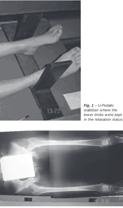
The patient w as positioned supine, w ith their knees in total ex-tension along w ith the U-podalic stabilizer, and each patient w as asked to keep her low er limbs in a relaxing status (figure 1).
Afterw ards, the same technician took radiographies in antero-posterior positioning using radiological 35 x 91 cm film, compris-ing a portion of the hip 15 cm below the previously market TAT using a lead film (4 cm2) fixed on the skin w ith adhesive tape, in
order to facilitate the visualization of the TAT after revealing the film, w hen the Q angle w as traced by the researcher using a con-ventional rule, a pen, and protractor (figure 2).
A similar procedure w as performed along w ith the request for a maximal voluntary isometric contraction of the quadriceps mus-cle.
Fig. 1 – U-Podalic
stabilizer w here the low er limbs w ere kept in the relaxation status
Fig. 2 – Radiological image w ith presence of the lead film (4 cm2) to facil-itate the TAT visualization after revealing the film, w here the Q angle w as traced by the researcher using conventional rule, pen and tractor
Statistical analysis
It w as calculated the averages of the collected values, follow -ing the statistical method, the t-Student test w ith a 0.05 signifi-cance level, and the test w as applied betw een the values of the Q angle betw een asymptomatic and symptomatic patients both in the relaxing status and in maximal voluntary isometric contrac-tion.
RESULTS
Rev Bras M ed Esporte _ Vol. 12, Nº 1 – Jan/Fev, 2006
7e
Next, it w as performed the statistical analysis using the t-Stu-dent test to analyze the equality betw een symptomatic and as-ymptomatic groups (figures 3 and 4). In the relaxation status, it w as attained a p = 0.004, w hile the maximal voluntary contraction presented p = 0.26.
function(16), especially having the low er limb in total extension (0o) w here it occurs the major articular instability due to the low er bone congruence(17,22), and it did not present any efficiency in restrain-ing the lateral traction of the patella. Thus, w ith the request on the maximal voluntary isometric contraction of the quadriceps mus-cle, it w as estimated that the patella w ould be lateralized, thus generating an increase in the Q angle.
But the reduction of the Q angle w hich w as seen can be justi-fied in studies analyzing the activation of the oblique medial and vastus lateralis muscles upon isometric exercises on the quadri-ceps having the knees in total extension, w here none of the mus-cular portions presented preferential activation(24,25).
Other electromyographic studies has examined variables such as the intensity and the time of the neuromuscular recruitment betw een the OVM and VL muscles w hile performing functional activities, such as ascending and descending stairs, and w alking in healthy and PFD bearer individuals, but it w as found no satisfac-tory results associated to the PFD(20,24,26).
Analyzing the patellar behavior in PFD bearer w omen through computerized tomography, no conclusion w as attained as to the effect of the quadriceps muscle contraction on the patellar decli-nation angles, but there w as a significant increase in the congru-ence angle, especially in the 0o and 20o flexion knee(27).
Therefore, the behavior in the reduction of the Q angle together w ith the maximal voluntary isometric contraction of the quadri-ceps muscle in PFD bearer individuals leads to a justification of the use of treatment w ays that use such kind of contraction.
The SLR, an exercise performed having the leg straight raised, is a w ay of treatment much employed in the PFD rehabilitation (28-31). Based on this study, w e believe that if it is associated to the maximal voluntary contraction during its execution, it can mini-mize the bad patellar alignment.
Besides, t he present t reat m ent proposals em phasize t he strengthening of the abductor muscles of the hip(32,33) due to the kinematical imbalance of the low er limbs, and thus, it is w ise to initiate a kinesiotherapeutic SLR exercising program associated to the hip abduction.
But regardless the form chosen to perform the SLR, physical exercises promote a sensorial reeducation through the motor ac-tivity(34), and this is extremely benefic to PFD bearer individuals, since in general they present proprioceptive abnormalities(3).
Despite this study presents significant data, it had some limita-tions, such as the cost involved in acquiring radiological images, as w ell as the positioning adopted to the procedure in supine, w hich w e believe that it does not reproduce the optimum func-tional measurement.
CONCLUSION
Considering the data attained in the present study, it w as veri-fied that in a relaxing status, there is significant difference be-tw een the value of the Q angle bebe-tw een symptomatic and asymp-tomatic individuals, and such difference is not present in a maximal isometric contraction of the quadriceps muscle, and there w as a reduction in the value of the angle in both individuals.
All the authors declared there is not any potential conflict of inter-ests regarding this article.
REFERENCES
1. Cabral CM , M onteiro-Pedro V. Recuperação funcional de indivíduos com disfun-ção femoropatelar por meio de exercícios em cadeia cinética fechada: revisão da literatura. Rev Bras Fisioter 2003;7:1-8.
2. Baker M M , Juhn M S. Patellofemoral pain syndrome in the female athlete. Clin Sports M ed 2000;19:315-29.
TABLE 1
Values of the angle Q means presented in the relaxation and maximal voluntary contraction status
Asymptomatic Symptomatic X D.P. X D.P.
Relaxation 17.15° 4.5 21.45° 3.9 Contraction 14.5°0 3.0 15.8°0 3.5
Fig. 3 – Graphic of the measurement of the Q angle attained in the
relax-ation status
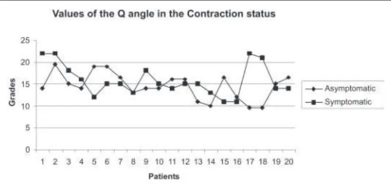
Fig. 4 – Graphic of the measurement of the Q angle attained in the
maxi-mal voluntary isometric Contraction
DISCUSSION
Follow ing the analysis of the results, it is possible to view the difference in the Q angle magnitude betw een symptomatic and asymptomatic individuals in the relaxation status (p = 0.004). The mean value found confirms the estimated values found in the lit-erature, w hich are the follow ing: 15o mean to asymptomatic indi-viduals, and 20o to symptomatic individuals.
The reduction in the Q angle value in the maximal voluntary isometric contraction for asymptomatic individuals w ere also found in a similar study analyzing the Q angle in mean 22 years old healthy w omen in the orthostatic position, w here the expressed values found w ere mean 13o(23).
8e
Rev Bras M ed Esporte _ Vol. 12, Nº 1 – Jan/Fev, 2006 3. Baker V, Bennell K, Stillman B, Cow an S, Crossley K. Abnormal knee jointposi-tion sense in individuals w ith patellofemoral pain syndrome. J Orthop Res 2002; 20:208-14.
4. Alaca R, Yilmaz B, Goktepe AS, M ohur H, Kalyon TA. Efficacy of isokinetic exer-cise on functional capacity and pain in patellofemoral pain syndrome. Am J Phys M ed Rehabil 2002;81:807-13.
5. Nissen CW, Cullen M C, Hew ett TE, Noyes FR. Physical and arthroscopic exami-nation techniques of the patellofemoral joint. J Orthop Sports Phys Ther 1998; 28:277-85.
6. Hung Y, Gross M T. Effect of foot position on electromyographic activity of the vastus medialis oblique and vastus lateralis during low er-extremity w eight-bear-ing activities. J Orthop Sports Phys Ther 1999;29:93-102.
7. Livingston LA. The quadriceps angle: a review of the literature. J Orthop Sports Phys Ther 1998;28: 105-9.
8. Thomeé P, Thomeé R, Karlsson J. Patellofemoral pain syndrome: pain, coping strategies and degree of w ell-being. Scand J M ed Sci Sports 2002;12: 276-81. 9. Andrade PH, Bevilaqua-Grosso D, Bérzin F, Gil I, M onteiro-Pedro V. Comparação
da atividade elétrica dos músculos vasto medial oblíquo e vasto lateral oblíquo em indivíduos com disfunção femoropatelar. Rev Fisioter Univ São Paulo 2001; 8:65-71.
10. Sheehy P, Burdett RG, Irrgang JJ, Vansw earingen J. An electromyographic study of vastus medialis oblique and vastus lateralis activity w hile ascending and de-scending steps. J Orthop Sports Phys Ther 1998;27:423-9.
11. Csintalan RP, Schulz M M , Woo J, M cM ahon PJ, Lee TQ. Gender differences in patellofemoral joint biomechanics. Clin Orthop Related Res 2002;402:260-9. 12. Fulkerson JP, Arendt EA. Anterior knee pain in females. Clin Orthop Related Res
2000;372:69-73.
13. Stiene HA, Brosky T, Reinking M F, Nyland JM , Beth M . A comparison of closed kinetic chain and isokinetic joint isolation exercises in patients w ith patellofemo-ral dysfunction. J Orthop Sports Phys Ther 1996;24:136-41.
14. Hubbard JK, Sampson HW, Elledge JR The vastus medialis oblique muscle and its relationship to patellofemoral joint deterioration in human cadavers. J Orthop Sports Phys Ther 1998;28:384-91.
15. Araujo RC, Amadio AC. Análise biomecânica da ativação das porções superficiais do m. quadríceps femoral durante contrações excêntrica e concêntrica. Rev Bras Fisiot 1996;1:13-20.
16. Sakai N, Luo Z, Rand JA, An K. The influence of w eakness in the vastus medialis oblique muscle on the patellofemoral joint: an in vitro biomechanical study. Clin Biomech 2000;15:335-9.
17. Corrêa JCF, Negrão Filho RF, Dócusse Filho AJ, Quialheiro JJA. Tratamento da instabilidade femoropatelar por meio da estimulação elétrica neuromuscular as-sociada a cinesioterapia. Rev Bras Fisiot 1996;1:37-43.
18. Tomsich DA, Nitz AJ, Threlkeld AJ, Shapiro R. Patellofemoral alignment: reliabil-ity. J Orthop Sports Phys Ther 1996;23:200-8.
19. M agee DJ. Avaliação músculo-esquelética. 3rd ed. São Paulo: M anole, 2002.
20. Tumia N, M affulli N. Patellofemoral pain in female athletes. Sports M edicine and Arthrosc Rev 2002;10:69-75.
21. Holmes SW, Clancy WGJr. Clinical classification of patellofemoral pain and dys-function. J Orthop Sports Phys Ther 1998;28:299-306.
22. Cow an SM , Bennell KL, Crossley KM , Hogdes PW, M acConnell J. Physical ther-apy alters recruitment of the vasti in patellofemoral pain syndrome. M ed Sci Sports Exerc 2002;34:1879-85.
23. Lathinghouse LH, Trimble M H. Effects of isometric quadriceps activation on the Q-angle in w omen before and after quadriceps exercise. J Orthop Sports Phys Ther 2000;30:211-6.
24. Pow ers CM , Landel R, Perry J. Timing and intensity of vastus muscle activity during functional activities in subjects w ith and w ithout patellofemoral pain. Phys Ther 1996;76:946-55.
25. Laprade J, Culham E, Brouw er B. Comparison of five isometric exercises in the recruitment of the vastus medialis oblique in persons w ith and w ithout patel-lofemoral pain syndrome. J Orthop Sports Phys Ther 1998;27:197-204.
26. Karen JM , Ronald SK, M arilyn M P, Bradley F, Jacquelin P. Electromyography of the quadriceps in patellofemoral pain w ith patellar subluxation. Clin Orthop Relat-ed Res 2003;415:261-71.
27. Jones S, Kumar A, Bickerstaff DR, Smith TWD. Axial computed tomography of the patello-femoral joint w ith and w ithout quadriceps contraction. J Bone Joint Surg 2000;82:21.
28. Blazer K. Diagnosis and treatment of patellofemoral pain syndrome in the female adolescent. Physician Assistant 2003;27:23-30.
29. Pow ers C. Rehabilitation of patellofemoral joint disorders: a critical review. J Or-thop Sports Phys Ther 1998;28:345-54.
30. Wilk KE, Reinold M M . Principles of patellofemoral rehabilitation. Sports M ed 2001;9:325-36.
31. Henry J. The patellofemoral joint. Southern M ed J 2004;97:757-61.
32. M ascal CL, Landel RF, Pow ers CM . M anagement of patellofemoral pain targeting hip, pelvis, and trunk muscle function: 2 cases reports. J Orthop Sports Phys Ther 2003;33:642-60.
33. Ireland M L, Willson JD, Ballantyne BT, Davis IM . Hip strength in females w ith and w ithout patellofemoral pain. J Orthop Sports Phys Ther 2003;33:671-6.