Comparative evaluation of cephalometric and occlusal
characteristics between the Long Face pattern and Pattern I
Elisa Gurgel Simas de Oliveira1, Célia Regina Maio Pinzan-Vercelino2
Objective: To compare the cephalometric and intraoral characteristics between Long Face pattern and Pattern I patients, besides evaluating associations between subjective facial patterns, cephalometric facial patterns and the intraoral charac-teristics. Methods: Through evaluation of frontal and right side extraoral photographs, three previously calibrated and experienced examiners selected 30 Long Face patients (Group 1) and 30 Pattern I patients (Group 2), aged between 9 and 19 years, of both genders. The cephalometric characteristics were assessed by the following variables: SN.GoGn, NS.Gn, AIFH, SNA, SNB, ANB, 1.1, 1.NA,1-NA, 1.NB, 1-NB, NA.Po, nasolabial angle and H-Nose. Clinical evaluations were also performed to determine the presence of posterior crossbite, anterior open bite and type of Angle’s malocclu-sion. The cephalometric data were compared by independent t test. The chi-square test was used to evaluate the associa-tion between qualitative variables. Results: Signiicant diferences were observed between groups regarding the variables SN.GoGn, NS.Gn, AIFH, ANB, NA.Pog, 1-NA, 1.NB and 1-NB, with an increase of these measures in Group 1. There were also signiicant diferences between groups on variable 1.1, being lower in Group 1 than in Group 2. Conclu-sions: The Long Face was associated to Angle Class II malocclusion, to the presence of posterior crossbite and to anterior open bite. The Long Face subjective facial pattern was associated to dolichofacial cephalometric pattern.
Keywords: Cephalometry. Orthodontics. Diagnosis.
How to cite this article: Oliveira EGS, Pinzan-Vercelino CRM. Comparative evaluation of cephalometric and occlusal characteristics between the Long Face Pattern and Pattern I. Dental Press J Orthod. 2013 May-June;18(3):86-93.
Submitted: January 22, 2010 - Revised and accepted: December 29, 2010 » The patients displayed in this article previously approved the use of their facial and intraoral photographs.
Contact address: Elisa Gurgel Simas de Oliveira Av. Dom Luís, 906/405 – Bairro Meireles – Brazil
CEP: 60.160-230 – Fortaleza/CE – E-mail: [email protected] » The authors report no commercial, proprietary or financial interest in the products
or companies described in this article.
1 MSc in Orthodontics, UniCEUMA
2 Professor, Post-Graduation Program – MSc in Orthodontics -UniCEUMA
Objetivo: comparar características cefalométricas e intrabucais entre pacientes Padrão Face Longa e Padrão I, além de avaliar as associações entre os padrões faciais subjetivos, os padrões faciais cefalométricos e as características intrabucais. Métodos: por meio da avaliação das fotograias extrabucais frontal e lateral direita, três examinadores experientes e previamente calibrados selecionaram 30 pacientes Padrão Face Longa (grupo 1) e 30 pacientes Padrão I (grupo 2), com idades entre 9 e 19 anos, de ambos os sexos. As características cefalométricas foram avaliadas por meio das seguintes variáveis: SN.GoGn, NS.Gn, AFAI, SNA, SNB, ANB, 1.1, 1.NA, 1-NA, 1.NB, 1-NB, NA.Pog, ângulo nasolabial e H-Nariz. Também foram realizadas avaliações clínicas para determinar a presença de mordida cruzada posterior, mordida aberta anterior e o tipo de má oclusão segundo Angle. Os dados cefalométricos obtidos foram comparados pelo teste t independente. Utilizou-se o teste χ2 para avaliar a associação entre as variáveis qualitativas. Resultados: foram
introduction
The long face is a deformity with skeletal
implica-tion and with unfavorable esthetics,8,10 that can be
ob-served in the three sagittal dental relations, being
how-ever, more associated to Class II sagittal discrepancies.7,10
The children, teenagers and adults that present this excessive vertical facial growth, have a peculiar characteristic, described in literature as “Long Face
syndrome”,4,30 hyperdivergent facial type16,24 and,
re-cently, as Long Face pattern.7-11
The diagnosis of Long Face pattern is based in evaluations of the face morphology and cephalometry. The facial analysis allows to verify several character-istics common to these individuals such as: Absence of passive lip seal and contraction of mentalis muscles
during labial closure,1 besides a great exposure of
up-per incisors when lips are resting and great gingival
exposure during smile,1,4,30 the nose is generally long,
with narrowing of the alar bases and the lower third of the face is increased, resulting in retrognathia
ap-pearance of the mandible.1,26,30
The cephalometry constitutes a necessary instru-ment to define, locate and quantify the skeletal dis-harmony present in patients with Long Face pattern, which can be associated to a horizontal growth of the
condyle6,26 and/or to an excessive posterior growth of
the maxilla.15,30
Regarding the cephalometric characteristics, it is observed an increase on the total anterior height of the
face and on the anterior and inferior facial height.15,17
The anterior and superior facial height is generally nor-mal,1,4,30 but the proportion between medium and lower
third is reduced.3 The angle of the mandibular plane is
increased,4,8,11,15,28,29 as well as the gonial angle.8,11 It is
ob-served a mandibular retropositioning in relation to the
skull base.1,11 The maxilla, however, is well positioned in
relation to the skull base.1,15
Lately, it is observed in literature13,21 that the
cephalometric means, used in several cephalometric tracings, cannot be generically applied for
diagno-sis and treatment. Several researchers5,20,23 concluded
that most cephalometric standards varies signifi-cantly, when compared to several facial patterns. Be-fore this, a more individualized interpretation of the cephalometry must become a rule, or rather, an as-sociation between the cephalometric analysis and the facial analysis must be standard for diagnosis,
plan-ning of treatment and orthodontic treatment. There-fore, the final decision of a planning must be taken based in cephalometric and facial findings. This way, this work had as objective to perform a comparative study of cephalometric and intraoral characteristics between Long Face patients and Pattern I, diagnosed by the subjective facial analysis, besides evaluating the associations between subjective facial patterns and cephalometric facial patterns.
MAtEriAL And MEtHodS Material
The sample was selected according to the follow-ing criteria of inclusion:
» Caucasians;
» Long Face pattern or Pattern I; » Full orthodontic records; » Ages between 9 and 19 years; » No previous orthodontic treatment.
Methods
Orthodontic records
Extraoral, frontal and right side photographs from the orthodontic records belonging to patients regis-tered for orthodontic treatment on the Specialization Course in Orthodontics, of the Odontology Acad-emy of Ceará were used. The sample was selected by three experienced and previously calibrated exami-nators, in distinct moments and each one individu-ally, to avoid that the evaluation of one examinator interfered on the others. Only the patients who had diagnostic concordance were selected.
All photographs were performed by the same ra-diology center (Prof. Perboyre Castelo), presenting a pattern. The patients during the photographs were in
natural position of the head (NPH):22 standing,with
the feet 10 cm apart from each other, lips at rest, looking to an oval mirror located 1 meter before the patient and instructed to observe their own eyes re-flection, keeping the pupils on the eye center.
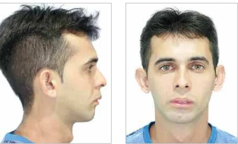
The patients classiied with Long Face pattern,7-11
The Pattern I patients7-11,13 were identiied by the
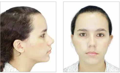
facial normality and characterized by balanced vertical and skeletal sagittal relations on the front and proile evaluations. In frontal photograph they had apparent symmetry, proportion between the facial thirds, pro-portional volume of the lips vermilion and passive lip seal (Fig 3). On the evaluation of the right side pho-tograph, Pattern I patients had mild facial convexity, expressive chin-neck line and parallel to Camper plane, and esthetically pleasant mentolabial sulcus, built with equal participation of the mentum and lower lip (Fig 4).
The patient’s full name, gender and date of birth were obtained also from the orthodontic records. The date of birth allowed the accurate calculus of initial ages of the patients.
All patients that agreed to participate in the re-search filled out a form with their data, as well as signed a free informed term of consent (FITC), ap-proved by the ethics committee in research (CER) of the University Center of Maranhão (UNICEUMA), protocol number 00469/08, according to standards of the resolution CNS 196/96.
Cephalometry
Ater evaluation of the photographs from orthodontic records for selection of the sample, the facial pattern was evaluated through cephalometry with variables that de-termined the facial growth pattern: SN.GoGn, NS.Gn (Axis Y) and AIFH (anterior inferior facial height).
Besides these variables, other measurements were per-formed of the skeletal components (SNA, SNB and ANB), dentoalveolar components (1.1, 1.NA, 1-NA, 1.NB, 1-NB) and bone and tissue proiles (NAPog, na-solabial angle, H-Nose).
The radiographs were performed by the digital pan-oramic x-ray device (Orthopantomograph OP100-D, Instrumentarium, Palodex group, Tuusula, Finland, 2006). This digital device used a sensor that sent the im-ages captured in the radiographic take to the computer screen. These images were saved in CD and printed in ilms by laser printer model Dryview 8150. The digi-tal radiographic images were inserted on the program Cef X (CDT Sotware, Dourados, Mato Grosso, Brazil, version 2.1.24, 1995) and the reference points, lines and planes were demarcated.
The cephalometric facial pattern was determined when the patient showed at least two measures alter-ated. It was considered: Dolichofacial the patient that
showed SN.GoGn with values higher than 34 degrees,19
NS.Gn (Axis Y) with values higher than 69.9 degrees19
and AIFH with values higher than 71 mm;27 mesofacial
the patient that showed SN.GoGn with values within
30 and 34 degrees,19 NS.Gn with values within 63.5
and 69.5 degrees19 and AIFH with values within 63
and 71 mm27 and brachyfacial the patient that showed
SN.GoGn with values lower than 30 degrees,19 NS.Gn
with values lower than 63.5 degrees,19 and AIFH with
values lower than 63 mm.27
Figure 1 and 2 - Frontal and right side
a group of 20% of the radiographs, that were digitalized again and had their points demarcated by the same re-searcher. It was applied the dependent Student’s t test in
order to assess the systematic error.18 For evaluation of
the random error it was used the Dahlberg’s formula.12
Statistical test
It was used the descriptive statistics (mean and standard deviation) for initial age and all cephalomet-ric measures used.
The independent t test was applied to verify the compatibility between the initial ages of studied groups and to compare the cephalometric variables between groups.
It was applied the Chi-square test to evaluate the compatibility of groups regarding the proportion be-tween genders, to verify the association bebe-tween sub-jective facial pattern and cephalometric facial pattern and to evaluate the association of Long Face pattern and intraoral characteristics (posterior crossbite, ante-rior open bite and Angle’s malocclusion).
Besides these tests, it was determined the preva-lences of cephalometric facial patterns, posterior crossbite, anterior open bite and the type of Angle’s malocclusion in relation to subjective facial pattern. Results with p < 0,05 were considered statistically significant. Tests were applied through BioEstat 5.0 (AYRES, Sociedade Civil Mamirauá, MCT-CNPq, Belém, PA, Brazil, 2005).
Clinical assessment
It was illed out a clinical form for each patient con-taining data obtained from orthodontic records and ob-servations such as: Initial age, presence of open bite and/ or crossbite and classiication of Angle’s malocclusion.
The patients classiied with presence of posterior crossbite, were those with abnormal buccolingual rela-tion between superior and inferior teeth, of at least three teeth, when the dental arches were in centric relation,
be-ing uni or bilateral25 and with presence of anterior open
bite when showed a negative overbite between the edges of anterior, superior and inferior teeth, with
measure-ment larger than 1 mm, obtained in millimetric ruler.25
On the investigation of the dental relation, it was ob-served a sagittal relation between the upper and lower
permanent irst molars.2 The patients were classiied as
Class I, when presented a molar relation with the upper permanent irst molar’s mesiobuccal cusp occluding the
lower permanent irst molar’s mesiobuccal sulcus,2 as
Class II when presented the lower permanent irst molar positioned distally in relation to the upper permanent irst molar and as Class III when presented the lower permanent irst molars situated mesially to the upper
permanent irst molars.2
Statistical analysis
Method error
To determine the reliability of the obtained cepha-lometric results, ater 15 days, it was randomly selected
Figure 3 and 4 - Right side and frontal
rESuLtS
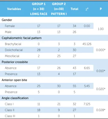
The sample consisted of photographs and telera-diographs of 60 patients, divided in two groups ac-cording to facial pattern (Table 1).
Regarding the method error, for the cephalometric variables it was not detected any random error and only one systematic error. For the variable NS.Gn, it was found, on the irst measurement, a mean value of 72.62 degrees, and for the second measurement a mean value of 73.97 degrees. Considering that it occurred only one systematic error (7.15%) with variation of 1.35 degrees between the measurements, and none random error, the obtained results can be considered reliable.
The groups were compatible in relation to age and distribution of genders (Tables 2 and 3).
Regarding cephalometric variables, the studied groups presented statistically significant differences in: NS.GoGn, NS.Gn, AIFH, ANB, 1.1, 1-NA, 1.NB, 1-NB, NA.Pog (Table 2).
The Long Face pattern was associated to dolicho-facial cephalometric dolicho-facial pattern, and the Pattern I was associated to mesofacial cephalometric facial pat-tern (Table 3).
The Long Face pattern patients were associated to posterior crossbite, anterior open bite and Angle Class II malocclusion (Table 3).
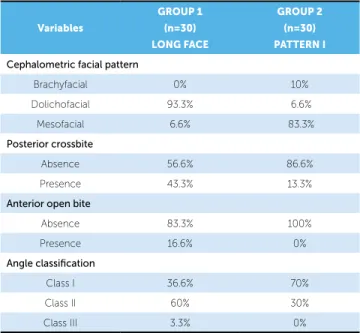
Among the Long Face pattern patients 93.3% were dolichofacial patients, 43.3% with presence of posteri-or crossbite, 16.6% with presence of anteriposteri-or open bite and 60% with Angle Class II malocclusion (Table 4).
Now, on the group of Pattern I patients 83.3% were mesofacial patients, 13.3% with presence of posterior
Variables GROUP 1 (n = 30) LONG FACE
GROUP 2 (n= 30) PATTERN I
Age
Mean ± SD 13.43 ± 2.95 12.83 ± 3.02
Maximum age 9 9
Minimum age 18 19
Gender
Male 13 13
Female 17 17
Table 3 - Analysis of the compatibility of genders, facial pattern, cephalometric facial pattern, posterior crossbite, anterior open bite and classiication of the Angle’s malocclusions between the groups, according to Chi-square test.
Table 1 - Classiication of the sample according to age and gender.
Table 2 - Comparative analysis of cephalometric variables and age between
groups, according to independent t test.
*Statistically signiicant (p < 0.05).
*Statistically signiicant (p < 0.05).
Variables
GROUP 1 (n = 30) LONG FACE
GROUP 2 (n= 30) PATTERN I p
Mean ± SD Mean ± SD
Skeletal components
SNA (degrees) 82.86 ± 4.29 82.59 ± 4.00 0.802
SNB (degrees) 78.00 ± 4.49 79.06 ± 3.92 0.333
ANB (degrees) 4.85 ± 2.22 3.57 ± 2.01 0.022*
Growth pattern
SN.GoGn (degrees) 41.80 ± 6.78 34.37 ± 6.77 0.000*
NS.Gn (degrees) 70.90 ± 4.64 67.76 ± 4.03 0.006*
AIFH (mm) 77.49 ± 8.88 68.64 ± 6.23 0.000*
Dentoalveolar components
1.1 (degrees) 117.11 ± 14.56 124.79 ± 10.58 0.022*
1.NA (degrees) 26.27 ± 10.24 23.02 ± 7.26 0.161
1-NA (mm) 6.27 ± 3.91 4.45 ± 2.33 0.033*
1.NB (degrees) 31.80 ± 7.39 27.80 ± 6.13 0.026*
1-NB (mm) 8.32 ± 2.87 5.78 ± 2.42 0.000*
Osseous profile x tissue profile
NA.Pog (degrees) 9.14 ± 4.91 6.07 ± 4.69 0.016*
ANL (degrees) 98.84 ± 13.54 101.24 ± 9.88 0.435
H-Nose (mm) 2.18 ± 4.62 2.99 ± 3.19 0.434
Compatibility between ages
Age 13.43 ± 2.95 12.83 ± 3.02 0.444
Variables
GROUP 1 (n = 30) LONG FACE
GROUP 2 (n=30) PATTERN I
Total χ2 P
Gender
Female 17 17 34 0.00
1.00
Male 13 13 26
Cephalometric facial pattern
Brachyfacial 0 3 3 45.126
0.000*
Dolichofacial 28 2 30
Mesofacial 2 25 27
Posterior crossbite
Absence 17 26 43 6.65
0.010*
Presence 13 4 17
Anterior open bite
Absence 25 30 55 5.45
0.020*
Presence 5 0 5
Angle classification
Class I 11 21 32 7.125
0.028*
Class II 18 9 27
crossbite, 70% with Angle Class I maloccusion and 30% with Angle Class II malocclusion. However, no Pattern I patient presented anterior open bite (Table 4).
diScuSSion
The subjective facial analysis represents an
impor-tant tool of orthodontic diagnosis,13 that can be easily
used for identification of Long Face pattern patients. These patients generally present several occlusal problems that may be associated to this facial growth pattern as: posterior crossbite, anterior open bite and Class II malocclusion. Many times, when the prob-lem of vertical growth is identified through the face, the problem is also cephalometrically confirmed, however, when the problem is diagnosed cephalo-metrically first, it is not necessarily confirmed on the face, ie, the facial analysis allows a more accurate di-agnosis of the Long Face pattern.
On the cephalometric assessment, the groups presented significative differences in relation to the following variables: SN.GoGn, NS.Gn, AIFH, ANB, 1.1, 1-NA, 1.NB, 1-NB and NA.Pog. The Long Face pattern patients presented increase on the angles: SN.GoGn (41.80±6.78), NS.Gn (70.90±4.64), indicating vertical growth pattern;
Table 4 - Prevalence of cephalometric facial patterns, posterior crossbite,
anterior open bite and type of Angle’s malocclusion on the diferent groups.
Variables
GROUP 1 (n=30) LONG FACE
GROUP 2 (n=30) PATTERN I
Cephalometric facial pattern
Brachyfacial 0% 10%
Dolichofacial 93.3% 6.6%
Mesofacial 6.6% 83.3%
Posterior crossbite
Absence 56.6% 86.6%
Presence 43.3% 13.3%
Anterior open bite
Absence 83.3% 100%
Presence 16.6% 0%
Angle classification
Class I 36.6% 70%
Class II 60% 30%
Class III 3.3% 0%
NA.Pog (9.14±4.91), showing increase of convexity of the bone profile; ANB (4.85±2.22), evidencing the increase on degree of sagittal discrepancy be-tween maxilla and mandible and 1.NB (31.80±7.39) indicating vestibularization of lower incisors in re-lation to bone base. The linear measures 1-NA (26.27±10.24), 1-NB (8.32±2.87) and AIFH (77.49±8.88) were increased; indicating protrusion of upper and lower incisors and increase of the lower third of the face. The measure 1.1, that also present-ed significative difference between groups, showpresent-ed decreased value for Long Face pattern patients, fac-tor indicative of maxillomandibular retrusion.
Cardoso et al11 also found significative differences
in relation to the variables ANB, AIFH, SN.GoGn and NA.Pog when compared the cephalometric
characteristics of Long Face pattern and of Pattern I.11
They observed that Long Face pattern individuals presented increase of the cephalometric measures
lo-cated below the palatal plane.11
Capelozza Filho et al8 evaluating the cephalometric
characteristics of Long Face pattern, did not observe signiicative diferences in relation to sexual dimor-phism and showed that male Long Face pattern pa-tients, when compared to Pattern I papa-tients, presented disparities in relation to magnitudes related to facial height, to growth pattern and to sagittal relation.
The assessment of maxillomandibular relation, based on value of angle ANB, was greater also in Long Face pattern individuals evaluated by Cardoso
et al9 and Capelozza Filho et al,8 showing the
ten-dency to Class II malocclusion that the carriers of this deformity present. The values for anterior and infe-rior facial height were higher for the Long Face pat-tern group with statistical significance, also observed
in studies performed by Cardoso et al9 and Capelozza
Filho et al.8 This result was also expected, since the
increase on anterior inferior facial height constitutes the essence of the studied disease, being frequently found in literature.
litera-ture4,14,15,16,20,28,29 as parameter to deine Long Face
pat-tern individuals. Although the values found in the
stud-ies performed by Capelozza Filho et al8 and Cardoso et
al9 overcome this value, they cannot be used separately.
In fact, the disease consists in an imbalance between the vertical components, and, therefore, a single parameter must not be used; therefore in this study it was associ-ated to the mandibular plane (SN.GoGn), the ment of the Y axis of growth (Ns.Gn) and the measure-ment of the anterior and inferior facial height for deter-mination of cephalometric facial pattern.
The mandibular retrusion was emphasized with the assessment of NAP angle which was significantly different between Long Face pattern and Pattern I
groups also in the study by Cardoso et al,10
show-ing greater mandibular retrognathia and convexity of bone profile in Long Face pattern patients.
As for the cephalometric growth pattern, it was ob-tained 93,3% of dolichofacial patients on Group 1 (Long Face pattern) and 83.3% of mesofacial patients on Group 2 (Pattern I). The Chi-square test showed pres-ence of association between the subjective facial pattern and the cephalometric facial pattern, which allows to conclude that the studied facial patterns, classiied by the subjective analysis, are associated to cephalometric clas-siication of the facial pattern, enabling the methodol-ogy used for identiication of patients by subjective facial analysis, besides allowing the comparison between results from this study and other results from works in literature. The patients on Group 1 presented a prevalence of 43.3% of posterior crossbite and the patients on Group 2 presented 13.3% of posterior crossbite. In relation to the Chi-square test it was observed an
association between facial pattern and posterior cross-bite. The prevalence of posterior crossbite in Long Face pattern patients is according to the observed in a
study by Cardoso et al10 in which the authors showed
a prevalence of 34.2% of this malocclusion.
Regarding the presence of anterior open bite, the patients on Group 1 presented a prevalence of 16.6%, while on Group 2, no patients showed anterior open bite. In relation to the Chi-square test, it was ob-served an association between the Long Face pattern and the presence of anterior open bite.
About Angle’s classiication, the patients on Group 1 presented: 36.6% Class I, 60% Class II and 3.33% Class III; now the patients on Group 2 presented: 70% Class I and 30% Class II. As result of the Chi-square test, it was found an association between the facial pattern and An-gle’s classiication. Patients on Group 1 were associated to Class II malocclusion and patients on Group 2 were associated to Class I malocclusion. This result agrees
with the study performed by Cardoso et al10 who found
in Long Face pattern patients the following prevalences in relation to Angle malocclusions: 13.2% Class I, 71% Class II and 15.8% Class III. Probably, the Long Face pattern is associated to Class II malocclusion for these patients present a clockwise mandibular rotation, which facilitate a Class II sagittal relation.
concLuSionS
1. Angelillo JC, Dolan EA. The surgical correction of vertical maxillary excess: long face syndrome. Ann Plast Surg. 1982;8(1):64-70.
2. Angle EH. Classiication of malocclusion. Dent Cosmos.
1899;41(3):248-64.
3. Bell WH. Correction of skeletal type of anterior open bite. J Oral Surg. 1971;29(10):706-14.
4. Bell WH, Creekmore TD, Alexander RG. Surgical correction of the long
face syndrome. Am J Orthod. 1977;71(1):40-67.
5. Bhat M, Enlow DH. Facial variations related to head form type. Angle Orthod. 1985;55(4):269-80.
6. Bjork A. Prediction of mandibular growth rotation. Am J Orthod.
1969;55(6):585-99.
7. Capelozza Filho L. Diagnóstico em Ortodontia. 1a ed. Maringá: Dental
Press; 2004.
8. Capelozza Filho L, Cardoso MA, An TL, Bertoz FA. Características cefalométricas do padrão face longa: considerando o dimorismo sexual. Rev Dental Press Ortod Ortop Facial. 2007;12(2):49-60.
9. Capelozza Filho L, Cardoso MA, An TL, Lauris JRP. Proposta para
classiicação, segundo a severidade, dos indivíduos portadores de más oclusões do padrão face longa. Rev Dental Press Ortod Ortop Facial. 2007;12(4):124-58.
10. Cardoso MA, Bertoz FA, Reis SAB, Capelozza Filho L. Estudo das características oclusais em portadores de padrão face longa com indicação de tratamento ortodôntico-cirúrgico. Rev Dental Press Ortod Ortop Facial. 2002;7(9):63-70.
11. Cardoso MA, Bertoz FA, Capelozza Filho L, Reis SAB. Características cefalométricas do padrão face longa. Rev Dental Press Ortod Ortop Facial. 2005;10(2):29-43.
12. Dahlberg G. Statistical methods for medical and biological students. New York: Interscience; 1940.
13. Feres R, Vasconcelos MHF. Estudo comparativo entre a análise facial subjetiva e a análise cefalométrica de tecidos moles no diagnóstico ortodôntico. Rev Dental Press Ortod Ortop Facial. 2009;14(2):81-9. 14. Fields HW, Proit WR, Nixon WL, Phillips C, Stanek E. Facial pattern
diferences in long-faced children and adults. Am J Orthod. 1984;85(3):217-23.
REFERENCES
15. Fish LC, Wolford LM, Epker BN. Surgical orthodontic correction of maxillary excess. Am J Orthod. 1978;73(3):241-57.
16. Fitzpatrick BN. The long face and V.M.E. Aust Orthod J. 1984;8:82-9. 17. Frost DE, Fonseca RJ, Turvey TA, Hall DJ. Cephalometric diagnosis
and surgical-orthodontic correction of apertognathia. Am J Orthod. 1980;78(6):657-69.
18. Houston WJ. Analysis of errors in orthodontics measurements. Am J Orthod. 1983;83(5):382-90.
19. Interlandi S. Ortodontia bases para iniciação. 5ª ed. São Paulo: Artes Médicas; 2002.
20. Isaacson JR, Isaacson RJ, Speidel TM, Worms FW. Extreme variations in vertical face growth and associated variation in skeletal and dental relations. Angle Orthod. 1971;41(3):219-29.
21. Jacobson A. Planning for orthognatic surgery-art or science? Int J Adult Orthodon Orthognath Surg. 1990;5(4):217-24.
22. Lundström F, Lundström A. Natural head position as a basis for cephalometric analysis. Am J Orthod Dentofacial Orthop. 1992;101(3):244-7.
23. Metzdorf DW. A cephalometric study of cranial, mandibular and lower incisor morphology in adult face. Angle Orthod. 1977;47(4):288-92. 24. Moloney F, West RA, McNeill RW. Surgical correction of vertical maxillary
excess: a re-evaluation. J Maxillofac Surg. 1982;10(2):84-91.
25. Moyers RE. Ortodontia. 4ª ed. Rio de Janeiro: Guanabara Koogan; 1991. 26. Nielsen IL. Vertical malocclusions: etiology, development, diagnosis and
some aspects of treatment. Angle Orthod. 1991;61(4):247-60. 27. Pinzan A, Pinzan-Vercelino CRM, Martins DR, Janson G, Henriques JFC,
Freitas MR, et al. Crescimento maxilomandibular no sentido antero-posterior e na altura anterior. In: Atlas de Crescimento Craniofacial. 10ª ed. São Paulo: Ed. Santos; 2006. p. 49-54.
28. Prittinen JR. Orthodontic diagnosis of long face syndrome. Gen Dent. 1996;44(4):348-51.
29. Schendel SA, Carlotti AE Jr. Variation of vertical maxillary excess. J Oral Maxillofac Surg. 1985;43(8):590-6.
30. Schendel SA, Eisenfeld J, Bell WH, Epker BN, Mishelevich DJ. The long face syndrome: vertical maxillary excess. Am J Orthod.