Radiol Bras vol.46 número6
Texto
Imagem
Documentos relacionados
Connection of the pulmonary veins to the left atrium, characterized by the flow of the upper pulmonary veins towards the superior vena cava and to the right atrium was identified1.
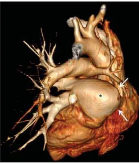
Slight tricuspid insufficiency was evidenced with significant pulmonary insufficiency, hypoplasia of the pulmonary trunk (PT) and pulmonary arteries with the right pulmonary
In case pulmonary embolism is clinically suspected and if there is unequivocal visualization of the thrombus in the atrium, in the right ventricle, or inside the pulmonary artery,
These questions could justify the drop in pulmonary artery pressure (regardless of whether the anomalous pulmonary artery is right or left) immediately after surgical
Total anomalous pulmonary venous drainage, congenital heart defect with increased pulmonary blood flow, pulmonary veins, adult.. patients decreased significantly in the last
In the absence of other associated heart defects the intraparenchymal pulmonary artery tree, ipsilateral to the absent right or left pulmonary artery is usually
The coronary angiographic findings in the patients with atrial septal defect and pulmonary hypertension (ASD + PH): compression of left main coronary artery by pulmonary
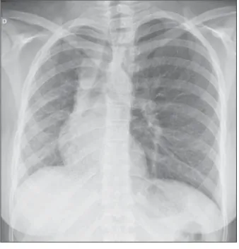
Subsequently he was catheterized and the pulmonary angiography showed agenesis of the right pulmonary artery (Figure 1), ruling out occult pulmonary branch.. The right lung was
