359 Radiol Bras. 2017 Nov/Dez;50(6):359–365
Magnetic resonance imaging indings in central
nervous system cryptococcosis: comparison between
immunocompetent and immunocompromised patients
Achados de ressonância magnética em criptococose do sistema nervoso central: comparação entre pacientes imunocompetentes e imunossuprimidos
Stenio Bruno Leal Duarte1, Mariana Mari Oshima1, João Vitor do Amaral Mesquita1, Felipe Barjud Pereira do Nascimento2, Paula Christina de Azevedo3, Fabiano Reis4
Duarte SBL, Oshima MM, Mesquita JVA, Nascimento FBP, Azevedo PC, Reis F. Magnetic resonance imaging indings in central nervous system crypto
-coccosis: comparison between immunocompetent and immunocompromised patients. Radiol Bras. 2017 Nov/Dez;50(6):359–365.
Abstract
Resumo
Objective: To assess the magnetic resonance imaging (MRI) patterns associated with central nervous system infection with Crypto-coccus sp. in relation to patient immune status.
Materials and Methods: This was a retrospective study of MRI data for 19 patients with neurocryptococcosis who underwent the examination between January 2000 and March 2014. The MRI characteristics examined included lesion topography, aspects of diffusion, T1-weighted images, T2-weighted images, and contrast enhancement patterns.
Results: In all cases, cryptococcal infection was conirmed by cerebrospinal luid analysis. Of the 19 patients, 10 were immuno
-compromised and 9 were immunocompetent. Abnormal imaging patterns occurred alone or in conjunction with other manifesta
-tions. The imaging patterns found in immunocompromised patients included the following: leptomeningeal enhancement, in 6; pachymeningeal enhancement, in 3 (due to intracranial hypotension in 2); perivascular space involvement, in 4; granulomas, in 2; hydrocephalus, in 2; miliary nodules, in 1; and plexitis, in 1. In immunocompetent patients, the following imaging patterns were observed: leptomeningeal enhancement, in 5; perivascular space involvement, in 3; granulomas, in 3; cryptococcoma, in 1; ven
-triculitis, in 1; and hydrocephalus, in 1. In 2 immunocompetent patients, diffusion-weighted imaging showed diffusion restriction in cerebral cryptococcal granuloma.
Conclusion: In both groups, the most common imaging inding was leptomeningeal enhancement, followed by dilatation of perivas
-cular spaces with the presence of mucoid material. Rare presentations, such as miliary nodules, plexitis, ventriculitis, and pachyme
-ningeal enhancement, were also observed. None of the imaging patterns common to immunocompetent and immunocompromised patients differed signiicantly in frequency between them.
Keywords: Cryptococcosis/diagnostic imaging; Central nervous system infections/diagnostic imaging; Brain injuries/pathology; Magnetic resonance imaging; Diagnostic imaging; Meninges/pathology.
Objetivo: Avaliar os padrões de ressonância magnética (RM) associados à infecção do sistema nervoso central por Cryptococcus sp. em relação ao status imunológico dos pacientes.
Materiais e Métodos: Dados de RM de 19 casos de neurocriptococose foram analisados retrospectivamente de janeiro de 2000 a
março de 2014. As características de RM examinadas incluíram: sítio da lesão, aspectos em difusão, imagens ponderadas em T1 e T2 e padrões de realce pelo contraste.
Resultados: A infecção por Cryptococcus sp. foi conirmada pela análise do liquor em todos os casos. Dos 19 pacientes, 10 eram imunossuprimidos e 9 eram imunocompetentes. Os padrões de imagem anormais ocorreram isoladamente ou em associação com outras manifestações. Os padrões de imagem encontrados nos pacientes imunossuprimidos incluíram: realce leptomeníngeo (n =
6), realce paquimeníngeo (n = 3; 2 devidos a hipotensão intracraniana), envolvimento do espaço perivascular (n = 4), granulomas (n = 2), hidrocefalia (n = 2), nódulos miliares (n = 1) e plexite (n = 1). Em pacientes imunocompetentes, os padrões de imagem incluíram: realce leptomeníngeo (n = 5), envolvimento do espaço perivascular (n = 3), granulomas (n = 3), criptococoma (n = 1), ventriculite (n = 1) e hidrocefalia (n = 1). As sequências ponderadas em difusão mostraram restrição em 2 pacientes imunocompe
-tentes com granulomas intracerebrais por criptococose.
Conclusão: O achado mais comum de imagem em ambos os grupos foi realce leptomeníngeo, seguido de dilatação dos espaços
perivasculares pela presença do material mucoide. Apresentações raras como nódulos miliares, plexite, ventriculite e realce paqui
-meníngeo foram também observadas. Nenhum dos padrões de imagem comuns aos pacientes imunocompetentes e imunossupri
-midos diferiu signiicativamente em frequência entre eles.
Unitermos: Criptococose/diagnóstico por imagem; Infecções do sistema nervoso central/diagnóstico por imagem; Lesões encefáli
munodeiciency syndrome (AIDS), after infection with human immunodeiciency virus (HIV) and Toxoplasma
gondii(3). The major environmental sources of C.
neofor-mans include soil contaminated with pigeon excreta (C. neoformans var. neoformans and C. neoformans var. grubii) and eucalyptus trees/decaying wood (C. neoformans var. gattii)(3). C. neoformans var. gattii is found mainly in tropi-cal and subtropitropi-cal regions, whereas C. neoformans var. neoformans is encountered worldwide. C. neoformans var.
neoformans usually infects immunodeicient individuals,
leading to acute diffuse meningitis or
meningoencepha-litis. In contrast, infection with C. neoformans var. gattii
more typically manifests as a granulomatous inlammatory
response in immunocompetent hosts(4).
The respiratory tract is the primary site of fungal in-fection in humans, and the yeast forms of fungi spread
he-matogenously from the lungs to the CNS(5,6), from which
they penetrate the meningeal vessel walls, migrating to the
Virchow-Robin (perivascular) spaces, which subsequently become dilated following the activation of inlammatory
cells and the deposition of mucoid material(7). Once the
fungus crosses the blood-brain barrier, the CNS provides an appropriate environment for fungal multiplication. C. neoformans has a predilection for the CNS because of
the presence of speciic neuronal substrates, especially
neurotransmitters, that can be used by the fungus to pro-duce melanin, which protects the fungus against oxida-tive stress, phagocytosis, and antifungal drugs, as well as
modifying the host immune responses(2).
The most common clinical indings in CNS cryptococ -cal infection are headache, nausea, and fever, less com-mon manifestations are meningism, confusion (altered mental state), seizures, visual symptoms, and focal
neu-rological deicit(6,8). A diagnosis of fungal CNS infection
spectrum of magnetic resonance imaging (MRI) patterns associated with CNS cryptococcal infection relects the
pathological behavior of the fungus.
The aim of this study was to examine the MRI pat -terns of CNS cryptococcal infection in immunocompetent and immunocompromised patients. This is of particular interest because differences have been observed between those two groups of patients in terms of the presentation
of this disease and have been associated with speciic viru
-lence factors, as well as with host-pathogen interactions(4).
MATERIALS AND METHODS
We retrospectively reviewed the cranial MRI scans of
19 patients with microbiologically proven CNS cryptococ-cosis. We excluded patients with other associated infec-tions (such as toxoplasmosis and tuberculosis) and those
without MRI follow-up. The structural images had been acquired in 1.5 T and 3 T MRI scanners (Achieva; Philips, Best, the Netherlands). The following MRI characteristics
were analyzed by an experienced neuroradiologist: lesion
topography; aspects of diffusion; T1- and T2-weighted images; and contrast enhancement patterns. The images were obtained between January 2000 and March 2014.
Because our study was retrospective, the image
acquisi-tion protocol was not the same for all patients. In this re -gard, leptomeningeal abnormalities were more
conspicu-ous when contrast-enhanced luid-attenuated inversion recovery (FLAIR) sequences were used.
RESULTS
The mean age of the subjects was 41 years (range, 20–58 years); 73.7% were male, and 26.3% were female. Of the 19 patients evaluated, 10 (52.6%) were immuno -compromised: 1 was a transplant recipient, and 9 had
AIDS.
Among the 10 immunocompromised patients, the following imaging patterns were identiied (Figures 1–4): leptomeningeal enhancement, in 6 (60%); pachymenin
-geal enhancement, in 3 (30%); perivascular space involve
-ment, in 4 (40%); cryptococcal granulomas, in 2 (20%); hydrocephalus, in 2 (20%); miliary nodules, in 1 (10%); and plexitis, in 1 (10%). None of the immunocompro -mised patients showed a normal imaging pattern. Five of
the patients (50%) had 2–3 concomitant MRI indings, the remaining 5 patients (50%) presenting with a single inding. Among the 3 patients with pachymeningeal en -hancement, it was secondary to intracranial hypotension
(with diffuse enhancement) in 2 and represented focal
pachymeningeal enhancement in 1 (Table 1). Study conducted in the Radiology Department of the Faculdade de Ciências
Médicas da Universidade Estadual de Campinas (FCM-Unicamp), Campinas, SP, Brazil.
1. MD, Resident in the Radiology Department, Faculdade de Ciências Médicas da Universidade Estadual de Campinas (FCM-Unicamp), Campinas, SP, Brazil.
2. MD, Attending Physician, Departamento de Radiologia e Diagnóstico por Ima -gem, Hospital Israelita Albert Einstein, São Paulo, SP, Brazil.
3. Master Degree, Neurologist, Head the Neuroinfectious Disease Clinic, Neu -rology Department, Faculdade de Ciências Médicas da Universidade Estadual de Campinas (FCM-Unicamp), Campinas, SP, Brazil.
4. PhD, Head of the Neuroradiology Sector, Professor in the Radiology Depart -ment, Faculdade de Ciências Médicas da Universidade Estadual de Campinas (FCM --Unicamp), Campinas, SP, Brazil.
Mailing address: Dr. Fabiano Reis. Departamento de Radiologia – FCM-Unicamp. Rua Tessália Vieira de Camargo, 126, Cidade Universitária Zeferino Vaz. Campinas, SP, Brazil, 13083-887. E-mail: [email protected].
Figure 2.Case 9. Contrast-enhanced axial T1-weighted image showing mild hydrocephalus. There is also focal pachymeningeal enhancement adjacent to the left parietal lobe.
Figure 1.Case 2. Contrast-enhanced coronal T1-weighted image showing su
-pratentorial and infratentorial focal enhancement (black arrows). In this case, there is also diffuse pachymeningeal enhancement (white arrow), due to intra
-cranial hypotension.
²
²
²
Figure 3.Case 5. Contrast-enhanced T1-weighted image showing miliary punc
-tate enhancement at the centrum semiovale and at the cortico-subcortical junction (arrows).
²
²
²
²
²
Figure 4.Case 4. Contrast-enhanced axial T1-weighted image showing bilater
²
²
²
²
Figure 5.Case 7. Axial T2-weighted image (A) and axial T1-weighted image (B) showing bilateral dilated perivascular spaces (arrows).
A B
A B
Figure 6.Case 18. Cryptococcoma. Sagittal T1-weighted image showing a lesion with a heterogeneous, hypointense signal in the superior vermis (A). Axial T2-weighted image of the same lesion (B) showing a heterogeneous hyperintense signal and perilesional edema.
Among the 9 immunocompetent patients, the imaging patterns identiied (Figures 5–7) included the following: leptomeningeal enhancement, in 5 (55.5%); perivascular space involvement, in 3 (33.3%); cryptococcal granulomas, in 3 (33.3%); cryptococcoma, in 1 (11.1%); ventriculitis,
in 1 (11.1%); and hydrocephalus, in 1 (11.1%). None of
the immunocompetent patients showed a normal imaging pattern. None of the patients in this group had miliary nodules, plexitis, or pachymeningeal enhancement. Four
MRI indings, the remaining 5 patients (55.6%) presenting with a single inding (Table 1). Diffusion-weighted imaging
of two immunocompetent patients showed restricted
dif-fusion in cerebral cryptococcal granulomas. In one of our
immunocompetent patients, spectroscopy showed a treha-lose and lipid/lactate peak in a brain granuloma.
In the study sample as a whole, the most common im
-aging inding was leptomeningeal enhancement, followed
by dilatation of the perivascular spaces. None of the imag-ing patterns common to immunocompetent and
immuno-compromised patients differed signiicantly in frequency,
as assessed by Fisher’s exact test.
DISCUSSION
CNS cryptococcosis produces a wide variety of MRI
features that may vary depending on the immunological
status of the patient. As shown here, the MRI indings
range from single to multiple alterations such as
hydro-cephalus, leptomeningeal/pachymeningeal enhancement, dilated perivascular spaces, miliary nodules, plexitis (via hematogenous dissemination), and pseudotumor (cryp-tococcoma), occurring in isolation or concomitantly with
other MRI indings.
Chronic granulomatous reactions caused by C. neo-formans are more common in immunocompetent hosts than in those with immunosuppression. On T1-weighted images, cryptococcal granulomas appear as hypointense
lesions, with or without homogenous enhancement(5). In
the present study, the most common MRI indings in im -munocompetent patients were variable-sized masses with low signal intensity on T1-weighted images and high
sig-nal intensity on T2-weighted images, accompanied by ring
or nodular enhancement and vasogenic edema.
Several authors have described the radiological
pat-terns in HIV-infected patients with CNS cryptococco -sis(2,3,6,8–12). The immunocompromised patients examined
Table 1—Characteristics of the 19 patients with neurocryptococcosis included in the study.
Case 1 2 3 4 5 6 7 8 9 10 11 12 13 14 15 16 17 18 19 Age (years) 55 40 33 37 43 41 35 48 44 50 32 20 30 58 55 29 41 26 39 Gender Male Female Female Female Male Male Male Male Male Male Male Male Male Male Male Male Male Female Female HIV status Negative/organ transplantation Positive Positive Positive Positive Positive Negative Negative Positive Negative Negative Negative Negative Negative Positive Positive Positive Negative Negative Findings Multiple Multiple Single Multiple Single Multiple Single Multiple Multiple Multiple Single Multiple Multiple Single Single Single Single Single Single Imaging pattern Granuloma/leptomeningeal/perivascular space Pachymeningeal*/leptomeningeal/granuloma Leptomeningeal Leptomeningeal/pachymeningeal*/perivascular space/plexitis Miliary Leptomeningeal/hydrocephalus Perivascular space Leptomeningeal/hydrocephalus Hydrocephalus/pachymeningeal Perivascular space/leptomeningeal/ventriculitis Granuloma Leptomeningeal/granuloma Perivascular space/leptomeningeal Leptomeningeal Perivascular space Perivascular space Leptomeningeal Cryptococcoma Granuloma
* Intracranial hypotension.
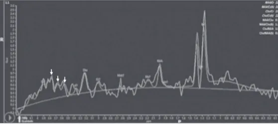
Figure 7. Case 19. Proton magnetic
resonance spectroscopy of the core of the lesion tissue showing a lactate peak at 1.3 ppm and multiple signals in the region of 3.6–3.8 ppm (arrows) corresponding to trehalose.
mature image acquisition after contrast administration
in those other studies; low CD4 expression in the HIV-infected group in those studies; and the enhanced immu -nological status among our patients following the
intro-duction of highly active antiretroviral therapy (HAART),
whereas the patients evaluated in other studies had no
access to HAART.
There was a time gap of 10–15 years between sev -eral important studies(1,2,5,9) and the present investigation. During that period, there were major advances in image
acquisition technology and clinical protocols that have markedly enhanced the diagnostic sensitivity of imaging
methods. Andreula et al.(11) detected leptomeningeal en-hancement in 7 of 8 HIV-infected patients with CNS cryp -tococcosis based on an analysis of T1-weighted sequences after delayed image acquisition and the use of double the
normal dose of contrast (20 mL of gadolinium, compared with the 10-mL dose used in the present study).
Dilatation of the perivascular spaces was a common inding in both groups in the present study. The perivas
-cular space is deined as a potential space that involves
a vessel and is an extension of the subarachnoid space. Most commonly located in the basal ganglia, white
mat-ter, cerebellum, and brainstem(13), with a “soap bubble
appearance”, gelatinous round masses within the peri-vascular spaces appear as round foci with intermediate to low signal intensity on T1-weighted images and high
signal intensity on T2-weighted images. Coalescence of
the perivascular spaces is often seen, leading to a mild
mass effect. Vasogenic edema is not present, and there
may be little or no enhancement at the periphery of these lesions(1–7,9,13).
In CNS cryptococcosis, the leptomeningeal involve
-ment and inlammatory reaction are mild and result in the
production of mucoid material within the subarachnoid space, a process that may extend to the perivascular spaces,
which typically become dilated and illed with mucoid material, inlammatory cells, and organisms(9).
Crypto-coccal meningitis is more common in the basal cisterns, although supratentorial leptomeningeal involvement was
more prevalent in the study conducted by Sarkis et al.(14).
FLAIR sequences were assessed in two of the patients in
our study. The value of contrast-enhanced T1-weighted images in detecting leptomeningeal disease is question-able, because cortical vessels can mimic meningeal en-hancement, leading to erroneous radiological
interpreta-tion and misdiagnosis(15,16). However, contrast-enhanced
FLAIR sequences are especially useful when the data ob -tained with T1-weighted images are inconclusive, because
contrast-enhanced FLAIR imaging should be included in the brain MRI protocol of HIV-infected patients and of
patients suspected of having leptomeningeal fungal
in-fection. Katchanov et al.(19) described a pattern of
lepto-meningeal enhancement and vasculitis of the small
per-forating arteries in HIV-infected patients under immune
reconstitution.
Cryptococcomas (accumulations of fungi, inlamma -tory cells, and gelatinous mucoid material) arise during in-fection and can extend to the parenchyma as focal masses,
having a tumor-like appearance(1,3,7,12). Again, our indings
differed from data reported in the literature. For example, whereas other studies reported no enhancement in
granu-lomas(1,2), 20% of the immunocompromised patients in
our study had granulomas with contrast enhancement.
An extensively immunocompromised system and poor in
-lammatory response in the host in the pre-HAART era could partially explain these divergent indings(19). Among the immunocompetent patients in our study, 44.4% had a
granulomatous reaction (cryptococcoma or granuloma), a
common inlammatory immune response in patients with
a preserved immune system.
Intraparenchymal cryptococcomas are mass-like le -sions that may mimic a brain tumor, as seen in one of
our patients (case 18). The correct diagnosis of crypto -coccomas, particularly in immunocompetent individuals, is challenging because these lesions may show a very high choline/N-acetylaspartate ratio in proton magnetic
reso-nance spectroscopy(20) and may require a biopsy to
con-irm the diagnosis (as was done here).
Choroid plexus disease (case 4) was characterized by
a multilobulated cystic appearance and abnormal
hyper-intense signal in T2-weighted FLAIR sequences, with in -tense enhancement on gadolinium-enhanced T1-weighted images. Choroid plexitis is a rare manifestation of CNS cryptococcal infection(10).
Restricted diffusion was observed in two cases of ce
-rebral cryptococcal granuloma, with a solid pattern. A sim
-ilar inding was reported by Kamezawa et al.(5), whereas Ho et al.(21) described indings contrary to this in the cen
-tral cavity of a cryptococcal lesion. Consequently, diffu-sion cannot differentiate between fungal and bacterial infection. Cerebral infarcts, as a cause of restricted
dif-fusion, were observed in 20% of the patients studied by
Loyse et al.(12). However, no cortical or lacunar infarcts
were observed in our patients.
On spectroscopy, one of our patients showed a
is speciic, although not highly sensitive, for fungal infec
-tion. Using spectroscopy, Luthra et al.(22) found a
treha-lose peak in cryptococcoma walls and cerebral
mucormy-cosis in 5 of the 8 patients evaluated, where it appeared as multiple signals ranging from 3.6 ppm to 3.8 ppm; this proile helps to distinguish fungal infection (which shows
a trehalose peak) from bacterial infection (which shows peaks in amino acid, acetoacetate, succinate, and alanine but no trehalose peak).
Fisher’s exact test was used in order to compare the
MRI indings between the two groups (immunocompe -tent and immunocompromised patients) in this study. No correlation was observed between the imaging param-eters and patient immune status, in contrast with other reports(1,6,11).
One limitation of the present study is the small num-ber of patients in the sample, which could explain the lack
of signiicant indings. None of the patients in our study had normal MRI results. That might relect a certain bias
in patient selection, because this study was conducted at a tertiary-care teaching hospital. Miliary nodules, plexitis, and pachymeningeal enhancement were found only in immunocompromised patients and at frequencies greater
than those reported in the literature(1–3,7–10). Pachyme
-ningeal enhancement was diffuse in two cases (patients with other features of intracranial hypotension) and focal, indicative of focal fungal disease (not usually reported in the literature), in one.
The principal limitation of this study was its small sample size. The retrospective nature of the investiga-tion also limited the possibilities for interveninvestiga-tion. Further studies, involving larger numbers of patients, standard-ized imaging protocols, and reliable data collection with
regard to the use of antiretroviral therapy and luconazole
maintenance therapy, could provide useful information on CNS cryptococcosis.
REFERENCES
1. Miszkiel KA, Hall-Craggs MA, Miller RF, et al. The spectrum of MRI indings in CNS cryptococcosis in AIDS. Clin Radiol. 1996;51:842–50.
2. Mathews VP, Alo PL, Glass JD, et al. AIDS-related CNS cryptococ
-cosis: radiologic-pathologic correlation. AJNR Am J Neuroradiol. 1992;13:1477–86.
3. Smith AB, Smirniotopoulos JG, Rushing EJ. From the archives of the AFIP: central nervous system infections associated with human immunodeiciency virus infection: radiologic-pathologic correla
-tion. Radiographics. 2008;28:2033–58.
4. Grosse P, Tintelnot K, Söllner O, et al. Encephalomyelitis due to Cryptococcus neoformans var gattii presenting as spinal tumour:
case report and review of the literature. J Neurol Neurosurg Psy
-chiatry. 2001;70:113–6.
5. Kamezawa T, Shimozuru T, Niiro M, et al. MRI of a cerebral crypto
-coccal granuloma. Neuroradiology. 2000;42:441–3.
6. Starkey J, Moritani T, Kirby P. MRI of CNS fungal infections: re -view of aspergillosis to histoplasmosis and everything in between.
Clin Neuroradiol. 2014;24:217–30.
7. Cheng YC, Ling JF, Chang FC, et al. Radiological manifestations
of cryptococcal infection in central nervous system. J Chin Med
Assoc. 2003;66:19–26.
8. Corti M, Villafañe MF, Negroni R, et al. Magnetic resonance imag
-ing ind-ings in AIDS patients with central nervous system crypto
-coccosis. Rev Iberoam Micol. 2008;25:211–4.
9. Berkefeld J, Enzensberger W, Lanfermann H. Cryptococcus menin
-goencephalitis in AIDS: parenchymal and meningeal forms. Neuro
-radiology. 1999;41:129–33.
10. Kumari R, Raval M, Dhun A. Cryptococcal choroid plexitis: rare imaging indings of central nervous system cryptococcal infection in an immunocompetent individual. Br J Radiol. 2010;83:e14–7. 11. Andreula CF, Burdi N, Carella A. CNS cryptococcosis in AIDS:
spectrum of MR indings. J Comput Assist Tomogr. 1993;17:438– 41.
12. Loyse A, Moodley A, Rich P, et al. Neurological, visual, and MRI brain scan indings in 87 South African patients with HIV-associ
-ated cryptococcal meningoencephalitis. J Infect. 2015;70:668–75. 13. Saigal G, Post MJD, Lolayekar S, et al. Unusual presentation of
central nervous system cryptococcal infection in an
immunocompe-tent patient. AJNR Am J Neuroradiol. 2005;26:2522–6.
14. Sarkis RA, Mays M, Isada C, et al. MRI indings in cryptococcal meningitis of the non-HIV population. Neurologist. 2015;19:40–5. 15. Galassi W, Phuttharak W, Hesselink JR, et al. Intracranial menin
-geal disease: comparison of contrast-enhanced MR imaging with luid-attenuated inversion recovery and fat-suppressed T1-weighted sequences. AJNR Am J Neuroradiol. 2005;26:553–9.
16. Mathews VP, Caldemeyer KS, Lowe MJ, et al. Brain: gadolinium-enhanced fast luid-attenuated inversion-recovery MR imaging. Ra
-diology. 1999;211:257–63.
17. Mamourian AC, Hoopes PJ, Lewis LD. Visualization of intrave
-nously administered contrast material in the CSF on luid-attenu
-ated inversion-recovery MR images: an in vitro and animal-model investigation. AJNR Am J Neuroradiol. 2000;21:105–11.
18. Jackson EF, Hayman LA. Meningeal enhancement of fast FLAIR images. Radiology. 2000;215:922–4.
19. Katchanov J, Branding G, Jefferys L, et al. Neuroimaging of
HIV-associated cryptococcal meningitis: comparison of magnetic
reso-nance imaging indings in patients with and without immune re
-constitution. Int J STD AIDS. 2016;27:110–7.
20. Batista RR, Gasparetto EL. Uncommon presentation of intracranial cryptococcoma in an immunocompetent patient. AJNR Am J Neu
-roradiol. 2012;33:E26.
21. Ho TL, Lee HJ, Lee KW, et al. Diffusion-weighted and conventional magnetic resonance imaging in cerebral cryptococcoma. Acta Ra
-diol. 2005;4:411–4.
22. Luthra G, Parihar A, Nath K, et al. Comparative evaluation of fun -gal, tubercular, and pyogenic brain abscesses with conventional and