Florida Elicits Molecular-Level Changes in Gene
Expression Indicative of Progesterone and Androgen
Exposure
Erica K. Brockmeier1*, B. Sumith Jayasinghe1, William E. Pine2, Krystan A. Wilkinson2,3, Nancy D. Denslow1,4*
1Department of Physiological Sciences, Center for Environmental and Human Toxicology, University of Florida, Gainesville, Florida, United States of America, 2Department of Wildlife Ecology and Conservation, University of Florida, Gainesville, Florida, United States of America,3Chicago Zoological Society, c/o Mote Marine Laboratory, Sarasota, Florida, United States of America,4Genetics Institute, University of Florida, Gainesville, Florida, United States of America
Abstract
Endocrine disrupting compounds (EDCs) are chemicals that negatively impact endocrine system function, with effluent from paper mills one example of this class of chemicals. In Florida, female Eastern mosquitofish (Gambusia holbrooki) have been observed with male secondary sexual characteristics at three paper mill-impacted sites, indicative of EDC exposure, and are still found at one site on the Fenholloway River. The potential impacts that paper mill effluent exposure has on theG. holbrookiendocrine system and the stream ecosystem are unknown. The objective of this study was to use gene expression analysis to determine if exposure to an androgen receptor agonist was occurring and to couple this analysis within vitro assays to evaluate the presence of androgen and progesterone receptor active chemicals in the Fenholloway River. Focused gene expression analyses of masculinizedG. holbrookifrom downstream of the Fenholloway River paper mill were indicative of androgen exposure, while genes related to reproduction indicated potential progesterone exposure. Hepatic microarray analysis revealed an increase in the expression of metabolic genes in Fenholloway River fish, with similarities in genes and biological processes compared toG. holbrookiexposed to androgens. Water samples collected downstream of the paper mill and at a reference site indicated that progesterone and androgen receptor active chemicals were present at both sites, which corroborates previous chemical analyses. Results indicate thatG. holbrooki downstream of the Fenholloway River paper mill are impacted by a mixture of both androgens and progesterones. This research provides data on the mechanisms of how paper mill effluents in Florida are acting as endocrine disruptors.
Citation:Brockmeier EK, Jayasinghe BS, Pine WE, Wilkinson KA, Denslow ND (2014) Exposure to Paper Mill Effluent at a Site in North Central Florida Elicits Molecular-Level Changes in Gene Expression Indicative of Progesterone and Androgen Exposure. PLoS ONE 9(9): e106644. doi:10.1371/journal.pone.0106644
Editor:Elizabeth Wilson, University of North Carolina at Chapel Hill, United States of America
ReceivedMay 28, 2014;AcceptedAugust 7, 2014;PublishedSeptember 8, 2014
Copyright:ß2014 Brockmeier et al. This is an open-access article distributed under the terms of the Creative Commons Attribution License, which permits unrestricted use, distribution, and reproduction in any medium, provided the original author and source are credited.
Data Availability:The authors confirm that all data underlying the findings are fully available without restriction. All relevant data are within the paper and its Supporting Information files, with the exception of microarray data which is held in the Gene Expression Ontology (GEO) database (accession number GSE49238).
Funding:This study was supported by the University of Florida College of Veterinary Medicine Faculty Grant. Additional support to EB was provided by the U.S. Environmental Protection Agency Science to Achieve Results (STAR) Fellowship 91728101-0. The funders had no role in study design, data collection and analysis, decision to publish, or preparation of the manuscript.
Competing Interests:The authors have declared that no competing interests exist.
* Email: [email protected] (EB); [email protected] (ND)
Introduction
In the early 1980s, female Eastern mosquitofish (Gambusia holbrooki) with elongated anal fins, mimicking the fins of adult males, were first documented from Elevenmile Creek, Florida [1]. This small stream served as the wastewater outfall of a large paper mill for several decades and has been known to be contaminated both ‘‘chemically and biologically’’ [2]. Additional surveys for masculinized femaleG. holbrookifound similar results downstream of other paper mill impacted sites in Florida including the St. John’s River at Rice Creek [3] and the Fenholloway River [4]. These findings are not limited to the US, as masculinized female Western mosquitofish (G. affinis) have recently been found downstream of an active paper mill in China [5]. Regulatory
policies in the US such as the Clean Water Act have led to improvements in toxicity of industrial effluents via reductions of chemicals such as dioxins, ammonia, and metals. However, endocrine disrupting chemicals (EDCs) are often still prevalent in this type of effluent, with aquatic wildlife residing downstream of paper mill-impacted areas exhibiting increased proportions of single sex embryos, abnormal sex steroid levels, modulations in egg yolk proteins, and reduced gonad sizes [6].
expression [7]. The compound androstenedione (AED), a precur-sor of testosterone in the steroidogenic pathway, was initially associated with the activation of the AR, and this chemical was found in Fenholloway River water and sediment. However, follow-up analysis demonstrated that AED was not in the individual HPLC fraction associated with the AR activation, as fractions were pooled before chemical identification was conducted [8,9]. Furthermore, a 6-week exposure ofG. affinisto the concentration of AED present in Fenholloway River water (0.04 ng/L) did not result in anal fin masculinization [10].
Other biological endpoints inG. holbrookifrom the Fenhollo-way River suggest larger liver masses and smaller body masses and lengths, as well as an increase in the number of oocytes in masculinized female G. holbrooki from the Fenholloway River [11]. Later studies found reductions in the number of early and late stage embryos in G. holbrooki residing downstream of the paper mill [12], potentially due to the lack of normalization of these endpoints to body mass [11,12].
Previous studies in the Fenholloway River succeeded in evaluating the physiological impacts of paper mill exposure on resident mosquitofish. However, better insights into the mecha-nisms of action of chemicals in paper mill effluent can be obtained by including molecular endpoints in these analyses [13]. mRNA levels of bone growth factor sonic hedgehog (shh) [14,15] and steroidogenic enzyme 17b-hydroxysteroid dehydrogenase 3
(17bhsd3) are potential biomarkers of androgen exposure in G. holbrooki [16,17]. In fish, vitellogenin (vtg) and zona pellucida
glycoproteins (zp) are crucial components of oocyte quality [18] and decreases in their expression often occur during androgen exposure [15,19], which can possibly lead to low-quality eggs. Hepatic transcriptome analysis can provide mechanistic informa-tion on the impacts of toxicant exposure, as the liver is the primary detoxification organ and comes into contact with any chemicals that fish are exposed to in the field.
Transcriptomics experiments of fish exposed to androgenic chemicals have revealed large impacts on the biological processes of metabolism [16,19] so focusing on the liver is of relevance for evaluating the androgenicity of paper mill effluents. In addition,in vitro methods in the lab can be used to help screen samples of unknown chemical contaminants for their ability to activate the transcription of genes via nuclear receptors. This has been previously demonstrated in the Fenholloway River below an active paper mill [7] and when data from these assays are coupled with molecular data from the fish impacted by the effluents, they can provide additional information for determining the type(s) of chemicals present and impacting aquatic organisms at this location.
The objective of this study was to evaluate changes in gene expression coupled with in vitro nuclear receptor assays to evaluate the androgenicity of water downstream of the paper mill on the Fenholloway River. Two specific aims were developed: (1) evaluate mRNA levels ofvtg,17bhsd3, andzp2in the liver,shhin the anal fin, and global hepatic gene expression profiles associated with paper mill exposure, and (2) determine if chemicals in the Fenholloway River could bind to the ligand binding domain of androgen and progesterone receptors. We hypothesized that modulations in gene expression patterns and in vitro analyses would be indicative of androgen exposure and that global gene expression analysis via microarrays would provide insights into the mode(s) of actions of the chemicals present in the effluent. This addresses an important gap in knowledge by evaluating the type(s) of EDC that impactG. holbrookiat this site.
Materials and Methods
Sample collection
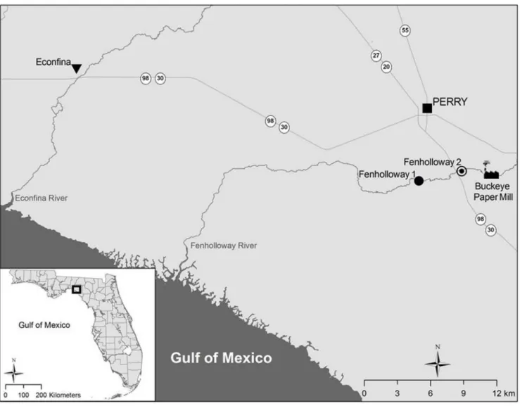
WildG. holbrookiwere captured from two sites downstream of the Buckeye Kraft Pulp and Paper Mill on the Fenholloway River as well as one site at the Econfina River which does not receive paper mill effluent and served as a reference site (Fig. 1). Only sexually matureG. holbrooki(females.15 cm standard length and with the presence of the gravid spot) were collected. In Summer 2012, three sampling events were conducted, once at the Fenholloway River downstream of the paper mill along County Road 361A (Fenholloway 1; GPS coordinates: N 30 058.3419, W 83 588.5699) and twice on the Econfina River (GPS coordinates: N 30 08.5499, W 83 51.9629). Additional sampling was conducted in 2013 to obtain fin samples from the Fenholloway River along US 98 (Fenholloway 2; GPS coordinates N 30 03.9259, W 83 33.4709) and the Econfina River. A 1/80mesh seine was used to collectG. holbrooki. This study was carried out in accordance with the recommendations laid out in ‘‘Guide for the Care and Use of Laboratory Animals’’ by the National Institutes of Health. The protocol was approved by the Institutional Animal Care and Use Committee of the University of Florida (Protocol 201105665). Field sampling was conducted under Florida Fish and Wildlife Commission fishing licenses; all water access points were public areas and no protected species were sampled.
Fish were transferred to 5 gallon aerated buckets filled with site water and were processed immediately after collection. Fish were euthanized using a lethal dose (300 mg/L) of Tricaine-S (Western Chemical, Ferndale, USA) and sacrificed via spinal transection. Anal fin elongation and oocyte stage, staged according to previously published criteria, [20] were assessed upon dissection. Livers and anal fins were excised and stored in RNAlater (Qiagen, Hilden, Germany) overnight at 4uC before long-term storage at
280uC. A subset of fin samples collected in 2012 (N = 4 per site) was used for evaluating the number of bone segments in the third ray of the anal fin using methods previously described [15].
For water samples, collection took place at each of the three sites previously indicated as well as at a site upstream of the Econfina River collection area (GPS coordinates N 30 08.5499, W 83 51.9629) in April 2013. EPA-approved 2.5 L amber bottles (Fisher Scientific, Hampton, USA) were filled with site water by placing the open beaker into the river using a 3 meter long net with only flowing surface water collected. Water was kept on ice immedi-ately after collection, brought to pH,4 to prevent bacterial growth after returning to the laboratory, and stored at 4uC until further processing.
RNA extraction
Hilden, Germany) for 15 seconds and 2 minutes at 4uC at 20,800 g were conducted before elution in 30mL RNase-free water. RNA quality and quantity of all samples were assessed using the Nanodrop (ThermoScientific, Waltham, USA). The A260/A280
values of all samples ranged from 1.75 to 2.18.
Quantitative polymerase chain reaction analysis
Real-time quantitative polymerase chain reaction (qPCR) analysis was performed using the myiQ single color real-time PCR detection system and analyzed by the iQ software (BioRad, Hercules, USA) as previously described [15]. 200 ng cDNA from livers and 25 ng cDNA from anal fins was amplified using SYBR Green (BioRad, Hercules, USA). Negative controls of water (no template control) and RNA (minus RT control) were analyzed on each plate. Gene copy number and qPCR efficiencies for all genes exceptshhwere determined using absolute standard curves [15]. For shh, serial dilutions of anal fin RNA were used to calculate reaction efficiency. The average qPCR reaction efficiency was 93.6%. Melt curve analysis revealed that all samples formed a single peak, with no primer dimers or alternate products formed.
RPL8 variability across all samples was 9.67%. qPCR primer
sequences can be found in the supporting information (Table S1 in File S1).
Microarray sample preparation
RNA integrity numbers (RINs) of stage-matched RNA samples (N = 4 per site) were determined with the 2100 BioAnalyzer (Agilent, Santa Clara, USA); samples had a range of RIN’s from 7.8 to 8.9. The Low RNA Input Amplification Kit for one color labeling (Cy3) (Agilent, Santa Clara, USA) was used to synthesize cRNA using procedures previously described [16]. Purified cRNA concentrations and specific activities were determined by Nano-drop (ThermoScientific, Waltham, USA) with a specific activity.
8 as the cut-off for further processing [16]. Hybridization of 600 ng cRNA and slide scanning was conducted as previously described [16]. The microarray data set (GSE49238) was submitted to the Gene Expression Omnibus (GEO) database, with all reports and deposits made according to the Minimum Information About a Microarray Experiment (MIAME) guidelines [21].
Figure 1. Spatial representation of the two sites where female Eastern mosquitofish (Gambusia holbrooki) were collected for this study.The paper mill-impacted sites include Fenholloway River sites 1 and 2 and the reference site in the Econfina River.G. holbrookiwere collected from Fenholloway site 1 on July 12th, 2012 and from Fenholloway site 2 on April 10th, 2013.G. holbrookiwere collected from the Econfina River on August 7thand October 4th, 2012. The map was created using ArcGIS v10.0 (Esri, Redlands, USA).
Water sample extraction andin vitroanalysis
Water (1.5 L) from each of the four collection sites was passed through Whatman filter paper (GE Healthcare, Maidstone, UK) via vacuum filtration and stored at 4uC until solid phase extraction (SPE). Water samples were extracted using 6 mL Oasis HLB columns (Waters, Milford, USA) after conditioning the columns with 5 mL of methanol and 5 mL of milli-Q water. Water samples were passed through columns using a vacuum manifold with a flow rate of 10 mL/min. After the entire sample was passed through the cartridge, organic constituents were eluted with 5 mL of methanol, evaporated to dryness with nitrogen, reconstituted in 100mL of dimethyl sulfoxide (DMSO), and stored at280uC until bioanalysis.
The GeneBLAzer progesterone receptor (PR) and androgen receptor (AR) assays (Life Technologies, Carslbad, USA) were conducted as previously described [22]. This assay functions in stably transfected mammalian cells with a chimera receptor gene that contains the human AR or PR ligand binding domain linked to the DNA binding domain of Gal 4. The cells also contain a stably transfected reporter gene with the DNA binding sites for Gal 4 in the promoter region upstream of the beta-lactamase gene. In the presence of ligands for the receptors, the chimera is transactivated and binds to sites on the promoter for the beta lactamase gene, resulting in transcription and translation of the beta lactamase enzyme. To measure beta lactamase activity, a substrate with two fluors is used; these fluors are in close proximity and exhibit fluorescence resonance energy transfer (FRET) at 520 nm (green spectrum) and upon hydrolysis the substrate fluoresces at 447 nm (blue spectrum).
To measure the amount of ligand in the water extract, the DMSO reconstituted samples were diluted 1:200, 1:400, 1:800 and 1:1600 in cell culture media. After an 16 h incubation, cells were loaded with LiveBLAzer-FRET B/G Substrate and incubated for 2 h. Fluorescence emission values at 460 nm and 530 nm were obtained using a standard fluorescence plate reader (Synergy H1 Hybrid Reader, Bio-Tek, Winooski, USA). The samples were analyzed on the same plate as a standard curve of levonorgestrel and R1881 for the PR and AR assays respectively. Bioanalytical equivalence quotients (BEQs) were calculated for the field samples by comparing the activity of the water samples to the standard curves following previously described methods [22] (Fig S3 in File S1). The EC10(10% effect concentration) value was
used and extrapolated back to the original 1.5 L of water collected at the sites to determine the chemical concentrations at each site. Data is presented as a ratio of blue spectrum values to green spectrum value, which is indicative of PR or AR transactivation.
Statistical analysis
Anal fin elongation was quantified by methods previously described [15] as a ratio of the measurement of the base of the anal fin (ray 6) to the total length of the fin (ray 4). Liver qPCR data were analyzed as the ratio of the gene copy number per ng RNA versus the RPL8 copy number per ng RNA multiplied by the averageRPL8value. All hepatic gene expression results were log-transformed before statistical analyses. Statistical analysis of shh
was conducted by the delta delta Ct method as previously described [16]. A Student’s t-test was used (after confirming normality and homogeneity of data distributions) to determine if there was a statistically significant difference between anal fin elongation, bone segment numbers, and gene expression levels between the Fenholloway River and the Econfina River. Results of all statistical tests were considered significant ata= 0.05.
Microarray data quality control analysis was completed as described previously [16] and data processing and analysis were
conducted with JMP Genomics 6.0 (SAS Institute, Cary, USA). A one-way analysis of variance (ANOVA) was conducted after LOcal regrESSion (loess) normalization to determine genes that were differentially expressed between sites. Genes that were statistically significantly differentially expressed with .1.5-fold change were subjected to hierarchical cluster analysis [16,22,23]. Changes in Gene Ontology (GO) Biological Processes were determined using the Fisher’s Exact test (Fisher raw p-value ,
0.05, FDR a= 0.05). Visualization of significant pathways and genes was conducted using PathwayStudio (Elsevier, Amsterdam, The Netherlands) based on ResNet 9.0.
Gene expression similarities were evaluated between femaleG. holbrooki downstream of the paper mill-impacted site and G. holbrooki that were exposed to the potent androgen 17b -trenbolone (TB) [16]. For this analysis, a one-way ANOVA was used to determine genes that were differentially expressed between samples from the Fenholloway River, the Econfina River, and the TB exposure versus the vehicle control. Only mRNAs that were altered in a statistically significant manner in any group (paper mill collection sites or laboratory TB treatment) compared to controls were used in further analysis. This focused list of genes from this set that were expressed in the same direction in the paper mill-impacted site fish and the TB-exposed fish were subjected to cluster analysis [16,23,24].
Results
Field sites and anal fin elongation
Since the Econfina River has been used in previous studies as a control site for the Fenholloway River [7,11,12,25] we selected the Econfina as the reference site due to the foundation of knowledge available for this location. The selection of a reference site in a separate system was further justified due to the reduced potential for complications from upstream migration of paper mill-impacted fish. A significant increase in anal fin elongation was seen inG. holbrookifrom the Fenholloway River (Student’s t-test, p,0.001) as well as the number of bone segments in the third ray of the anal fin (Student’s t-test, p = 0.029) (Fig. 2A) which was absent in females from the Econfina River. The distribution of anal fin elongation size classes based on site and collection season is also provided, demonstrating an increase in the number of masculin-ized females in the Fenholloway River during both collection seasons (Fig. 2B).
Focused Gene Expression Analyses
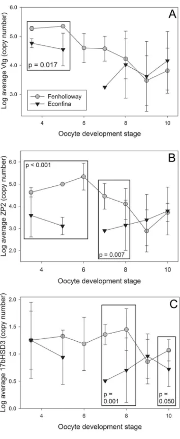
An increase in the mRNA levels of hepatic vtg, zp2, and
17bhsd3was seen in femaleG. holbrookifrom the Fenholloway River (Fig. 3). Due to differences in gene expression based on oocyte development stage [26], apost-hocanalysis was conducted. Forvtg, significantly increased expression was observed at early oocyte development stages (Student’s t-test, p = 0.017 stages 3 through 5) (Fig. 3A), for zp2 at both early and mid-stages of development (Student’s t-test, p,0.001 stages 3 through 5; p = 0.007 stages 7 through 8), (Fig. 3B), and for17bhsd3at mid-stages of oocyte development (Student’s t-test, p = 0.001 mid-stages 7 through 8; p = 0.050 stage 10) (Fig. 3C). There was also significantly increased expression ofshhin masculinized Fenhollo-way River femaleG. holbrookianal fins (Student’s t-test, p = 0.012, N = 22 Econfina River and 16 Fenholloway River) (Fig. 4).
Hepatic Transcriptome and Pathway Analysis
genes downregulated in the Fenholloway River (One-way ANOVA, p,0.05, FDR a= 0.05, fold change .61.5) (Fig. S1 in File S1). A complete list of all statistically significant transcripts with .61.5 fold change can be found in the supporting information (File S2).
In total, 22 gene ontology (GO) biological processes were significantly enriched inG. holbrookifrom the Fenholloway River (Fisher’s Exact test, p,0.05) (Table 1). Of note are pathways related to metabolism (isoprenoid biosynthetic process, energy reserve metabolic process, cyclic nucleotide biosynthetic process), reproduction (gonad development, cell migration involved in gastrulation), and nuclear processes (rRNA processing, mRNA
transport, DNA topological change).G. holbrookiexposed to the potent androgen receptor agonist 17b-trenbolone (TB) in the lab [16] and G. holbrooki from the Fenholloway River share some similarly enriched GO biological processes, including regulation of protein metabolic process (go:0051246) and mRNA transport (go:0051028). Enriched processes and their connection to genes were visualized by PathwayStudio (Fig. S2 in File S1) and these results corroborate the findings of the Fisher’s Exact test while revealing interactions between key metabolic pathways and the differential regulation of several genes linked to these processes.
We found similarity between hepatic gene expression data sets inG. holbrookifrom the paper mill-impacted site andG. holbrooki
Figure 2. Anal fin elongation and bone segment formation in anal fin ray 3 of female Eastern mosquitofish (Gambusia holbrooki) collected from the Fenholloway and Econfina Rivers.Data are repsresented as (A) Mean (6standard deviation) of all collection events and, (B) Distribution of anal fin elongation classes between both paper mill exposed (Fenholloway) and reference (Econfina) field sites. An asterisk indicates statistical significance between the two groups as determined using a Student’s t-test with p,0.05. For anal fin elongation levels there was an N of 46 and 50 from the Fenholloway and Econfina rivers respectively and a subset N of 4 from both sites for the bone segment evaluation.
exposed to 1mg TB/L for 14 days when compared to those from the reference site or exposed to a vehicle control (Fig. 5) [16]. In a set of 62 similarly regulated genes, TB-exposed and PME-exposed
G. holbrookihepatic expression profiles cluster together, and the reference site and vehicle control samples cluster separately (Fig. 5). A complete list of these genes can be found in the supporting materials (File S3).
In vitroassay results
Fig. 6A demonstrates the PR ligand transactivation of chemicals present in water collected from both the reference and paper mill impacted field sites. All water samples had detectable levels of PR activation as determined by the GeneBLAzer assay. The levonorgestrel BEQs of the field site water are as follows: Econfina creek, 3.33 ng/L (10.65 pM); Econfina boat ramp, 4.24 ng/L (13.56 pM); Fenholloway Hwy 98, 3.21 ng/L (10.28 pM); and Fenholloway Co Rd 361A, 3.28 ng/L (10.48 pM). Both sites also had detectable AR transactivation (Fig. 6B) but at a reduced concentration as compared to the PR assay. The R1881 BEQs of these sites are: Econfina creek, 2.24 ng/L (7.86 pM); Econfina boat ramp, 2.58 ng/L (9.07 pM); Fenholloway Hwy 98, 1.94 ng/ L (6.82 pM); and Fenholloway Co Rd 361A, 0.804 ng/L (2.83 pM). Using this assay, levonorgestrel was also demonstrated to activate the AR in the GeneBLAzer assay more strongly than P4 (Fig. 6C).
Discussion
We found that masculinization of femaleG. holbrookicontinues to occur in the Fenholloway River. Paper mill effluent exposure is associated with both anal fin elongation as well as with significantly increased bone segment formation at this site. Additionally, we found an increase in the mRNA levels ofvtg, zp2, 17bhsd3, and
shhin Fenholloway RiverG. holbrooki. Through comparison of hepatic gene expression patterns to data from laboratory exposures, we found that paper mill effluent exposure resulted in an increase of genes associated with metabolic pathways, with 62 genes similarly expressed byG. holbrookiexposed to androgens, indicating a similarity between impacts at the molecular level between paper mill and androgen exposure. We also found detectable levels of both AR and PR ligands in the transactivation Figure 3. Focused hepatic gene expression patterns in female
Eastern mosquitofish (Gambusia holbrooki) using qualitative polymerase chain reaction analysis. Gene expression of (A) vitellogenin (vtg), (B) zona pellucida glycoprotein 2 (zp2), (C) 17b -hydroxysteroid dehydrogenase 3 (17bhsd3) was analyzed between paper mill exposed (Fenholloway) and reference (Econfina) collection sites. Each point represents the mean of each group and error bars represent standard deviation; time points with no standard deviation have an N of 1. A box indicates statistical significance between groups at the selected oocyte developmental stages as determined using a Student’s t-test with p,0.05.
Figure 4. Anal fin gene expression patterns of sonic hedgehog (shh) in female Eastern mosquitofish (G. holbrooki).An asterisk indicates statistical significance between the paper mill exposed (Fenholloway) and reference (Econfina) rivers as determined using a Student’s t-test with p,0.05.
Table 1.Significantly differentially regulated biological processes in femaleG. holbrookiresiding downstream of a paper mill as compared to a control site as determined by gene set enrichment and gene ontology (GO) analyses.
Gene Ontology Biological Process Fisher’s Raw p-value Type of regulation Dif. rega Not dif. regb
go:0006511; ubiquitin-dependent protein catabolic process 0.00659 Enriched 0.950% 0.472%
go:0006816; calcium ion transport 0.00968 Enriched 0.712% 0.328%
go:0051603; proteolysis involved in cellular protein catabolic processes
0.00014 Enriched 0.626% 0.144%
go:0006364; rrna processing 0.02156 Enriched 0.561% 0.246%
go:0051246; regulation of protein metabolic process 0.02663 Enriched 0.453% 0.185%
go:0044419; interspecies interaction between organisms 0.01696 Enriched 0.410% 0.144%
go:0001947; heart looping 0.04791 Enriched 0.388% 0.164%
go:0051028; mrna transport 0.00914 Enriched 0.367% 0.103%
go:0009615; response to virus 0.04432 Enriched 0.259% 0.082%
go:0042074; cell migration involved in gastrulation 0.01105 Enriched 0.237% 0.041%
go:0008299; isoprenoid biosynthetic process 0.00075 Enriched 0.216% 0.00%
go:0008045; motor axon guidance 0.00526 Enriched 0.216% 0.021%
go:0018149; peptide cross-linking 0.01959 Enriched 0.216% 0.041%
go:0009887; organ morphogenesis 0.00994 Enriched 0.194% 0.021%
go:0006890; retrogrAED vesicle-mediated transport, golgi to er 0.00318 Enriched 0.173% 0.000%
go:0048738; cardiac muscle tissue development 0.01865 Enriched 0.173% 0.021%
go:0008406; gonad development 0.00652 Enriched 0.151% 0.000%
go:0006265; dna topological change 0.03469 Enriched 0.151% 0.021%
go:0007266; rho protein signal transduction 0.03469 Enriched 0.151% 0.021%
go:0006112; energy reserve metabolic process 0.02748 Enriched 0.108% 0.000%
go:0009190; cyclic nucleotide biosynthetic process 0.02748 Enriched 0.108% 0.000%
go:0016339; calcium-dependent cell-cell adhesion 0.02748 Enriched 0.108% 0.000%
aPercentage of genes within the GO category that were significantly differentially regulated between Econfina and Fenholloway groups. bPercentage of genes within the GO category that were not significantly differentially regulated between Econfina and Fenholloway groups.
doi:10.1371/journal.pone.0106644.t001
assay in concentrated water samples collected from both the paper mill impacted and reference sites.
Anal fin ratios correlate with earlier observations of elongation in field samples collected in 2010 (Brockmeier, unpublished results) and the increase in the level of bone segmentation is also similar to previous findings [7,12]. Because the mechanism of anal fin growth in mosquitofish is associated with androgen concentration [14,27,28], these data appear to support an ongoing androgen exposure occurring at this site. In addition to anal fin growth, the mRNA levels of shh were upregulated in females from the Fenholloway River (Fig. 4). This gene is expressed in mosquitofish fry and adult females during androgen exposure [14,15], and is constitutively expressed in adult male anal fins (Erica K. Brockmeier, Ph.D. dissertation, University of Florida, 2013). Other androgenic gene expression responses have also been seen downstream of other paper mills, such asspigginexpression in the kidneys of female three-spined stickleback (Gasterosteus aculeatus) exposed to paper mill effluent [29]. This gene is, however, only usable in members of theGasterosteusgenus, which is not found in Florida. The benefit of measuringshhor other biomarker genes to evaluate androgenicity, in addition to anal fin elongation, is that significant changes in gene expression occurs at time points of exposure earlier than physiological responses [14,15]. This sensitivity in terms of exposure timing can serve as an ‘early warning’ biomarker, indicating exposure to an androgenic compound before the physiological effects of anal fin growth manifest.
We found significant differences in hepatic mRNA expression forvtgandzp2gene (Fig. 3A and 3B). Differences invtgoccurred for oocyte development stages where active vitellogenesis occurs [20] and zp2 differences were apparent during later stages of oocyte development, corresponding to this gene’s role in oocyte maturation [18]. Previous laboratory studies in fish have indicated that exposure to androgenic chemicals negatively impactvtgand
zpgene expression inG. holbrooki[15,16], as well as in other fish species [19,30]. Our findings of increases in these mRNAs did not support the initial hypothesis. However, our findings match results from other fish species exposed to paper mill effluent extracts. The
Vtg protein was significantly increased in rainbow trout ( Onco-rhynchus mykiss) after intraperitoneal (IP) injection with primary extracts [31] and after exposure to 100% combined mill outfall (CMO) and in 10% untreated kraft effluent exposures to fathead minnow [32]. While gene and protein levels are not always directly correlated, the upregulation of this protein in rainbow trout and fathead minnow and the gene in mosquitofish may be due to the presence of phytoestrogens in these effluents such as b-sitosterol [6]. This supports the findings of paper mill-specific gene expression patterns, as both androgenic and estrogenic responses were observed.
Hepatic 17bhsd3 mRNA was significantly increased in G. holbrookiresiding downstream of the pulp and paper mill plant on the Fenholloway River (Fig. 3C). This increase was seen at advanced oocyte development stages [20,26]. Other forms ofhsd
proteins, including the protein 3bhsd, have also been found upregulated in the livers of male fathead minnows (Pimephales promelas) after exposure to CMO [33]. Previous work conducted at the Fenholloway River indicated that males from both the paper mill impacted and the reference sites had similar levels of both
testosterone and E2, as was the case for females [11]. Due to the lack of differences in sex steroid levels amongG. holbrookifrom these sites, it is possible that induced expression of genes for steroidogenic enzymes such as17bhsd3may modulate abnormal steroid levels as a homeostatic response to EDC exposure [34]. In addition, other enzymes related to steroid synthesis, such as aromatase, also have significantly increased activities in G. holbrookiin the Fenholloway River [25].
A distinct pattern of differential gene expression was present in the livers of female G. holbrooki from the Econfina and the Fenholloway rivers (Fig. S1 in File S1). Using gene ontology analysis, numerous pathways and processes were found to be enriched in the livers of the Fenholloway River G. holbrooki
(Table 1, Fig. S2 in File S1). Notable pathways include nuclear processes such as mRNA transport and rRNA processing as well as several pathways linked to metabolism, including regulation of protein metabolic process, isoprenoid biosynthetic process, and energy reserve metabolic process. Similar results were found in studies conducted on fathead minnows exposed to the CMO of an androgenic paper mill effluent in Canada, another paper mill with processes similar to those used at the Fenholloway River site [33]. In male fathead minnows, the isoprenoid biosynthetic process was also upregulated in the liver, along with several pathways linked to steroid and cholesterol metabolic processes. Female fathead minnows exposed to CMO experienced enrichment in the proteolysis pathway [33], similar to our results of significantly increased ubiquitin-dependent protein catabolic process. Isopren-oid biosynthesis is the necessary precursor for cholesterol synthesis, which serve as templates for sex steroid synthesis. Genes upstream of the synthesis of cholesterol have previously been found to be upregulated by androgen exposure [35], lending support to the hypothesis that chemicals present in paper mill effluents are acting to disrupt the normal metabolism of steroids and hormones in a manner similar to androgen exposures [31].
We found a similarity in hepatic gene expression between TB-exposedG. holbrookiandG. holbrookifrom the Fenholloway River (Fig. 5). Within this list of genes, GO biological processes for metabolic process, biosynthetic process, regulation of transcrip-tion, and steroid biosynthetic process are present, providing additional evidence for the importance of changes to metabolic pathways—notably steroid biosynthesis—as the toxic mechanism of action of androgen and paper mill effluent exposure.
Progesterone and androgen receptor active chemicals were found to be present at two sites downstream of the paper mill-impacted area of the Fenholloway and Econfina rivers as determined by the GeneBLAzer assay (Fig. 6). While this is not a direct measurement of chemicals (e.g. GC-MS), we were able to determine a concentration of chemicals which could bind to the ligand binding domain of both the PR and AR and transactivate the receptors. We report their bioanalytical equivalency quotients (BEQs), calculated based on the positive standards R1881 and levonorgestrel as known AR and PR activators respectively. These values were between 3.28 and 4.24 ng/L levonorgestrel equiva-lencies and between 0.804 and 2.58 ng/L R1881 equivaequiva-lencies respectively. Based on these results, it appears that natural progesterones are not the causative chemical(s) in inducing abnormal anal fin elongation in the Fenholloway River, as PR-positive activity is present both in the Fenholloway River Figure 6. Results of progesterone receptor (PR) and androgen receptor (AR) GeneBLAzer assays for concentrated water samples collected downstream of the Fenholloway River paper mill and the Econfina River conservation area.The graph bars represent the mean of three replicates from each dilution for the PR (A) and AR (B) assays. Error bars represent standard deviation of the three replicates per assay. The dose-responses of levonorgestrel, progesterone, and 17b-trenbolone (C) were also evaluated by the AR assay.
downstream of the paper mill and at the Econfina River. In previous studies, both progesterone (P4) and the weak androgen androstenedione (AED) were present in HPLC fractions that induced androgen receptor-mediated activity using an in vitro
assay [7,9]. AED was found in the water column and sediment of the Fenholloway River at 0.04mg/L and 0.7mg/L respectively, whereas P4 was present at much higher levels at 2.06mg/L (water) and 48.8mg/L (sediment) [36]. Compared to these concentra-tions, the effective concentrations found in the water samples in this study are reduced by,100-fold and,4-fold, as determined
by levonorgestrel and R1881 equivalencies respectively (Fig. 6). The presence of low levels of AR-active chemicals at both sites may be due to the breakdown of this chemical from high levels of progesterones in the sediments via microorganisms [37]. The source of these progesterones is most likely the loblolly pine (Pinus taeda L.) which is highly prevalent along this entire area [38]. As anal fin elongation is found only at the Fenholloway River, this suggests that the causative chemical is specific to that site or may be related to a combined effect from both progesterone and androgen loads. While chemicals in the sediment may not normally be available to G. holbrooki, as they generally feed off organisms that reside towards the water surface, abiotic pressures present in the Fenholloway River, such as very low dissolved oxygen, may drive G. holbrookiin this area to alternative food sources at the river bottom (i.e. Nematocera) [39]. This would result in theG. holbrookidownstream of the Fenholloway being exposed to a greater load of progesterones and androgens, which could explain the increases of expression of reproductive genes [40] as well as anal fin elongation via breakdown of P4 into androgenic compounds [37].
At other paper mill sites, the phytoestrogen b-sitosterol is thought to be the causative estrogenic chemical [41], and this chemical has been found (but not quantified) at the Fenholloway River paper mill-impacted site [39]. The presence of phytoestro-gens is an area of future work towards determining the causative chemicals and impacts of paper mill exposure in the Fenholloway River ecosystem. In addition, there may be synergistic interactions between non-androgenic chemicals present in the paper mill effluent, as these effluents represent complex mixtures with a diversity of effects [32] that may be causing the manifestation of abnormal secondary sexual characteristics at this site.
While AED was initially thought to be the causative masculin-izing chemical, a follow-up exposure ofG. affinisto doses of AED present in the Fenholloway River (0.04mg/L) showed no statistically significant increase in anal fin elongation after 6 weeks of exposure [10]. These impacts were not evaluated on early life stages, which may be more sensitive to androgen exposure, but nonetheless it appears that AED may not be the masculinizing chemical in this system, and other currently unknown androgenic and progestagenic chemical(s) may be present. Additional studies
which focus on the role of sediments as well as the transferability of chemicals from adult femaleG. holbrookito their offspring during oocyte development would help address the role of soil contact and maternal transfer of paper mill exposure in this environment.
Conclusions
Our findings indicate that a mixture of progesterones and androgens may be driving changes in gene expression at this paper mill-impacted site. These findings provide the insights into the complex nature and persistence of gene expression patterns that coincide with male phenotypic characteristics, and can serve as an initial research focus towards efforts to examine exposure routes and possible chemical sinks in field samples at a site with a population ofG. holbrookithat has exhibited abnormal physiology for over 30 years.
Supporting Information
File S1 File S1 contains qPCR primer sequences (Table S1), hierarchical cluster analysis for the paper mill and reference site females (Fig. S1), PathwayStudio analysis of metabolic gene changes during paper mill exposure (Fig. S2), and standard curves for both the AR and PR GeneBLAzer assays are provided (Fig. S3).
(DOCX)
File S2 File S2 contains fold change levels of all transcripts that were significantly differentially regulat-ed from the paper mill-impactregulat-ed and reference sites. (XLSX)
File S3 File S3 is a list of genes with similar regulation between the paper mill-impacted G. holbrooki and G. holbrooki exposed to the potent androgen receptor agonist 17b-trenbolone.
(XLS)
Acknowledgments
The authors would like to thank J. Bisesi, V. Dang, N. Cole, Z. Slagle, I. Rodriguez, and F. Hayes for their invaluable assistance with fish captures and dissections. The contents of this article are solely the responsibility of the authors and do not necessarily represent the views of any of the funding agencies.
Author Contributions
Conceived and designed the experiments: EB WP ND. Performed the experiments: EB BSJ. Analyzed the data: EB BSJ KW. Contributed reagents/materials/analysis tools: WP ND. Contributed to the writing of the manuscript: EB BSJ WP KW ND.
References
1. Howell WM, Black DA, Bortone SA (1980) Abnormal expression of secondary sex characters in a population of mosquitofish,Gambusia affinis holbrooki -Evidence for Environmentally-Induced Masculinization. Copeia 1980(4): 676– 681.
2. Friends of Perdido Bay homepage, ‘‘A Brief History of Perdido Bay by Jim Lane’’. Available: http://www.friendsofperdidobay.com/hispb.htm#N_3_. Ac-cessed 2013 Nov 9.
3. Bortone SA, Cody RP (1999) Morphological masculinization inpoeciliidfemales from a paper mill effluent receiving tributary of the St. Johns River, Florida, USA. B Environ Contam Tox 63(2): 150–156.
4. Bortone SA, Drysdale DT (1981) Additional evidence for environmentally-induced intersexuality inPoeciliidfishes. Association of Southeastern Biological Bulletin 28: 67.
5. Hou LP, Xie YP, Ying GG, Fang ZQ (2011) Developmental and reproductive characteristics of western mosquitofish (Gambusia affinis) exposed to paper mill
effluent in the Dengcun River, Sihui, South China. Aquat Toxicol 103(3–4): 140–149.
6. Hewitt LM, Kovacs TG, Dub MG, MacLatchy DL, Martel PH, et al. (2008) Altered reproduction of fish exposed to pulp and paper mill effluents: Roles of individual compounds and mill operating procedures. Environ Toxicol Chem 27(5): 1226–1226.
7. Parks LG, Lambright CS, Orlando EF, Guillette LJ, Ankley GT, et al. (2001) Masculinization of female mosquitofish in kraft mill effluent-contaminated Fenholloway River water is associated with androgen receptor agonist activity. Toxicol Sci 62(2): 257–267.
9. Jenkins R, Angus RA, McNatt H, Howell WM, Kemppainen JA, et al. (2001) Identification of androstenedione in a river containing paper mill effluent. Environ Toxicol Chem 20(6): 1325–1331.
10. Stanko JP, Angus RA (2007) In vivo assessment of the capacity of androstenedione to masculinize female mosquitofish (Gambusia affinis) exposed through dietary and static renewal methods. Environ Toxicol Chem 26(5): 920– 926.
11. Toft G, Baatrup E, Guillette LJ (2004). Altered social behavior and sexual characteristics in mosquitofish (Gambusia holbrooki) living downstream of a paper mill. Aquat Toxicol 70(3): 213–222.
12. Orlando EF, Bass DE, Caltabiano LM, Davis WP, Gray LE, et al. (2007) Altered development and reproduction in mosquitofish exposed to pulp and paper mill effluent in the Fenholloway River, Florida USA. Aquat Toxicol 84(4): 399–405. 13. Denslow ND, Garcia-Reyero N, Barber DS (2007) Fish ‘n’ chips: the use of
microarrays for aquatic toxicology. Mol Biosyst 3(3): 172–177.
14. Ogino Y, Katoh H, Yamada G (2004) Androgen dependent development of a modified anal fin, gonopodium, as a model to understand the mechanism of secondary sexual character expression in vertebrates. Febs Lett 575(1–3): 119– 126.
15. Brockmeier EK, Ogino Y, Iguchi T, Barber DS, Denslow ND (2013) Effects of 17beta-trenbolone on Eastern and Western mosquitofish (Gambusia holbrooki
andG. affinis) anal fin growth and gene expression patterns. Aquat Toxicol 128–129: 163–70.
16. Brockmeier EK, Yu F, Moraga D, Bargar TA, Denslow ND (2013) Custom microarray construction and analysis for determining potential biomarkers of subchronic androgen exposure in the Eastern Mosquitofish (Gambusia holbrooki). BMC Genomics 14(1): 660.
17. Mindnich R, Moller G, Adamski J (2004) The role of 17 beta-hydroxysteroid dehydrogenases. Mol Cell Endocrinol 218(1–2): 7–20.
18. Arukwe A, Goksoyr A (2003) Eggshell and egg yolk proteins in fish: hepatic proteins for the next generation: oogenetic, population, and evolutionary implications of endocrine disruption. Comp Hepatol 2(1), 4.
19. Dorts J, Richter CA, Wright-Osment MK, Ellersieck MR, Carter BJ, et al. (2009) The genomic transcriptional response of female fathead minnows (Pimephales promelas) to an acute exposure to the androgen, 17beta-trenbolone. Aquat Toxicol 91(1): 44–53.
20. Haynes JL (1995) Standardized classification of poeciliid development for life-history studies. Copeia 1995(1): 147–154.
21. Brazma A, Hingamp P, Quackenbush J, Sherlock G, Spellman P, et al. (2001) Minimum information about a microarray experiment (MIAME) - toward standards for microarray data. Nat Genet 29(4): 365–371.
22. Escher BI, Mayumi A, Altenburger R, Bain PA, Patrick B, et al. (2014) Benchmarking organic micropollutants in wastewater, recycled water and drinking water with in vitro bioassays. Environmental Science and Technology 48: 1940–1956.
23. Eisen MB, Spellman PT, Brown PO, Botstein D (1998) Cluster analysis and display of genome-wide expression patterns. Proc Natl Acad Sci U S A 95(25): 14863–8.
24. Saldanha AJ (2004) Java Treeview–extensible visualization of microarray data. Bioinformatics 20(17): 3246–8.
25. Orlando EF, Davis WP, Guillette LJ (2007) Aromatase activity in the ovary and brain of the eastern mosquitofish (Gambusia holbrooki) exposed to paper mill effluent. 2 Health Persp 110: 429–433.
26. Kristensen T, Edwards TM, Kohno S, Baatrup E, Guillette LJ (2007) Fecundity, 17 beta-estradiol concentrations and expression of vitellogenin and estrogen receptor genes throughout the ovarian cycle in female Eastern mosquitofish from three lakes in Florida. Aquat Toxicol 81(3), 245–255.
27. Ogino Y, Miyagawa S, Katoh H, Prins GS, Iguchi T, et al. (2011) Essential functions of androgen signaling emerged through the developmental analysis of vertebrate sex characteristics. Evol Dev 13(3): 315–325.
28. Turner CL (1941) Gonopodial characteristics produced in the anal fins of females of Gambusia affinis affinis by treatment with ethinyl testosterone. Biol Bull-Us 80(3): 371–383.
29. Wartman CA, Hogan NS, Hewitt LM, McMaster ME, Landman MJ, et al. (2009) Androgenic effects of a Canadian bleached kraft pulp and paper effluent as assessed using threespine stickleback (Gasterosteus aculeatus). Aquat Toxicol 92(3): 131–139.
30. Ankley GT, Jensen KM, Makynen EA, Kahl MD, Korte JJ, et al. (2003) Effects of the androgenic growth promoter 17-beta-trenbolone on fecundity and reproductive endocrinology of the fathead minnow. Environ Toxicol Chem 22(6): 1350–1360.
31. Orrego R, Guchardi J, Krause R, Holdway D (2010) Estrogenic and anti-estrogenic effects of wood extractives present in pulp and paper mill effluents on rainbow trout. Aquat Toxicol 99(2): 160–167.
32. Werner J, Ouellet JD, Cheng CS, Ju YJ, Law RD (2010) Pulp and paper mill effluents induce distinct gene expression changes linked to androgenic and estrogenic responses in the fathead minnow (Pimephales Promelas). Environ Toxicol Chem 29(2): 430–439.
33. Costigan SL, Werner J, Ouellet JD, Hill LG, Law RD (2012) Expression profiling and gene ontology analysis in fathead minnow (Pimephales promelas) liver following exposure to pulp and paper mill effluents. Aquat Toxicol 122: 44– 55.
34. Villeneuve DL, Knoebl I, Kahl MD, Jensen KM, Hammermeister DE, et al. (2006) Relationship between brain and ovary aromatase activity and isoform-specific aromatase mRNA expression in the fathead minnow (Pimephales promelas). Aquat Toxicol 76(3–4): 353–368.
35. Schirra F, Richards SM, Sullivan DA (2007) Androgen influence on cholesterogenic enzyme mRNA levels in the mouse Meibomian gland. Curr Eye Res 32 (5): 393–398.
36. Jenkins RL, Wilson EM, Angus RA, Howell WM, Kirk M (2003) Androstene-dione and progesterone in the sediment of a river receiving paper mill effluent. Toxicol Sci 73(1): 53–59.
37. Jenkins RL, Wilson EM, Angus RA, Howell WM, Kirk M, et al. (2004) Production of androgens by microbial transformation of progesteronein Vitro: A model for androgen production in rivers receiving paper mill effluent. Environmental Health Perspectives 112(15): 1508–1511.
38. Carson JD, Jenkins RL, Wilson EM, Howell WM, Moore R (2008) Naturally occurring progesterone in loblolly pine (Pinus taeda L.): A major steroid precursor of environmental androgens. Environ Toxicol Chem 27: 1273–1278. 39. Garcia-Berthou E (2005) Food of introduced mosquitofish: ontogenetic diet shift
and prey selection. Journal of Fish Biology 55(1): 135–147.
40. Mori T, Matsumoto H, Yokota H (1998) Androgen-induced vitellogenin gene expression in primary cultures of rainbow trout hepatocytes. The Journal of steroid biochemistry and molecular biology 67(2): 133–41.