Pedicle Screw Fixation Study in Immature
Porcine Spines to Improve Pullout Resistance
during Animal Testing
Sophie Le Cann1,2,3*, Thibaut Cachon4, Eric Viguier4, Lotfi Miladi5, Thierry Odent5, Jean-Marie Rossi1,3, Patrick Chabrand1,3
1Aix-Marseille Université, CNRS, ISM UMR 7287, 13288, Marseille cedex 09, France,2EUROS, Z.E.
Athélia III, 824 Voie Antiope, 13600, La Ciotat, France,3APHM, Hôpital Sainte-Marguerite, Institute for
Locomotion, 13009, Marseille, France,4ICE UPSP, VetAgro Sup, Campus vétérinaire de Lyon, University
of Lyon, F-69280, Marcy l'Étoile, France,5Hôpital Universitaire Necker Enfants malades, Assistance
Publique Hôpitaux de Paris, Faculté de médecine Paris Descartes, Université Sorbonne Paris Cité, Paris, France
Abstract
The porcine model is frequently used during development and validation of new spinal devices, because of its likeness to the human spine. These spinal devices are frequently composed of pedicle screws with a reputation for stable fixation but which can suffer pull-outs during preclinical implantation on young animals, leading to high morbidity. With a view to identifying the best choices to optimize pedicle screw fixation in the porcine model, this study evaluates ex vivo the impact of weight (age) of the animal, the level of the vertebrae (lumbar or thoracic) and the type of screw anchorage (mono- or bi-cortical) on pedicle screw pullouts. Among the 80 pig vertebrae (90- and 140-day-old) tested in this study, the average screw pullout forces ranged between 419.9N and 1341.2N. In addition, statistical differ-ences were found between test groups, pointing out the influence of the three parameters stated above. We found that the the more caudally the screws are positioned (lumbar level), the greater their pullout resistance is, moreover, screw stability increases with the age, and finally, the screws implanted with a mono-cortical anchorage sustained lower pullout forces than those implanted with a bi-cortical anchorage. We conclude that the best anchorage can be obtained with older animals, using a lumbar fixation and long screws traversing the vertebra and inducing bi-cortical anchorage. In very young animals, pedicle screw fixations need to be bi-cortical and more numerous to prevent pullout.
Introduction
New medical devices require not only biocompatibility but also“biofunctionality”, with a
proof of concept. Thus, the pre-clinical phase of development frequently involves the use of animal experimentation. However, this raises the familiar issue of the pain and distress
caused to the animals used for research. The“Three Rs”ethical concept, standing for
a11111
OPEN ACCESS
Citation:Le Cann S, Cachon T, Viguier E, Miladi L, Odent T, Rossi J-M, et al. (2015) Pedicle Screw Fixation Study in Immature Porcine Spines to Improve Pullout Resistance during Animal Testing. PLoS ONE 10(10): e0127463. doi:10.1371/journal. pone.0127463
Editor:Carlos E. Ambrósio, Faculty of Animal Sciences and Food Engineering, University of São Paulo, BRAZIL
Received:July 29, 2014
Accepted:April 15, 2015
Published:October 9, 2015
Copyright:© 2015 Le Cann et al. This is an open access article distributed under the terms of the
Creative Commons Attribution License, which permits unrestricted use, distribution, and reproduction in any medium, provided the original author and source are credited.
Data Availability Statement:All relevant data are within the paper.
Funding:This work was supported by Association Française contre les Myopathies (AFM), grant number 16457,www.afm-france.org. Authors EV and PC received the funding. The funders had no role in study design, data collection and analysis, decision to publish, or preparation of the manuscript.
Replacement, Reduction and Refinement, was first described by Russel and Burch in 1959 [1] and aims to protect animals through useful and appropriate experimentation. The porcine
model is commonly used to validate spinal implants [2,3] because it most closely matches the
pediatric spine in terms of size and shape of vertebrae [4]. Pedicle screws are widely accepted
as an anchorage system in many surgical vertebral stabilization systems for their safety and
stability. Furthermore they have several advantages over other fixation methods [5–7].
Tra-versing all three columns of the vertebrae, they can rigidly stabilize both the ventral and dor-sal aspects of the spine. Pedicle screw fixation does not require intact dordor-sal elements; it can be used after a laminectomy or traumatic disruption of lamina, spinous processes and/or fac-ets and does not violate the vertebral canal when inserted correctly. Used in conjunction with metallic systems, pedicle screws enhance fusion and decrease the number of vertebral
fixa-tions along the spine compared to other fixation systems (hooks for example) [7]. The pedicle
represents the strongest point of attachment of the vertebra, and allows significant multi-planar forces to be applied to the spine through pedicle screws without failure of the bone-metal junction.
Pedicle screws are widely used in treating spinal diseases such as scoliosis [6–9] and other
vertebral pathologies [10–12]. In animal research, pedicle screws are commonly used in
scolio-sis models aimed at developing human therapeutic treatments [13,14]. Creating scoliosis in the
porcine model has been achieved in the past [13,15–18] through the use of flexible tethers or
stainless steel cables. The anchorage for a pedicle screw is commonly mono-cortical, with an
ideal screw penetration of around 70% of the vertebral body [6]. However, to enhance stability
for animal experiments, bi-cortical anchorage may be used with long screws traversing the
ver-tebra. Although this anchorage was tested on pigs without any resulting vascular issues [17],
during previous animal studies the veterinary community noted screw pullouts. Those
observa-tions may be explained by the soft bone of young animals [16,17], small vertebrae size, and
high loads sustained by the screws; forces above 750N have recently been observed during the
creation of scoliotic deformities [19]. Fixation of pedicle screws depends on various parameters
such as bone quality and screw-bone interface [20]; if pedicle screws are not well-anchored,
animals may sustain more pain. At worst, pullouts could induce animal death, and the ensuing need for additional animals has obvious ethical as well as financial implications for develop-mental models. In the interests of efficiency, therefore, better knowledge of the resistance of the screws used in animal models for spinal device development is vital, so as to keep morbidity to a minimum.
Although the pedicle fixation technique is not new, the literature contains little informa-tion on the pullout resistance of pedicle screws inserted in young pigs. Existing studies
evalu-ate the impacts either of screw design [21] or of insertion technique [4], or of type of
anchorage system (screws or hooks [22]) on pedicle screw stability. It is important to study
and take into account new parameters that could influence pedicle screw stability. To the
authors’knowledge, no comparison has been made of the pullout resistance of pedicle screws
inserted in porcine spines related to age of the animal, level of the vertebrae or type of anchor-age of the screws. Yet this information would clearly help determine the best options for implantation of pedicle screws.
Materials and Methods
Sample preparation
We chose as animal model the landrace pig, known to most closely approximate the size and
shape of human vertebrae [22]. Eight immature cadaveric porcine spines were collected
follow-ing previous studies not affectfollow-ing their spines: 4 of 30kg (approximate age 90 days) and 4 of 50kg (approximate age 140 days) at the Institut Claude Bourgelat in Lyon. We were not the actors of those previous in vivo studies; they were realized at the Institut Claude Bourgelat, and approved by the National Institutes of Health, with ethic projects number 1065 and 1341 (Institutional Study Committee approved by French Education and Research Ministry). This guarantees that the previous euthanasias were carried out in strict accordance with the recom-mendations in the Guide for the Care and Use of Laboratory Animals. From each spine, 5 lum-bar (L1–L6) and 5 thoracic (T5–T9) vertebrae were extracted. All 80 vertebrae were frozen at -20°C immediately after extraction.
The pedicle dimensions of the animal model at these ages are given inTable 1(anatomical
measurements, Osirix Imaging Software). We based our choices of screw dimensions on a
review by Suk, advising a maximum of 80% of the pedicle diameter for the screw diameter [6],
which here limits the diameter to 6 mm. The pedicle screws tested (EUROS SAS, La Ciotat, France) are therefore 4.35 mm diameter, 1.95 mm thread pitch and 0.6 mm thread depth, based on existing designs. Two screw lengths were used for this study so as to induce two dif-ferent cortical anchorages: and bi-cortical. For the first anchorage, the standard mono-cortical pedicle insertion, Suk advises an ideal length of 70% penetration of the vertebral body
[6], which here represents a range between 19.9 mm and 21.4 mm (Table 1); we used 20 mm
long screws (Fig 1A and 1B). For the bi-cortical anchorage, we used 40 mm long screws to
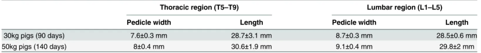
Table 1. Pedicle dimensions for porcine vertebrae according to weight (age) of animal, vertebra level and type of measurements.
Thoracic region (T5–T9) Lumbar region (L1–L5)
Pedicle width Length Pedicle width Length
30kg pigs (90 days) 7.6±0.3 mm 28.7±3.1 mm 8.7±0.3 mm 28.5±0.6 mm
50kg pigs (140 days) 8±0.4 mm 30.6±1.9 mm 9.1±0.4 mm 29.8±2 mm
Length is sum of pedicle and vertebral body lengths. Values are mean±SD, averaged from 50 measurements.
doi:10.1371/journal.pone.0127463.t001
Fig 1. X-rays of thoracic (A, C) and lumbar (B, D) vertebrae of a 90-day-old pig (30kg), with implanted screws 20mm (A, B) and 40mm (C, D) long.
ensure that they traversed the vertebral body, crossing the anterior cortical part of the vertebral
body on a perpendicular line (Fig 1C and 1D).
All pedicle screw insertions were performed with a free-hand technique, with a convergent
direction (Magerl insertion technique [23]) by the same senior veterinary surgeon. The entry
point was chosen at the cross section between the bottom of the articular process and a
hori-zontal line drawn in the middle of the transverse process [24]. After determination of this entry
point, the cortical was perforated with a small awl. The hole was then prepared with a pedicle finder, with no pre-tapping. Each screw was gently inserted with a progressive insertional tor-que until immediate cortical contact of the screw head to avoid micro fracture of bone cortex.
X-rays were performed after insertion to assess the position of the screws (Fig 1). No vertebra
was excluded due to malposition (no medial cortical wall violation or breach in the foramen was observed).
Each vertebra was then casted in polyurethane resin (F1, Axson) in a PVC circular container 100mm in diameter and between 25mm and 35mm high, depending on the size of the vertebra. The heating associated with the hardening of this particular resin was previously proved not to
degrade bone [25]. Playdough was used to cover the tips of the screws (for the bi-cortical
con-figuration), to avoid contact between the screw and the resin, and was also placed inside the vertebral canal. The vertical alignment of the screws was checked during resin hardening. Each embedded vertebra was then inspected to ensure that the resin did not interfere with the screw.
Pullout tests
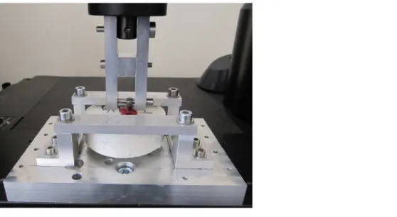
The screws were tested on a mechanical testing machine (MTS INSTRON 5566A), using a load cell with maximum capacity 10 kN and precision ±0.5%. A special assembly was developed to
firmly maintain the embedded vertebra with kind of metallic shelf brackets (seeFig 2). The
head screw was attached to the load cell of the testing machine. A preload of 10 N was applied to set up the system and then a constant ascendant displacement speed of 3mm/min was
applied in line with the screw axis until the screws were extracted. We monitored forcesvs
dis-placements during the tests, and noted the maximum pullout force for each tested vertebra. Mean values and standard deviations (SD) were calculated for each set of 10 repeats. A total of 8 different configurations of type of anchorage, vertebra level and weight of animal were
Fig 2. Pullout assembly.
tested. A non-parametric statistical study was realized on the results in pairs, using the Mann Whitney test.
Results
During the tests, two vertebrae were eliminated because of poor resin casting (the vertebra slipped), in the 50kg, bi-cortical, lumbar configuration. At the end of the tests, 78 vertebrae (thus 78 screws) were available for analysis. Pullout forces were averaged from the ten repeats for each level (all thoracic and all lumbar vertebrae), weight and type of anchorage; results are
shown inTable 2. The average pullout forces range between 419.9 and 1341.2N.
Results of the statistical tests on the pullout forces are presented inTable 3through p-values.
Statistical differences were found between most tested groups; only 5 comparisons returned
non-statistical differences, which are in italic font inTable 3. When we compared young and
aged animals, we found statistical differences on the pullout forces between 30kg’s and 50kg’s
animals (4 groups, p<.01). Concerning the vertebral level, statistical differences were also
found between lumbar and thoracic vertebrae (4 groups, p<.01) as well as for the type of
anchorage, between mono- and bi-cortical implantation (4 groups, p<.01).
The following three graphs highlight the respective influence of vertebra level (Fig 3), weight
(age) (Fig 4) and type of anchorage (Fig 5) on the averaged pullout forces. For both types of
anchorage (mono- and bi-cortical) and for each weight we found that thoracic vertebrae
sus-tained significantly lower pullout forces than lumbar vertebrae (p<.01). For both levels
(tho-racic and lumbar), the vertebrae of the 30kg pigs were found to sustain lower pull-out forces
than those of the 50kg pigs (p<.01). When types of anchorage are compared, for any weight
Table 2. Pullout forces averaged for 10 repeats.
Pig weight 30kg (approximate age 90 days) 50kg (approximate age 140 days)
Vertebra level Lumbar Thoracic Lumbar Thoracic
Mono-cortical Average pullout forces (in N) 761.1±55.1 419.9±105.8 1076.7±84 594.3±106.7 Bi-cortical Average pullout forces (in N) 993±126.2 682.2±127.7 1341.2±199.6ª 954.2±180.4
Values are mean±SD. aOnly 8 repeats.
doi:10.1371/journal.pone.0127463.t002
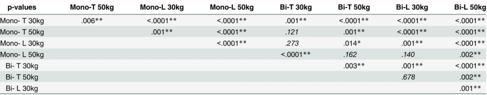
Table 3. P-values for pullout forces from Mann Whitney statistical test.
p-values Mono-T 50kg Mono-L 30kg Mono-L 50kg Bi-T 30kg Bi-T 50kg Bi-L 30kg Bi-L 50kg
Mono- T 30kg .006** <.0001** <.0001** .001** <.0001** <.0001** <.0001**
Mono- T 50kg .001** <.0001** .121 .001** <.0001** <.0001**
Mono- L 30kg <.0001** .273 .014* .001** <.0001**
Mono- L 50kg <.0001** .162 .140 .002**
Bi- T 30kg .003** .001** <.0001**
Bi- T 50kg .678 .002**
Bi- L 30kg .001**
*Means p<.05 **p<.01.
In italic font, non-statistically different results (p>.05). Mono- and Bi- stand for Mono-cortical and Bi-cortical anchorage. T and L stand for Thoracic and Lumbar level.
and vertebra level the mono-cortical anchorage induced lower pullout forces than the bi-cortical anchorage (p<.01).
Discussion
Current medical device development processes frequently require an experimental phase using animals, to assess efficacy before implantation in humans. During previous studies on the pig model to validate spinal devices, some veterinarians and researchers noted screw pullouts
[16,17] leading to high animal morbidity. In line with the ethical concept of the“Three Rs”,
this study investigated how certain parameters (pig weight (age), vertebra level and type of cor-tical anchorage) impact pedicle fixation in a young porcine model, in order to optimize pedicle fixation. We observed the highest pullout forces for the lumbar level, the bi-cortical anchorage and the oldest animals.
Fig 3. Impact of vertebra level, lumbar or thoracic, on the averaged pullout forces.Mono- and Bi-respectively stand for Mono-cortical and Bi-cortical anchorage of the screws.
doi:10.1371/journal.pone.0127463.g003
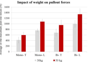
Fig 4. Impact of animal’s weight: 30kg (90 days) or 50kg (140 days) on the averaged pullout forces.
Mono- and Bi- respectively stand for Mono-cortical and Bi-cortical anchorage of the screws. T and L stand for Thoracic and Lumbar vertebrae.
Yazici et al [4] studied the effect of dilatation of the pedicles on screw stability using two-month-old pigs, and found a mean pullout force of 408.1±102N for non-dilated pedicles (tho-racic and lumbar vertebrae, with screws of respectively Ø 3.5 mm and Ø 4 mm). Abshire et al
[21] studied the impact of the geometry of the screws (conical vs cylindrical, respectively Ø 7.5
mm and Ø 6.5 mm, and 40 mm long), but using mature pigs (70kg–90kg); they found a mean pullout force of 2634.1N for lumbar vertebrae. Our study using three- and five-month-old pigs (30kg and 50kg) yields values consistent with the literature: higher than for 2-month-old pigs and lower than for pigs weighing 70–90kg. These are the first average values for ultimate pull-out forces of pedicle screws on such young animals, ranging between 420N and 1340N.
When we investigated how the level of the vertebra affects screw pullout forces, the results for a given weight and anchorage were statistically different between the thoracic and lumbar
level (p<.01) (Fig 3). We found that the more caudally the screws are positioned (lumbar
level), the greater their pullout resistance is, with an increase of approximately 80% for mono-cortical anchorage and 45% for bi-mono-cortical anchorage. This could be explained by the size of the vertebrae: lumbar vertebrae are wider than thoracic, especially in terms of pedicle dimensions (Table 1): lumbar pedicle width is around 14% higher than thoracic at 30kg, and 13% higher at 50kg.
Looking at how animal weight (age) of the animals affects pedicle screw pullout resistance, results for a given level and anchorage were statistically different between 30kg and 50kg
(p<.01) (Fig 4). Screw stability increases with the age of the animals. Between 3 and 5 months
of age the maximum pullout values for the lumbar and thoracic vertebrae increased respec-tively by 32% and 78% for bi-cortical anchorage and by 52% and 39% for mono-cortical anchorage. This may be explained by the increasing bone mineral density of the vertebrae as
pigs grow. It has been reported to rise by around 10% in 2 months of growth [26]; bone is
fre-quently found to be softer by veterinary surgeons implanting screws in very young animals.
Moreover, growth also affects the dimensions of the vertebrae (Table 1), increasing both the
length of the pedicles (5% to 7% increase in 2 months) and their width (around 5% to 6% increase). These vertebra modifications could explain the increase in primary stability of the screws observed between 90 and 140 days.
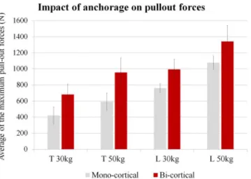
Examining the impact of type of cortical anchorage, we obtained results that, for a given age and vertebra level, were statistically different between mono- and bi-cortical anchorage Fig 5. Impact of type of screw anchorage, mono- or bi-cortical, on the averaged pullout forces.T and L stand for Thoracic and Lumbar vertebrae.
(p<.01) (Fig 5). The screws implanted with a mono-cortical anchorage sustained lower pullout
forces than those implanted with a bi-cortical anchorage. Costa et al [27] studied the impact of
misalignment of pedicle screws on their pullout resistance in lumbar porcine spines (age of the animals not provided), with different degrees of cortical violation (superior, inferior, medial and lateral). For standard insertions, they obtained forces around 1400N, while cortical viola-tion reduced forces to as low as 600N. In our study, pullout force increased when a second cor-tical anchorage (anterior aspect of the vertebral body) was added. For the lumbar vertebrae, the increase was 25% and 30% (respectively for 50kg and 30kg), and for the thoracic vertebrae it was 60% and 62% (respectively for 50kg and 30kg). However, the main difference is the orien-tation of the cortical bone relative to the screw axis. In our study, bi-cortical insertion induces a fairly perpendicular crossing of the anterior cortical part of the vertebral body. When screws are misaligned, the cortical crossing is more tilted and the axis of the screws is not perpendicu-lar to the cortical bone. This underlines the importance for screw stability of whether screws are anchored to one or to two cortical bones, but even more importantly, of the position of the screw crossing the cortical bone. Any bi-cortical insertion must be correctly performed to ensure a cortical crossing perpendicular to the screw axis, thus enhancing the pullout resistance of the screw threads.
This study sought the most relevant parameters for pedicle fixation in a porcine model being used to develop new human implant systems. We found statistical evidence of the impact of age of pigs, level of the implanted vertebra and type of screw anchorage. It would be interest-ing to extend these tests to osteointegrated screws, to assess the influence of bone healinterest-ing on their stability. Previous work on the creation of scoliotic deformities suggests that the conserva-tion of an osteointegraconserva-tion period before applying stresses to screws limits the risk of pullouts [17].
One limitation of our study is that pure axial loading does not faithfully represent the in vivo loading sustained by screws used in spinal systems. However, axial loading is commonly
used to evaluate screw stability [28]. Moreover, the use of long screws to induce a bi-cortical
anchorage may be limited by the risk of vascular issues during the insertion. We only tested here one design of pedicle screw; results may be different with other types of pedicle screws.
Other methods could also be used to enhance pedicle fixations such as specific screws [21,29]
or augmentation techniques [4].
Conclusions
To limit screw pullouts, we conclude that the best choices are implantation in older animals, using a lumbar fixation and long screws traversing the vertebra and inducing bi-cortical anchorage. However, bi-cortical anchorage implies that the screw goes beyond the anterior aspect of the vertebral body, and has to be performed with care to avoid any vascular issue. If very young animals are required, pedicle screw fixations need to be bi-cortical to enhance sta-bility, and as many screws as possible should be implanted to prevent avulsion.
Acknowledgments
The authors thank Marjorie Sweetko for English language editing.
Author Contributions
References
1. Russell WMS, Burch RL. The principles of humane experimental technique. Methuen; 1959.
2. Kim WJ, Lee SH, Shin SW, Rivard CH, Coillard C, Rhalmi C. The influence of fixation rigidity on
inter-vertebral joints. An experimental comparison between a rigid and a flexible system. J Korean Neuro-surg Soc. 2005; 37: 364–369.
3. Akbarnia BA, Mundis GM, Salari P, Yaszay B, Pawelek JB. Innovation in Growing Rod Technique: A
Study of Safety and Efficacy of a Magnetically Controlled Growing Rod in a Porcine Model. Spine. 2012; 37: 1109–1114. doi:10.1097/BRS.0b013e318240ff67PMID:22146279
4. Yazici M, Pekmezci M, Cil A, Alanay A, Acaroglu E, Oner FC. The effect of pedicle expansion on
pedi-cle morphology and biomechanical stability in the immature porcine spine. Spine. 2006; 31: E826–829. doi:10.1097/01.brs.0000240759.06855.e6PMID:17047529
5. Yilmaz G, Demirkiran G, Ozkan C, Daglioglu K, Pekmezci M, Alanay A, et al. The effect of dilation of
immature pedicles on pullout strength of the screws: Part 2: In vivo study. Spine. 2009; 34: 2378–2383. doi:10.1097/BRS.0b013e3181b0bb9bPMID:19829250
6. Suk SI, Kim JH, Kim SS, Lim DJ. Pedicle screw instrumentation in adolescent idiopathic scoliosis (AIS). European Spine Journal. 2011; doi:10.1007/s00586-011-1986-0
7. Ledonio CGT, Polly DWJ, Vitale MG, Wang Q, Richards BS. Pediatric pedicle screws: comparative
effectiveness and safety: a systematic literature review from the Scoliosis Research Society and the Pediatric Orthopaedic Society of North America task force. J Bone Joint Surg Am. 2011; 93: 1227–1234. doi:10.2106/JBJS.J.00678PMID:21776576
8. Cuartas E, Rasouli A, O’Brien M, Shufflebarger HL. Use of all-pedicle-screw constructs in the treatment of adolescent idiopathic scoliosis. J Am Acad Orthop Surg. 2009; 17: 550–561. PMID:19726739 9. Liljenqvist UR, Halm HF, Link TM. Pedicle screw instrumentation of the thoracic spine in idiopathic
sco-liosis. Spine. 1997; 22: 2239–2245. PMID:9346144
10. Boos N, Webb JK. Pedicle screw fixation in spinal disorders: a European view. Eur Spine J. 1997; 6: 2–18. PMID:9093822
11. Kim YM, Kim DS, Choi ES, Shon HC, Park KJ, Cho BK, et al. Nonfusion method in thoracolumbar and lumbar spinal fractures. Spine. 2011; 36: 170–176. doi:10.1097/BRS.0b013e3181cd59d1PMID:
20671591
12. Ruf M, Harms J. Pedicle screws in 1- and 2-year-old children: technique, complications, and effect on further growth. Spine. 2002; 27: E460–466. doi:10.1097/01.BRS.0000030203.48525.E3PMID:
12438997
13. Janssen M, de Wilde RF, Kouwenhoven JWM, Castelein RM. Experimental animal models in scoliosis research: a review of the literature. The Spine Journal. 2011; 11: 347–358. doi:10.1016/j.spinee.2011.
03.010PMID:21474088
14. Roth AK, Bogie R, Jacobs E, Arts JJ, van Rhijn LW. Large animal models in fusionless scoliosis correc-tion research: a literature review. The Spine Journal. 2013; 13: 675–688. doi:10.1016/j.spinee.2013.
02.043PMID:23582429
15. Newton PO, Upasani VV, Farnsworth CL, Oka R, Chambers RC, Dwek J, et al. Spinal growth modula-tion with use of a tether in an immature porcine model. J Bone Joint Surg Am. 2008; 90: 2695–2706.
doi:10.2106/JBJS.G.01424PMID:19047716
16. Schwab F, Patel A, Lafage V, Farcy JP. A Porcine Model for Progressive Thoracic Scoliosis. Spine. 2009; 34: E397–E404. doi:10.1097/BRS.0b013e3181a27156PMID:19444053
17. Odent T, Cachon T, Peultier B, Gournay J, Jolivet E, Elie C, et al. Porcine model of early onset scoliosis
based on animal growth created with posterior mini-invasive spinal offset tethering A preliminary report. European Spine Journal. 2011; doi:10.1007/s00586-011-1830-6
18. Upasani VV, Farnsworth CL, Chambers RC, Bastrom TP, Williams GM, Sah RL, et al. Intervertebral
disc health preservation after six months of spinal growth modulation. J Bone Joint Surg Am. 2011; 93: 1408–1416. doi:10.2106/JBJS.J.00247PMID:21915546
19. Cachon T, Odent T, Chuzel T, Viguier E. In vivo measurement of tensile force applied to the spine
dur-ing induction of scoliosis like deformities in a porcine model. Computer Methods in Biomechanics and Biomedical Engineering; In Press.
20. Gao M, Lei W, Wu Z, Liu D, Shi L. Biomechanical evaluation of fixation strength of conventional and expansive pedicle screws with or without calcium based cement augmentation. Clin Biomech (Bristol, Avon). 2011; 26: 238–244. doi:10.1016/j.clinbiomech.2010.10.008
21. Abshire BB, McLain RF, Valdevit A, Kambic HE. Characteristics of pullout failure in conical and
22. Mahar AT, Bagheri R, Oka R, Kostial P, Akbarnia BA. Biomechanical comparison of different anchors (foundations) for the pediatric dual growing rod technique. Spine J. 2008; 8: 933–939. doi:10.1016/j.
spinee.2007.10.031PMID:18082463
23. Magerl FP. Stabilization of the lower thoracic and lumbar spine with external skeletal fixation. Clin Orthop Relat Res. 1984; 125–141. PMID:6478690
24. McLain RF, Yerby SA, Moseley TA. Comparative morphometry of L4 vertebrae: comparison of large
animal models for the human lumbar spine. Spine. 2002; 27: E200–206. PMID:11935119
25. Subit D. Modélisation de la liaison os-ligament dans l’articulation du genou [Internet]. Université
Aix-Marseille 2. 2004. Available:http://www.theses.fr/2004AIX22099
26. Guillot M, Alexander K, Pomar C, del Castillo JRE. In growing pigs, chlortetracycline induces a revers-ible green bone discoloration and a persistent increase of bone mineral density dependent of dosing regimen. Research in Veterinary Science. 2011; 90: 484–490. doi:10.1016/j.rvsc.2010.06.022PMID:
20723952
27. Costa F, Villa T, Anasetti F, Tomei M, Ortolina A, Cardia A, et al. Primary stability of pedicle screws
depends on the screw positioning and alignment. Spine J. 2013; 13: 1934–1939. doi:10.1016/j.spinee. 2013.03.046PMID:23684239
28. ASTM F543—Specification and Test Methods for Metallic Medical Bone Screws [Internet]. ASTM
Inter-national; 2007. Available:http://www.astm.org/Standards/F543.htm
29. Lill C, Schneider E, Goldhahn J, Haslemann A, Zeifang F. Mechanical performance of cylindrical and dual core pedicle screws in calf and human vertebrae. Arch Orthop Trauma Surg. 2006; 126: 686–694.