27
Pakistan Veterinary Journal
ISSN: 0253-8318 (PRINT), 2074-7764 (ONLINE)Accessible at: www.pvj.com.pk
Day-3 Medium Changes can Affect Developmental Potential of Porcine Somatic Cell Nuclear
Transfer and Parthenogenesis Embryos
In Vitro
Dibyendu Biswasand Sang Hwan Hyun*
Laboratory of Veterinary Embryology and Biotechnology, College of Veterinary Medicine, Chungbuk National University, Cheongju 361-763, South Korea
*Corresponding author: [email protected]
A R T I C L E H I S T O R Y A B S T R A C T Received:
Revised: Accepted:
July 16, 2010 August 08, 2010 August 14, 2010 Key words:
Monoculture Perthenogenesis Porcine
Somatic cell nuclear transfer
The aim of the present study was to compare the developmental competence of porcine parthenotes and somatic cell nuclear transfer (SCNT) embryos after day-3 medium change with fresh embryo culture medium to that of embryos that did not have a medium change (monoculture system). The parthenogenetic and SCNT blastocyst formation rates were significantly (P<0.05) higher in the no-medium-change group (43.3±2.3, 18.5±1.1%, respectively) compared with the day-3 medium-change group (35.9±2.4, 7.9±0.9%, respectively). Total cell number in parthenotes and SCNT blastocysts was also significantly (P<0.05) higher in the no-change group (92.0±4.2, 66.9±7.7, respectively) compared with the media-change group (81.5±3.1, 46.6±4.9, respectively). No significant difference in cleavage rate was found in either group for parthenotes or SCNT embryos. This result suggests that day-3 medium changes have negative effects on porcine parthenotes and SCNT embryos in vitro.
©2011 PVJ. All rights reserved To cite this article: Biswas Dand SH Hyun, 2011. Day-3 medium changes can affect developmental potential of porcine somatic cell nuclear transfer and parthenogenesis embryos in vitro. Pak Vet J, 31(1): 27-30.
INTRODUCTION
Competence of any pre-implantation embryo through development up to blastocyst formation resulting from in vitro maturation, in vitro fertilization, and in vitro culture is affected by factors in the culture environment during the production of embryos in the laboratory. It has been suggested that a variety of molecules that are secreted by the embryo itself (e.g., ions, energy substrates, amino acids, vitamins, growth factors, cytokines, and hormones) play important roles in early embryonic development (Kuran et al., 2002). The secreted growth factors are essential for further growth and development, and their absence can lead to implantation failure or, in rare cases, severe developmental abnormalities (Richter, 2008). Evidence also suggests that the addition of exogenous growth factors during in vitro embryo culture can influence developmental potential. Some genes [e.g., leukemia inhibitory factor (LIF) and interferon-tau (IFN-T)] are expressed by the early embryo of several species, and it has been shown that successful development and blastocyst quality is dependent upon the action of these growth factors (Stewart, 1994). Studies have shown that pre-implantation development-related gene knockout can result in embryonic lethality or severe post-implantation
abnormalities. Injection of secreted factors can be a useful method to rescue the defects that cause embryonic lethality (Uehara et al., 2000). Constant secretion and growth factor level in the medium are essential for successful in vitro embryo development. Alteration or changes in embryo-secreted growth factor levels could lead to developmental retardation. During in vitro embryo development, medium changes might alter developmental potential. Recently, sequential culture media have been used for in vitro porcine embryo development (Swain et al., 2001). This technique is well documented in human in
vitro embryo culture systems. The implantation and
Pak Vet J, 2011, 31(1): 27-30. 28
cell nuclear transfer (SCNT) technology using a day-3 porcine zygotic medium-3 (PZM-3)-change group and a no-medium-change group.
MATERIALS AND METHODS
Ovary collection, recovery and in vitro oocyte maturation
Ovaries of pre-pubertal gilts were collected from a commercial abattoir. The follicular contents were aspirated from 3- to 7-mm superficial antral follicles with a 10-ml disposable syringe and 20-gauge needle. Cumulus-oocyte complexes (COC) with at least three layers of compact cumulus cells and with homogenous cytoplasm were selected and a group of 50-60 COCs were cultured in tissue culture medium-199 (M-199) (Invtrogen, Grand Island, NY) supplemented with 0.6 mM cysteine, 0.91 mM pyruvate, 15 ng/ml epidermal growth factor, 75 µg/ml kanamycin, 1 µg/ml insulin and 10% porcine follicular fluid in each well of four-well multi dish (Nunc, Roskilde, Denmark). The COCs were then statically cultured at 39°C in a humidified atmosphere containing 5% CO2 with 10 IU/ml eCG (Intervet
International BV, Boxmeer, Holland) and 10 IU/ml hCG (Intervet International BV). After 20–22 h of maturation with hormones, the oocytes were washed two times in fresh maturation medium before being cultured in hormone-free medium for an additional 18 h for SCNT and 22 h for PA. The pFF was prepared according to Hyun et al. (2003) and stored at -20ºC until use.
Donor cells preparation
Porcine ear skin fibroblasts from adult female pigs were seeded in to four-well plates and were grown in Dulbecco’s modified Eagle medium (DMEM) with 1 mM sodium pyruvate, 1% (v/v) non-essential amino acids (Invitrogen, USA), and 10 µg/ml penicillin–streptomycin solution, which was supplemented with 10% (v/v) fetal bovine serum from a single batch until a complete monolayer of cells had formed. The donor cells were synchronized at the G0/G1 stage of the cell cycle by contact inhibition for 3–4 days. The cells of the same passage were used in each replicate for the various treatments. The individual cells were retrieved from the monolayer by trypsinization for ~1 min and subsequently used for SCNT.
SCNT and parthenogenesis
After 40 h of IVM, cumulus-cell-free oocytes were incubated for 2 min in manipulation medium (calcium free TLH-BSA) containing 5 µg/ml Hoechst 33343 and 7.5 µg/ml cytoclasin B (Sigma-Aldrich Co). Following incubation, the oocytes were transferred into a drop of manipulation medium containing 7.5 µg/ml cytoclasin B and were overlaid with warm mineral oil. The zona pellucida was partially dissected with a fine glass needle near the first polar body (PB). The first PB and adjacent cytoplasm (~10%), presumably containing the metaphase-II chromosomes, were extruded by squeezing the oocytes with the same needle. Enucleation was confirmed under an epifluorescence microscope (TE 300, Nikon, Tokyo, Japan). Using a injecting pipette, a 12–15-µm trypsinized fetal fibroblast with a smooth surface was transferred into the periviteline space through the same slit of an enucleated oocyte. The reconstructed couplets were
equilibrated with 0.26 M mannitol containing 0.5 mM HEPES, 0.001 mM CaCl2, and 0.05 mM MgSO4 for 2–3
min and transferred to a 1 mm fusion chamber containing overlaid with same mannitol solution. Membrane fusion and activation were done according to Song et al. (2009). Activated oocytes were washed and cultured in PZM-3 medium supplemented with 3 mg/ml fatty-acid free BSA and placed in humidified incubator at 39°C under 5% CO2.
For PA, the MII oocytes at 42 h of IVM were activated using a pulse sequence identical to that used to activate SCNT oocytes. The culture procedures of PA embryos were similar to SCNT embryos. Cleavage and blastocyst formation were evaluated at 48 and 168 h post activation, respectively, with the day of SCNT or PA designated Day 0.
Embryo evaluation and nuclear staining
Blastocysts considered viable were washed with 1% PVA in Dulbecco’s phosphate buffered saline (DPBS) for 1 min and then fixed with 100% ethanol containing 10
µg/ml Hoechst for 5 min. Then, the blastocysts were mounted on glass slides in a drop of 100% glycerol and squashed gently with a coverslip. The nuclei were counted using fluorescence microscopy.
Embryo culture procedure
Following activation, the culture medium was changed on day 3 with fresh PZM-3 medium and cultured for further development. In the other group, the medium was unchanged up to day 7. At day 4 in both in vitro cultures (IVC), medium was supplemented with 10% FBS (final concentration) (Invitrogen, Carlsbad, CA).
The data were statistically analyzed by student t-test using GraphPad Version 5.0 with a probability level of P<0.05 being considered significant.
RESULTS
A total of 613 parthenogenetic activated embryos were randomly allocated into two groups of six replicates and were examined for developmental potential (Table 1). The parthenogenetic blastocyst formation rate and total cell number per blastocyst (Fig. 1: D) was significantly higher (43.3 ± 2.3%, 92.0 ± 4.2, respectively) in the no-medium-change group as compared with the medium change group (35.9 ± 2.4%, 81.5 ± 3.1, respectively). No significant difference in cleavage rate was observed between the two groups.
After SCNT, a total of 829 oocytes were injected in five replicates (Table 2). As shown in Table 2, the blastocyst formation rate and total cell number per blastocyst (Fig. 1: B) significantly increased (18.5±1.1%, 66.9±7.7, respectively) in the no-medium-change group compared with the medium change group (7.9±0.9%, 46.6±4.9, respectively). No significant difference in cleavage rate was found in either group for parthenotes or SCNT embryos.
DISCUSSION
Pak Vet J, 2011, 31(1): 27-30. 29
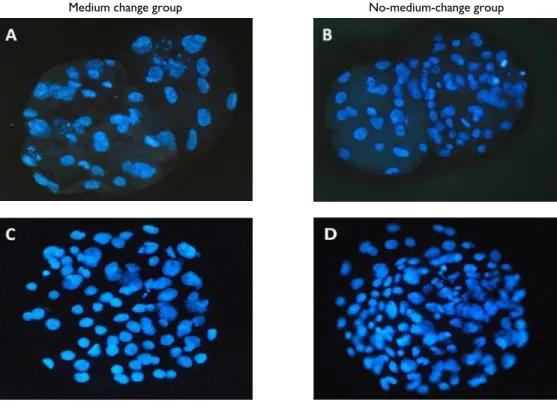
Medium change group No-medium-change group
Fig. 1: Nuclei of blastocyst stained with Hoechst at 168 h after activation in medium change (A: SCNT and C: parthenotes) and no-medium-change group (B: SCNT and D: parthenotes) (x400).
Table 1: Effects of medium changes at day-3 on developmental potential of porcine in vitro parthenogenetic embryos
Group Replications Total oocytes
cultured
Cleavage (%)
Blastocyst (%)*
Total cell number/BL (Lowest-Highest)
Medium change 6 314 257 (85.4±2.7) 92 (35.9±2.4)a 81.5±3.1a (36-134)
No medium change 6 299 258 (86.2±1.6) 111 (43.3±2.3)b 92.0±4.2b (41-167)
*Percentage of cleaved embryos. a,bValues with different superscripts in the same column are significantly different (p<0.05).
Table 2: Effects of medium changes at day 3 on developmental potential of porcine in vitro SCNT produced embryos.
Group
Repli-cations
Total oocytes injected
Fusion (%)
Cleavage (%)
Blastocyst (%)*
Total cell number/BL (Lowest-Highest)
Medium change 5 420 328 (77.9±1.7) 205 (62.6±3.2) 16 (7.9±0.9 )a 46.6±4.9a (20-73)
No medium change
5 409 302 (73.1±1.8) 194 (63.8±3.5) 36 (18.5±1.1)b 66.9±7.7b (32-121)
* Percentage of cleaved embryos. a,b Values with different superscripts in the same column are significantly different (P<0.05).
methods are widely and successfully used under in vitro embryo culture, i.e., monoculture and sequential culture systems and a debate about the relative merits of in vitro culture systems continues. In vitro embryo culture medium was developed according to embryo metabolism. Sequential embryo culture mediums were used according to embryo requirements. Before embryo compaction, embryos have low metabolic activity and need a low level of glucose in a pyruvate-preferred nutrient medium (Flood and Wiebold, 1988). After compaction, embryos need a glucose-preferred nutrient medium, as well as an essential and non-essential amino acid-based medium (Lane and Gardner, 1997).
During in vitro embryo culture, the post-activation environment in which embryos are cultured is critical for normal development. This includes i) the first cleavage division, ii) the activation of the embryonic genome at the 8–16 cell stage, iii) compaction of the morula on day 5 and iv) blastocyst formation on day 6–7, which involves the trophoectoderm and the inner cell mass formation (Lonergan et al., 2006). During this period, the embryo
IGF-Pak Vet J, 2011, 31(1): 27-30. 30
2, insulin, platelet-derived growth factor-alpha, TGF-beta, EGF) to cultured pre-implantation embryos stimulates DNA and RNA synthesis and increases cell number (Richter, 2008). On the other hand, culture of mouse pre-implantation embryos at high density improves their developmental processes, perhaps due to the stimulatory effect of an increase in the local concentration of endogenously produced growth factors (Paria and Dey, 1990). Concentration of such autocrine growth factors is believed to be the primary reason for improved embryonic development with group culture and with minimization of medium volume (Jin et al., 2001). This hypothesis is in agreement with our result that when the medium was changed at day 3 without adding any growth factor, the blastocyst formation rate and cell number per blastocyst in both cases declined compared with the no-medium-change condition.
During pre-implantation, developmental gene expression has a fundamental role in the coordination of homeostatic and metabolic mechanisms throughout life. In domestic species, some evidence suggests that culture environments post-fertilization or activation can perturb the gene expression pattern in pre-implantation embryos. During embryo culture, several genes known to be involved in important developmental process are expressed, including genes involved in apoptosis (Bax), gap junction formation (Cx43), and differentiation (LIF and LIF-Rβ) (Rizos et al., 2002). The post-activation culture environment can have a dramatic effect on the pattern of mRNA abundance of many developmentally important genes in the embryos. Supplementation of growth factors like IGF-I (Makarevich and Markkula, 2002) and EGF (Cui and Kim, 2003) in IVC culture medium has been shown to reduce apoptosis and increase cell number in porcine diploid parthenotes (Cui and Kim, 2003). This is similar to our results under the no-medium-change condition; total cell number increased in both cases. Even though, pre-implantation embryos express many growth factors, e.g. EGF, TGF-α, TGF-β, IGF-I, and II, VEGF, PDGF, PAF and fibronectin (Rappolee et al., 1988; Richter, 2008), expression of a subset of growth factors in embryos suggests that they were involved in the growth and differentiation of early mammalian embryos (Rappolee et al., 1988). In conclusion, our results suggest that using a monoculture system improved the developmental potential of porcine parthenogenetic and SCNT in vitro embryo production.
Acknowledgement
This research was supported by a grant (#20070301034040) from the BioGreen 21 program, Rural Development Administration, South Korea.
REFERENCES
Blake DA, M Proctor and NP Johnson, 2004. The merits of blastocyst versus cleavage stage embryo transfer: a cochrane review. Hum Reprod, 19: 795-807.
Cui XS and NH Kim, 2003. Epidermal growth factor induces Bcl-xL gene expression and reduces apoptosis in porcine parthenotes developing in vitro. Mol Reprod Dev, 66: 273-278.
Flood MR and JL Wiebold, 1988. Glucose metabolism by preimplantation pig embryos. J Reprod Fertil, 84: 7– 12.
Hyun SH, GS Lee, DY Kim, HS Kim, SH Lee, DH Nam, YW Jeong, S Kim, SC Yeon, SK Kang, JY Han, BC Lee and WS Hwang, 2003. Production of nuclear transfer-derived piglets using porcine fetal fibroblasts transfected with the enhanced green fluorescent protein. Biol Reprod, 69: 1060-1068.
Jin Y, XZ Guo, L Li, CY Xie and LL Tan, 2001. The effect of autocrine factors on development of early embryos of mouse. Shi Yan Sheng Wu Xue Bao, 34: 77-80.
Kuran M, JJ Robinson, DS Brown and TG Mcevoy, 2002. Development, amino acid utilization and cell allocation in bovine embryos after in vitro production in contrasting culture systems. Reprod, 124: 155-165. Lane M and DK Gardner, 1997. Non-essential amino
acids and glutamine decrease the time of the first cleavage divisions and increase compaction of mouse zygotes in vitro. J Ass Reprod Genet, 14: 398-403. Lonergan P, T Fair, D Corcoran and ACO Evans, 2006.
Effect of culture environment on gene expression and developmental characteristics in IVF-derived embryos. Theriogenology, 65: 137-152.
Makarevich AV and M Markkula, 2002. Apoptosis and cell proliferation potential of bovine embryos stimulated with insulin-like growth factor I during in vitro maturation and culture. Biol Reprod, 66: 386-392.
Paria BC and SK Dey, 1990. Preimplantation embryo development in vitro: Cooperative interactions among embryos and role of growth factors. Proc Natl Acad Sci, 87: 4756–4760.
Rappolee DA, CA Brenner, R Schultz, D Mark and Z Werb, 1988. Developmental expression of PDGF, TGF-alpha and TGF-beta genes in preimplantation mouse embryos. Science, 241: 1823-1825.
Richter KS, 2008. The importance of growth factors for preimplantation embryo development and in vitro culture. Curr Opin Obstet Gynecol, 20: 292-304. Rizos D, P Lonergan, MP Boland, R Arroyo-García, B
Pintado, J de la Fuente and A Gutiérrez-Adán, 2002. Analysis of differential messenger rna expression between bovine blastocysts produced in different culture systems: Implications for blastocyst quality. Biol Reprod, 66: 589-595.
Song KY, SH Hyun, T Shin and ES Lee, 2009. Post-activation treatment with demecolcine improves development of somatic cell nuclear transfer embryos in pigs by modifying the remodeling of donor nuclei. Mol Reprod Dev, 76: 611-619.
Stewart CL, 1994. Leukaemia inhibitor factor and the regulation of preimplantation development of the mammalian embryo. Mol Reprod Dev,39: 233-238. Swain JE, CL Bormann and RL Krisher, 2001.
Development and viability of in vitro derived porcine blastocysts cultured in NCSU 23 and G1.2/G2.2 sequential medium. Theriogenology, 56: 459-469. Uehara Y, C Mori, T Noda, K Shiota and N Kitamura,