R E S E A R C H A R T I C L E
Open Access
Characterization of B cells in healthy
pregnant women from late pregnancy to
post-partum: a prospective observational
study
Jorge Lima
1,2*, Catarina Martins
2, Maria J. Leandro
3, Glória Nunes
2, Maria-José Sousa
4,5, Jorge C. Branco
6and Luís-Miguel Borrego
7,2Abstract
Background:B cells play a role in pregnancy due to their humoral and regulatory activities. To our knowledge, different maturational stages (from transitional to memory) of circulating B cell subsets have not yet been characterized (cell quantification and phenotype identification) in healthy pregnant women. Thus, the objective of our study was to characterize these subsets (as well as regulatory B cells) from late pregnancy to post-partum and to compare them with the circulating B cells of non-pregnant women.
Methods:In all of the enrolled women, flow cytometry was used to characterize the circulating B cell subsets according to the expression of IgD and CD38 (Bm1-Bm5 classification system). Regulatory B cells were characterized based on the expression of surface antigens (CD24, CD27, and CD38) and the production of IL-10 after lipopolysaccharide stimulation.
Results:Compared to the absolute counts of B cells in the non-pregnant women (n= 35), those in the pregnant women (n= 43) were significantly lower (p< 0.05) during the 3rd trimester of pregnancy and on delivery day (immediately after delivery). The percentages of these cells on delivery day and at post-partum were significantly lower than those in the non-pregnant women.
In general, the absolute counts and percentages of the majority of the B cell subsets were significantly lower in the 3rd trimester of pregnancy and on delivery day than in the non-pregnant women. However, these counts and percentages did not differ significantly between the post-partum and the non-pregnant women.
The most notable exceptions to the above were the percentages of naïve B cells (which were significantly higher in the 3rd trimester and on delivery day than in the non-pregnant women) and of CD24hiCD38hiregulatory B cells (which were significantly higher in the post-partum than in the non-pregnant women).
Conclusion:According to our study, the peripheral B cell compartment undergoes quantitative changes during normal late pregnancy and post-partum. Such findings may allow us to better understand immunomodulation during human pregnancy and provide evidence that could aid in the development of new strategies to diagnose and treat pregnancy-associated disturbances. Our findings could also be useful for studies of the mechanisms of maternal responses to vaccination and infection.
Keywords:B cell subsets, Flow cytometry, Human pregnancy, Obstetrics
* Correspondence:jorgeramoslima@sapo.pt
1Department of Obstetrics and Gynecology, CUF Descobertas Hospital,
Lisbon, Portugal
2CEDOC, Chronic Diseases Research Center, Immunology, NOVA Medical
School, Faculty of Medical Sciences, Lisbon, Portugal
Full list of author information is available at the end of the article
and infectious agents during the course of normal human pregnancy has also been reported [4].
B cells have a role in pregnancy because of their humoral activity (i.e., the production of protective antibodies against paternal antigens during pregnancy and the production of auto-antibodies that may lead to pregnancy complications) [5]. Normally, B cells leave the bone marrow and enter the circulation as immature transitional B cells, which later ma-ture into naïve B cells. Finally, when naïve B cells encounter their cognate antigens in secondary lymphoid organs, these cells become activated and mature into memory B cells and plasma cells [6, 7]. B cell subsets of different maturational stages, from transitional to memory B cells, have been iden-tified in peripheral blood using the mature B (Bm)1-Bm5 classification system. This classification system has proven to be effective in the identification of disturbances in the proportions of peripheral blood B cell subsets in patients with autoimmune diseases (e.g., Lupus or Sjögren’s syn-drome) [8–10] and in those undergoing therapy (e.g., with biological agents), by assessing the depletion and repopula-tion of B cells [11].
Furthermore, it has been suggested that, in addition to their humoral activity, specific B cells can also have a regulatory function although this is still controversial. According to recent studies, regulatory B cells (Bregs) can inhibit pro-inflammatory responses by secreting the anti-inflammatory cytokine IL-10 [12, 13]. Breg counts increase in the first trimester of pregnancy, suppressing unwanted immune responses of maternal effector T cells, protecting against pregnancy loss [14]. While the pheno-type and function of regulatory T cells has been extensively studied [15], further studies are needed to investigate the mechanisms behind the activation and expansion of Bregs and other B cell subsets in pregnancy.
These regulatory functions have been attributed to dif-ferent B cell subsets, and despite some controversy, great progress has been made in the characterization of Bregs. The inability to identify a Breg-specific transcription fac-tor, together with the phenotypic heterogeneity of Bregs, supports the idea that Bregs are not lineage specific and that they may expand in response to inflammation when immunosuppression is necessary [16]. It remains unclear whether the regulatory B cell function is a specific role of a particular subset or whether it is a reflection of their
defined by the expression of CD20 or CD19 with older studies [18, 25, 26] using either expression of Ia (HLA-DR) or surface immunoglobulin. A few studies [17, 19, 24, 27] have reported on the frequency of B cell subsets expressing CD5 with majority describing lower frequency or lower total numbers of this subset during pregnancy, at delivery or early in the postpartum period. One study [19] reported lower CD21 and CD23 frequencies at delivery. However, peripheral B cells have not been characterized in human pregnancy while considering the different maturational stages, from transitional to memory B cells (using CD38 and immunoglobulin IgD as differentiation markers). Con-sequently, the objective of our study was to characterize these specific peripheral blood B cell subsets (transitional, naïve, unswitched memory, post-germinal, and resting memory B cells as well as plasmablasts) and Bregs (CD24hiCD27+, CD24hiCD38hi and IL-10 regulatory B cells) from late pregnancy to post-partum and compare them with those in non-pregnant women.
Methods
Study population
This prospective observational study followed healthy pregnant women over time to characterize (i.e., cell quanti-fication and phenotype identiquanti-fication) their peripheral blood B cell subsets from late pregnancy to post-partum. This characterization of B cells in the pregnant women was also compared with the characterization of single samples of peripheral blood B cells from a control group of healthy non-pregnant women to investigate changes associated with pregnancy.
Sequential non-laboring healthy women with singleton pregnancies who were attending an outpatient clinic (routine obstetrical care) during the 3rd trimester were recruited for participation. None of the pregnancies had complications prior to recruitment. Furthermore, all of the fetuses exhibited appropriate growth (as measured by uterine fundal height and by ultrasound performed after 28 weeks of gestation).
For all of the women, the exclusion criteria were a his-tory of diabetes, hypertension, or autoimmune disease and smoking during the 6 months prior to peripheral blood sample collection. Additional exclusion criteria for the pregnant women included prenatal use of any medi-cation (other than vitamins and iron supplements) and ongoing complications in the pregnancy. Non-pregnant women taking oral contraceptives were also excluded, as these drugs affect circulating B cells [28].
All of the women were recruited at theHospital CUF Descobertas in Lisbon (Portugal) between July 2013 and March 2014. The Ethics Committee of this hospital ap-proved the study protocol. All of the recruited women provided written informed consent before the start of the study.
Study visit procedures
Three visits were planned for the pregnant women: visit 1 was planned for the 3rd trimester of pregnancy (3rd trimester); visit 2, for the day of delivery; and visit 3, for post-partum (at least 6 weeks after delivery). A single visit was planned for the non-pregnant controls.
To characterize B cell subsets from late pregnancy to post-partum, peripheral blood samples were collected from all of the pregnant women at each planned visit: the “3rd trimester” sample was collected at visit 1, the“on delivery day” sample was collected at visit 2 (immediately after de-livery, within 15 min after placental expulsion and oxytocin administration), and the “post-partum” sample was col-lected at visit 3. A peripheral blood sample was colcol-lected from the non-pregnant women at the planned visit, which took place during the follicular phase of their menstrual cycle because hormone status during the luteal phase is similar to that during pregnancy [29].
The baseline data collected for all women at the time of enrollment included demographics (age and ethnicity), anthropometrics [body mass index (BMI)], obstetric his-tory, and systolic and diastolic blood pressures. The data collected for the pregnant women on the day of delivery in-cluded gestational age, type of analgesia and/or anesthesia, and mode of delivery. The data collected for the newborns included gender, weight, and 1-min and 5-min Apgar scores.
Flow cytometry analysis and laboratory measurements Peripheral blood samples were collected into EDTA-coated and heparinized tubes. These samples were analyzed by four-color flow cytometry (BD FACSCalibur, BD Biosci-ences, San Jose, CA, USA) to characterize B cell subsets and their maturation profiles. MultisetTM and CellQuest 3.3TM (BD Biosciences) software were used for both acqui-sition and analysis.
To obtain absolute counts of B cells (CD19+), a single-platform strategy was used. EDTA samples were assayed
using a lyse-no-wash technique, with a BD IMK Kit with BD Trucount™ Tubes (BD Biosciences). The assay was performed according to the manufacturer’s instructions. In brief, 50 μL of blood were incubated for 15 min in
the dark, at room temperature, with the monoclonal antibodies provided in the kit, in Trucount™ tubes con-taining a calibrated number of microbeads for counting purposes. Red blood cells were then lysed with the lysing solution (also provided with the BD IMK Kit), for 15 min and finally samples were acquired. The cells were gated on CD45/SSC, and a minimum of 2500 lympho-cyte events were acquired. Multiset software provided percentage and absolute counts of B cells using the number of microbeads in each Trucount™ tube, along with the number of microbead and lymphocyte events acquired in each tube.
To study the surface B cell markers, a modified lyse-wash protocol was used. EDTA samples were washed twice in phosphate-buffered saline (PBS) to lower background stain-ing. The washed cells were then stained with a panel of monoclonal antibodies (mAbs) that were conjugated with different fluorochromes: anti-CD19 PerCPCy5.5 (clone HIB19, Biolegend), anti-CD24 PE (clone ML5, Biolegend), anti-CD27 FITC (clone O323, Biolegend), CD38 APC (clone HIT2, Biolegend), and anti-IgD PE (clone IA6-2, BD Pharmingen). Red blood cells were incubated for 15 min at room temperature in the dark. The red cells were then lysed with BD FACS lysing solution (BD Biosciences) according to the manufacturer’s instructions. After a wash step with PBS, events were acquired. For the characterization of IL-10-producing Bregs, heparin sam-ples were incubated for 5 h at 37 °C in a 5 % CO2
atmos-phere with phorbol 12-myristate 13-acetate (PMA) (50 ng/mL, Sigma Aldrich), calcium ionophore (1μg/mL,
Sigma Aldrich), and lipopolysaccharide (LPS) (10μg/mL,
Sigma Aldrich) in the presence of Brefeldin A (1.0μg/ml,
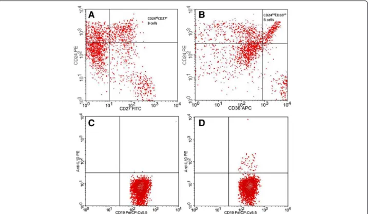
BD Pharmingen) [13, 30]. After the stimulation, the red blood cells were lysed via the addition of BD FACS lysing solution and were stained for surface markers with anti-CD3 FITC (clone SK7, BD Biosciences), anti-CD19 PerCPCy5.5 (clone HIB19, Biolegend), and anti-CD8 APC (clone SK1, Biolegend) mAbs. The Cytofix-Cytoperm kit (BD Pharmingen) was used for cell fixation and permeabilization according to the manufacturer’s instruc-tions. To assess the cytoplasmic expression of IL-10 in the B cells, a final intracellular staining step with an anti-IL-10 PE mAb (clone JES3-19F1, Biolegend) was performed be-fore cell acquisition. A minimum of 2000 B cells (CD19+) were acquired in all tubes (gate in CD19/SSC). The analysis strategies are presented in Figs. 1 and 2. The flow cytometry results are presented as a percentage of total B cells and as absolute cell counts (cells/μL).
of IgD/CD38 phenotypic markers. The cells were charac-terized as follows: transitional B cells (Bm2’: IgD+CD38hi), naïve B cells (Bm2: IgD+CD38+), unswitched memory B cells (Bm1: IgD+CD38−), and switched memory B cells
(Bm5: IgD−CD38+/−) and were subsequently divided into
post-germinal memory B cells (early Bm5: IgD−CD38+
), resting memory (late Bm5: IgD−CD38−) B cells, and
plasmablasts (Bm3 + Bm4: IgD−CD38hi
) [8–10, 14]. Bregs were evaluated in three different populations: CD24hiCD27+, CD24hiCD38hiand IL-10-producing B cells.
Our laboratory measurement included both absolute counts and percentages of total B cells and the different B cell subsets, as we feel that the two types of data are complementary. Percentages were measured as these allow interpreting the relative fluctuations in distinct B cell sub-sets from pregnancy to post-partum. Absolute counts were also measured and reported, although we are aware that pregnancy is characterized by variable degrees of hemodilu-tion, and that changes in these counts may not reflect true variations in the total numbers of circulating cells.
Fig. 1Identification of B cell subsets according to Bm1-5 classification system.aandbGating strategy for CD19+B cells using an initial CD19/SSC plot and refinement of the gate using a plot of FSC vs SSC.cBm1-5 classification from double staining for IgD and CD38 (unswitched memory Bm1: IgD+CD38−; naïve Bm2: IgD+CD38+; transitional Bm2′: IgD+CD38hi; plasmablasts Bm3 + Bm4: IgD−CD38hi; post-germinal memory/early
eBm5: IgD−CD38+; and resting memory/late Bm5: IgD−CD38−)
Statistical analysis
If the baseline data were normally distributed, they were presented as means (±standard deviations); otherwise, these data were presented as medians and ranges. Cat-egorical variables were described as absolute and relative frequencies and were expressed as percentages.
Cell counts and percentages were presented as me-dians and ranges. If normally distributed, 2 independent groups were compared using Student’s t-tests; otherwise, Mann–Whitney U tests were used. If normally distrib-uted, pairs of samples were compared using paired Student’s t-tests; otherwise, Wilcoxon signed-rank tests were used. For normally distributed data, comparisons between more than 2 groups were performed using ANOVA I; otherwise, Kruskal-Wallis tests were used. Statistical significance was defined by a P-value <0.05. The P-values for the comparisons of B cells between the non-pregnant women and pregnant women at differ-ent visits, as well as for the comparisons of the B cells of the pregnant women between visits, were adjusted for multiplicity using the Benjamini and Yekutieli method [31]. All of the data were analyzed using R software, ver-sion 3.12 for Windows.
Results
Baseline characteristics
A total of 78 women were enrolled in the study (43 preg-nant and 35 non-pregpreg-nant). The characteristics of these women and of their newborns are presented in Table 1. The mean BMI of the non-pregnant women was 21.5 (±2.8) Kg/m2, while for the pregnant, it was 26.2 (±2.8) Kg/m2. All of the women were normotensive [mean sys-tolic blood pressure for the non-pregnant was 119.8 (±10.5) mmHg, while for the pregnant, it was 115.7 (±9.3) mmHg; mean diastolic blood pressure for the non-pregnant was 74.7 (±7.4) mmHg, while for the non-pregnant, it was 67.4 (±7.4) mmHg]. Among the non-pregnant women, the median number of weeks since the last pregnancy (regardless of whether the pregnancies were interrupted or resulted in a live birth) was 169 (23–449). The me-dian gestational age in the 3rd trimester of pregnancy was 33.0 (31–35) weeks, while it was 39.0 (37–41) weeks on the day of delivery. The pregnant group was significantly younger (p= 0.016) and included signifi-cantly more nulliparous women (p< 0.001) than the non-pregnant group. All of the pregnant women, regard-less of the mode of delivery, received regional analgesia and/or anesthesia. No general anesthesia was administered to these women. All of the pregnant women were dis-charged from the hospital 2 days after a vaginal delivery or 3 days after a cesarean section. Final post-partum mea-surements were carried out a median of 45 (41–58) days after delivery.
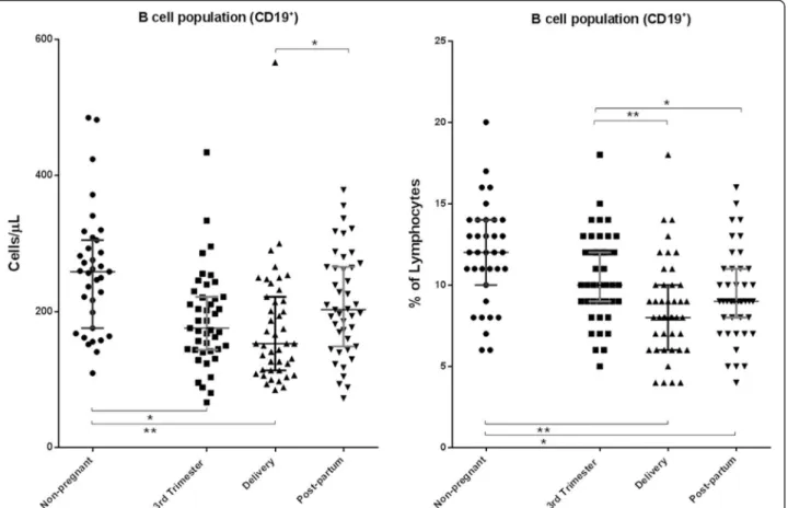
Characterization of the B cell population (CD19+)
The characterization of the B cell population for all of the enrolled women is presented in Fig. 3. The median absolute numbers (259 [110–485]) and percentages (12 [6–20]) of these cells in the non-pregnant were within the expected normal ranges of our protocol (absolute count: 80–616 cells/μL; percentage: 5–22 %). The
abso-lute counts of B cells at delivery were significantly lower (p< 0.05) than those at post-partum and in the non-pregnant women. Furthermore, the absolute counts of these cells during the 3rd trimester were also significantly lower (p< 0.05) than those in the non-pregnant. The percentages of B cells at delivery and at post-partum were significantly lower than those in the pregnant women during the 3rd trimester of pregnancy and in the non-pregnant.
Table 1Characteristics of the women enrolled in the study and of their newborns
Non-pregnant women (n= 35)
Pregnant women (n= 43)
Age in years, median (range) 35.0 (20–40) 32.0 (25–41)*
Ethnicity,n(%)
White 35 (100) 42 (97.8)
Black 0 1 (2.2)
Gestational age in weeks, median (range)
3rd trimester 33.0 (31–35)
Day of delivery 39.0 (37–41)
Parity,n(%)
Nulliparous 5 (14.3) 24 (55.8)*
Primiparous 14 (40) 18 (41.9)
Multiparous 16 (45.7) 1 (2.3)
Mode of delivery,n(%)
Vaginal 18 (41.8)
Cesarean 25 (58.2)
Elective cesareana 14 (55.6)
Intrapartum cesareanb 11 (44.4)
Newborns
Birth weight in grams, mean (± SD)
3265.0 (±393.5)
Gender,n(%)
Male 22 (51)
Female 21 (49)
APGAR score, median (range)
1-min Apgar score 9 (6; 10)
5-min Apgar score 10 (9; 10)
5-min Apgar score less than 7
0 (0)
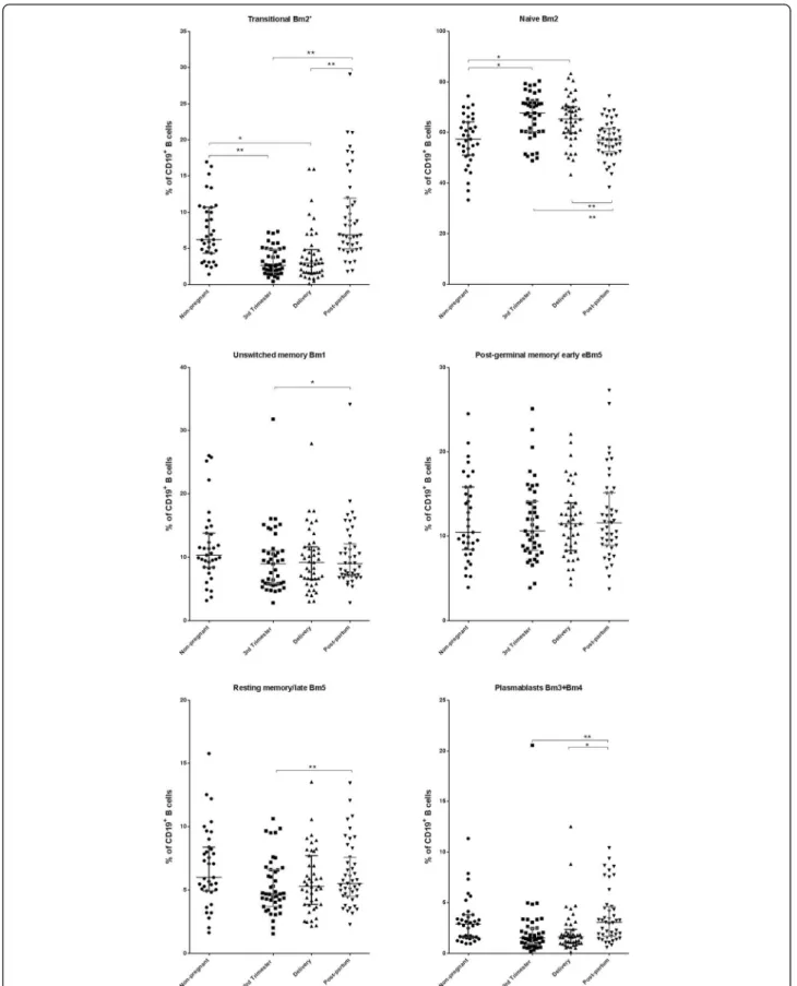
Characterization of maturational stages of B cells
The characterization of specific B cell subsets (transi-tional, naïve, unswitched memory, post-germinal, and resting memory B cells as well as plasmablasts) for all of the enrolled women is presented in Fig. 4 (absolute counts) and Fig. 5 (percentages). The absolute counts of transitional B cells, unswitched memory B cells, resting memory B cells, and plasmablasts during the 3rd trimes-ter of pregnancy and on delivery day were significantly lower (p< 0.05) than the corresponding counts in the non-pregnant. The absolute counts of naïve and of post-germinal memory/early B cells did not significantly differ (p≥0.05) between the pregnant and non-pregnant women at any of the study visits. The absolute counts of all of the B cell subsets, excluding naïve B cells, were significantly higher (p< 0.05) at post-partum compared to those during the 3rd trimester of pregnancy and on delivery day.
The percentages of transitional B cells in the 3rd trimes-ter of pregnancy and on delivery day were significantly lower (p< 0.05) than those in the non-pregnant and post-partum women. Conversely, the percentages of naïve B cells in the 3rd trimester of pregnancy and on delivery day
were significantly higher (p< 0.05) compared those in both the non-pregnant and post-partum women. No significant differences (p< 0.05) in the percentages of unswitched memory, post-germinal memory, and resting memory B cells as well as of plasmablasts were identified between the pregnant and non-pregnant women at any of the study visits. However, the percentages of unswitched memory and resting memory B cells and plasmablasts were signifi-cantly lower (p< 0.05) during the 3rd trimester compared to post-partum. Furthermore, the percentages of plasma-blasts were also significantly lower (p< 0.05) on delivery day compared to post-partum.
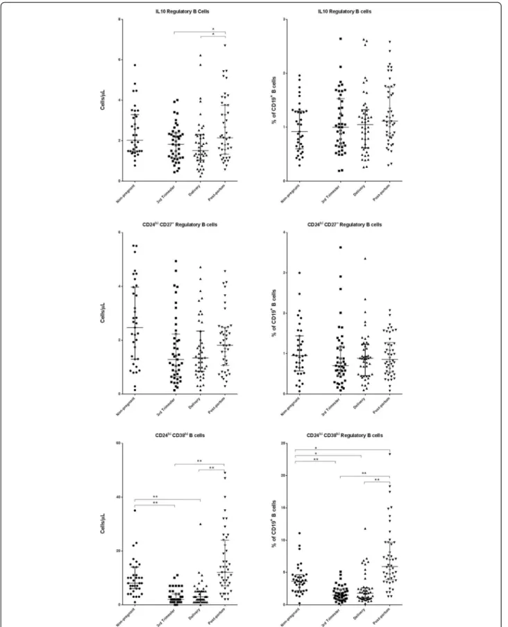
Characterization of Breg
The characterization of Breg is presented in Fig. 6. The absolute counts of IL-10 regulatory B cells and CD24hiCD38hiBregs during the 3rd trimester of pregnancy and on delivery day were significantly lower (p< 0.05) than those in the post-partum women. Additionally, the absolute counts of CD24hiCD38hiBregs were also significantly lower (p< 0.05) during the 3rd trimester of pregnancy and on de-livery day compared to the corresponding counts in the non-pregnant women. The absolute counts of CD24hiCD27+
women or between study visits.
Discussion
According to our study, late-stage pregnancy (between the 3rd trimester and delivery) is associated with periph-eral blood B cell lymphopenia. Indeed, the absolute counts and percentages of most B cell subsets in the 3rd trimester of pregnancy and on delivery day were signifi-cantly lower compared to the corresponding counts and percentages in the non-pregnant. However, these differ-ences did not significantly differ between the post-partum and non-pregnant women, suggesting that at this later time point, the absolute counts and percentages of most B cell subsets revert (or at least partially revert) to normal values. The most notable exceptions to this observation were identified for naïve B cells (whose percentages were significantly higher during the 3rd trimester and on deliv-ery day than in the non-pregnant) and for CD24hiCD38hi Bregs (whose percentages were significantly higher in the post-partum compared to the non-pregnant women).
To our knowledge, this is the first study to characterize the circulating B cell compartment in pregnancy while taking into account the maturational stages of the different B cell subsets. Furthermore, the study was conducted pro-spectively, from the 3rd trimester of pregnancy to post-partum, in a sample of 43 pregnant women.
The pregnancy-associated B lymphopenia that we iden-tified in our study has already been described in animal models [32, 33] and in humans [17–27]. According to Medina et al. [32], B lymphopoiesis in bone marrow is selectively reduced during normal pregnancy because of hormonal influences. Furthermore, Muzzio et al. [33] demonstrated that B lymphopoiesis is reduced in late pregnancy, when estradiol levels are high. Cellular migra-tion is another mechanism that can contribute to B cell lymphopenia. Studies of animal models have shown that monocytes and other immune cells migrate to the uterus during the later stages of pregnancy due to changes in the expression of chemokines [34, 35]. Furthermore, small populations of B cells have been identified in the decidua, suggesting leucocyte recruitment into the maternal-fetal interface [36]. The biological meaning of this suppression of B lymphopoiesis in normal pregnancy is uncertain but is probably related to the physiological immune tolerance.
shown). Nonetheless, B cell percentages in pregnant also seem to decrease during pregnancy, which does not hap-pens with either T cells or NK cells.
Compared to the percentages of peripheral blood naïve B cells in the non-pregnant women, we found higher values during the 3rd trimester and on delivery day, but no differences were observed in absolute counts. This relative increase in naïve B cells may be a consequence of decreased differentiation of B cells into memory cells and/or plasmablasts. In fact, Muzzio et al. [33] reported the expansion of naïve B cells in pregnant mice. The high levels of progesterone present in late pregnancy may poten-tially explain this, as high progesterone levels inhibit B cell activation in mice [37]. Our results may also be explained by the mobilization of more differentiated B subsets from peripheral blood to other body tissues.
Normal pregnancy has been compared to a state of quiescent systemic inflammation, while parturition has been likened to an immunological reaction that results in the recruitment of immune cells not only to the ma-ternal-fetal interface but also to the systemic circulation [38]. The results of our study support this idea, as we identified higher counts and percentages of CD24hiCD38hi Bregs post-partum relative to during the 3rd trimester and on delivery day. This observation may represent a regulatory mechanism for the suppression of immune cell activation events and may also explain the increased susceptibility to infections that occurs during the post-partum period and the altered clinical outcomes of some autoimmune diseases.
Unlike for the CD24hiCD38hiBregs, no significant dif-ferences between the non-pregnant and the pregnant or from the 3rd trimester of pregnancy to post-partum were identified for IL-10 Bregs (cell percentages) or for CD24hiCD27+ Bregs (cell percentages and counts). This heterogeneity of Breg subsets has been reported in other studies with humans [39]. Furthermore, CD24hiCD27+ Bregs, an activated memory subset, are more mature than transitional CD24hiCD38hi Bregs; thus, it is more likely for them to develop into antibody-producing cells that no longer possess a regulatory function [40].
The differences between pregnant and non-pregnant women identified for age, and parity are not likely to bias our results. In fact, among all of the women who were included in our study, counts and percentages of B cell subsets were not significantly associated with age, as demonstrated by the non-significant Spearman correl-ation coefficients between these variables (see Additional file 1). Furthermore, in the vast majority of cases, there were no statistically significant differences in counts and percentages of B cell subsets among women, despite parity (see Additional file 2). Finally, in the vast majority of cases, we have also not found significant associations be-tween counts (and percentages) of B cell subsets and gesta-tional age at the 3rd trimester of pregnancy (see Addigesta-tional file 3), gestational age at delivery (see Additional file 4), length of time post-partum until the collection of the final blood samples (see Additional file 5), and time since last pregnancy in the non-pregnant (see Additional file 6).
Previous studies have identified differences in the counts and percentages of B cells (total and subsets) be-tween neonates and individuals of up to 50 years of age [41]. However, to the best of our knowledge, there are no data regarding B cell variation in women over short periods of time, such as that of our study.
Although the pregnant women received analgesia and/ or anesthesia, which may cause temporary changes in maternal blood pressure, this is unlikely to cause import-ant changes in B cell counts because regional administra-tion is generally associated with low plasma levels of these drugs. Ideally, samples collected before pregnancy would have been compared with samples collected during preg-nancy in the same individuals; however, this would have been very difficult for us from a practical point of view. The fact that several of the changes that were observed during the 3rd trimester of pregnancy seem to be reversed during the post-partum period suggests that comparisons with non-pregnant women were adequate.
In future research, it is important to investigate whether B cell subset characterization could help to identify risk markers for the development of obstetric complications in pregnant women with or without autoimmune diseases. In this context, it would also be important to clarify the role of B-cell activating factor (BAFF), an essential survival
factor for transitional B cells, and of CD23, a B-lymphocyte differentiation marker.
Conclusion
According to our study, the characteristics of peripheral B cell compartment differ significantly between pregnant and non-pregnant women and vary over time from late pregnancy to post-partum. Such findings may allow us to recognize normal fluctuations in B cell subsets to better understand immune regulation during human pregnancy and to identify new strategies for the diagnosis and treat-ment of pregnancy-associated disturbances as well as the mechanisms of maternal responses to vaccination and infection.
Additional files
Additional file 1:Correlation coefficients between counts (and percentages) of B cell subsets and age, both for pregnant and non-pregnant women.aHealthy non-pregnant women;bThird trimester of pregnancy;cWithin 15 min after placental expulsion;dAt least 6 weeks after delivery;eSpearman correlation coefficient. (XLSX 11 kb)
Additional file 2:Comparison of counts (and percentages) of B cell subsets between nulliparous, primiparous, and multiparous women, both for pregnant and non-pregnant women. Values presented as median (Interquartile range); Three classes were compared using one-way ANOVA, or Kuskal-Wallis tests; Two classes were compared with t-Student or Wilcoxon tests; There was only one multiparous women among the pregnant, and this woman was excluded from this analysis.aNon-pregnant women;bThird trimester of pregnancy;cWithin 15 min after placental expulsion;dAt least 6 weeks after delivery;eKuskal-Wallis test; f Wilcoxon test; *p< 0.05. (XLSX 46 kb)
Additional file 3:Correlation coefficients between counts (and percentages) of B cell subsets and gestational age at the 3rd trimester of pregnancy. Corr. Coef., Spearman correlation coefficient. (XLSX 9 kb)
Additional file 4:Correlation coefficients between counts (and percentages) of B cell subsets and gestational age at delivery. Corr. Coef., Spearman correlation coefficient. (XLSX 9 kb)
Additional file 5:Correlation coefficients between counts (and percentages) of B cell subsets and length of time post-partum until the collection of the final blood samples. Corr. Coef., Spearman correlation coefficient. (XLSX 9 kb)
Additional file 6:Correlation coefficients between counts (and percentages) of B cell subsets and time since last pregnancy, in the non-pregnant women. Corr. Coef., Spearman correlation coefficient. (XLSX 9 kb)
Abbreviations
Bm, mature B cells; Bregs, regulatory B cells
Acknowledgements
The authors would like to thank José de Mello Saúde for partially funding the research.
Funding
This study was partially funded by José de Mello Saúde.
Authors’contributions
Author details
1Department of Obstetrics and Gynecology, CUF Descobertas Hospital,
Lisbon, Portugal.2CEDOC, Chronic Diseases Research Center, Immunology, NOVA Medical School, Faculty of Medical Sciences, Lisbon, Portugal.3Center for Rheumatology Research, Department of Medicine, University College London, London, UK.4Centro de Medicina Laboratorial Germano Sousa, Lisbon, Portugal.5Department of Clinical Pathology, Hospital Prof. Fernando Fonseca, E.P.E., Amadora, Portugal.6Obstetrics and Gynecology, Lisbon, Portugal.7Department of Immunoallergy, CUF Descobertas Hospital, Lisbon, Portugal.
Received: 30 July 2015 Accepted: 31 May 2016
References
1. Nahmias AJ, Schollin J, Abramowsky C. Evolutionary-developmental perspectives on immune system interactions among the pregnant woman, placenta, and fetus, and responses to sexually transmitted infectious agents. Ann N Y Acad Sci. 2011;1230:25–47.
2. Perricone C, de Carolis C, Perricone R. Pregnancy and autoimmunity: a common problem. Best Pract Res Clin Rheumatol. 2012;26(1):47–60. 3. Yasumizu T. Influenza complicating pregnancy. Nihon Rinsho. 2006;64(10):1930–3. 4. Birkeland SA, Kristoffersen K. Lymphocyte transformation with mitogens and
antigens during normal human pregnancy: a longitudinal study. Scand J Immunol. 1980;11(3):321–5.
5. Muzzio D, Zenclussen AC, Jensen F. The role of B cells in pregnancy: the good and the bad. Am J Reprod Immunol. 2013;69(4):408–12.
6. Bemark M, Holmqvist J, Abrahamsson J, Mellgren K. Translational mini-review series on B cell subsets in disease. Reconstitution after haematopoietic stem cell transplantation - revelation of B cell developmental pathways and lineage phenotypes. Clin Exp Immunol. 2012;167(1):15–25.
7. Marie-Cardine A, Divay F, Dutot I, Green A, Perdrix A, Boyer O, et al. Transitional B cells in humans: characterization and insight from B lymphocyte reconstitution after hematopoietic stem cell transplantation. Clin Immunol. 2008;127(1):14–25.
8. Bohnhorst JO, Bjorgan MB, Thoen JE, Natvig JB, Thompson KM. Bm1-Bm5 classification of peripheral blood B cells reveals circulating germinal center founder cells in healthy individuals and disturbance in the B cell subpopulations in patients with primary Sjogren’s syndrome. J Immunol. 2001;167(7):3610–8.
9. Sanz I. Rationale for B cell targeting in SLE. Semin Immunopathol. 2014;36(3):365–75.
10. Sims GP, Ettinger R, Shirota Y, Yarboro CH, Illei GG, Lipsky PE. Identification and characterization of circulating human transitional B cells. Blood. 2005;105(11):4390–8.
11. Guzman Moreno R. B-cell depletion in autoimmune diseases. Advances in autoimmunity. Autoimmun Rev. 2009;8(7):585–90.
12. Blair PA, Norena LY, Flores-Borja F, Rawlings DJ, Isenberg DA, Ehrenstein MR, et al. CD19(+)CD24(hi)CD38(hi) B cells exhibit regulatory capacity in healthy individuals but are functionally impaired in systemic Lupus Erythematosus patients. Immunity. 2010;32(1):129–40.
13. Iwata Y, Matsushita T, Horikawa M, Dilillo DJ, Yanaba K, Venturi GM, et al. Characterization of a rare IL-10-competent B-cell subset in humans that parallels mouse regulatory B10 cells. Blood. 2011;117(2):530–41. 14. Rolle L, Memarzadeh Tehran M, Morell-Garcia A, Raeva Y, Schumacher A,
Hartig R, et al. Cutting edge: IL-10-producing regulatory B cells in early human pregnancy. Am J Reprod Immunol. 2013;70(6):448–53.
lymphocyte subsets in normal pregnant and postpartum women: postpartum increase in NK/K (Leu 7) cells. Am J Reprod Immunol Microbiol. 1988;18(2):52–5. 21. Kraus TA, Engel SM, Sperling RS, Kellerman L, Lo Y, Wallenstein S, et al.
Characterizing the pregnancy immune phenotype: results of the viral immunity and pregnancy (VIP) study. J Clin Immunol. 2012;32(2):300–11. 22. Kuhnert M, Strohmeier R, Stegmuller M, Halberstadt E. Changes in
lymphocyte subsets during normal pregnancy. Eur J Obstet Gynecol Reprod Biol. 1998;76(2):147–51.
23. Mahmoud F, Abul H, Omu A, Al-Rayes S, Haines D, Whaley K. Pregnancy-associated changes in peripheral blood lymphocyte subpopulations in normal Kuwaiti women. Gynecol Obstet Investig. 2001;52(4):232–6. 24. Matthiesen L, Berg G, Ernerudh J, Hakansson L. Lymphocyte subsets and
mitogen stimulation of blood lymphocytes in normal pregnancy. Am J Reprod Immunol. 1996;35(2):70–9.
25. Moore MP, Carter NP, Redman CW. Lymphocyte subsets defined by monoclonal antibodies in human pregnancy. Am J Reprod Immunol. 1983;3(4):161–4. 26. Valdimarsson H, Mulholland C, Fridriksdottir V, Coleman DV. A longitudinal
study of leucocyte blood counts and lymphocyte responses in pregnancy: a marked early increase of monocyte-lymphocyte ratio. Clin Exp Immunol. 1983;53(2):437–43.
27. Watanabe M, Iwatani Y, Kaneda T, Hidaka Y, Mitsuda N, Morimoto Y, et al. Changes in T, B, and NK lymphocyte subsets during and after normal pregnancy. Am J Reprod Immunol. 1997;37(5):368–77.
28. Auerbach L, Hafner T, Huber JC, Panzer S. Influence of low-dose oral contraception on peripheral blood lymphocyte subsets at particular phases of the hormonal cycle. Fertil Steril. 2002;78(1):83–9.
29. Shinoda R, Watanabe M, Nakamura Y, Maruoka H, Kimura Y, Iwatani Y. Physiological changes of Fas expression in peripheral lymphocyte subsets during the menstrual cycle. J Reprod Immunol. 2003;60(2):159–68. 30. Yanaba K, Bouaziz JD, Haas KM, Poe JC, Fujimoto M, Tedder TF. A regulatory
B cell subset with a unique CD1dhiCD5+ phenotype controls T cell-dependent inflammatory responses. Immunity. 2008;28(5):639–50. 31. Benjamini Y, Yekutieli D. The control of the false discovery rate in multiple
testing under dependency. Ann Statist. 2001;29(4):1165–88. 32. Medina KL, Smithson G, Kincade PW. Suppression of B lymphopoiesis
during normal pregnancy. J Exp Med. 1993;178(5):1507–15.
33. Muzzio DO, Soldati R, Ehrhardt J, Utpatel K, Evert M, Zenclussen AC, et al. B cell development undergoes profound modifications and adaptations during pregnancy in mice. Biol Reprod. 2014
34. Gomez-Lopez N, Tanaka S, Zaeem Z, Metz GA, Olson DM. Maternal circulating leukocytes display early chemotactic responsiveness during late gestation. BMC Pregnancy Childbirth. 2013;13 Suppl 1:S8.
35. Tessier DR, Raha S, Holloway AC, Yockell-Lelievre J, Tayade C, Gruslin A. Characterization of immune cells and cytokine localization in the rat utero-placental unit mid- to late gestation. J Reprod Immunol. 2015;110:89–101. 36. Hussein MR, Abd-Elwahed AR, Abodeif ES, Abdulwahed SR. Decidual
immune cell infiltrate in hydatidiform mole. Cancer Invest. 2009;27(1):60–6. 37. Zhang L, Chang K-K, Li M-Q, Li D-J, Yao X-Y. Mouse endometrial stromal cells
and progesterone inhibit the activation and regulate the differentiation and antibody secretion of mouse B cells. Int J Clin Exp Pathol. 2014;7(1):123–33. 38. Christiaens I, Zaragoza DB, Guilbert L, Robertson SA, Mitchell BF, Olson DM.
Inflammatory processes in preterm and term parturition. J Reprod Immunol. 2008;79(1):50–7.
40. Quan C, ZhangBao J, Lu J, Zhao C, Cai T, Wang B, et al. The immune balance between memory and regulatory B cells in NMO and the changes of the balance after methylprednisolone or rituximab therapy. J Neuroimmunol. 2015;282:45–53.
41. Morbach H, Eichhorn EM, Liese JG, Girschick HJ. Reference values for B cell subpopulations from infancy to adulthood. Clin Exp Immunol. 2010;162(2):271–9.
• We accept pre-submission inquiries
• Our selector tool helps you to find the most relevant journal
• We provide round the clock customer support
• Convenient online submission
• Thorough peer review
• Inclusion in PubMed and all major indexing services
• Maximum visibility for your research
Submit your manuscript at www.biomedcentral.com/submit