965
Arq Neuropsiquiatr 2012;70(12):960-966 1. Tulving E. Concepts of human memory. In: Squire L, Lynch G,
Weinberger NM, McGaugh JL (editors). Memory: organization and locus of change. New York: Oxford Univ. Press, 1991.
2. Brandt J, Benedict RHB. Hopkins verbal learning test – revised. Odessa: Psychological Assessment Resource, 2001.
3. Benedict RHB. Brief visuospatial memory test – revised. Odessa:
Psychological Assessment Resource, 1997.
4. Brucki S, Nitrini R, Caramelli P, Bertolucci PHF, Okamoto IH. Sugestões para o uso do mini-exame do estado mental no Brasil. Arq Neuropsiquiatr 2003;61:777-781.
5. Zigmond AS, Snaith RP. The hospital anxiety and depression scale. Acta Psychiatr Scand 1983;67:361-370.
References
Reversible parkinsonism associated with
neurocysticercosis
Parkinsonismo reversível associado com neurocisticercose
Plínio M. G. de Lima, Renato P. Munhoz, Hélio A. G. Teive
Movement Disorders Unit, Neurology Service, Internal Medicine Department, Hospital de Clínicas, Federal University of Paraná, Curitiba PR, Brazil. Correspondence: Hélio A. G. Teive; Rua General Carneiro 1.103 / 102; 80060-150 Curitiba PR - Brasil; E-mail: [email protected]
Conflict of interest: There is no conflict of interest to declare.
Received 23 May 2012; Received in final form 04 June 2012; Accepted 11 June 2012
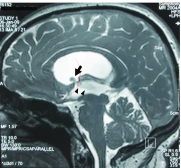
A 38-year-old female was assessed in a county hospi-tal with a four-month history of severe throbbing morning headaches. Due to the syndromic diagnosis of intracranial hypertension, a brain magnetic resonance imaging (MRI) was performed showing evidence of supratentorial non-communicating hydrocephalus because of an intraven-tricular cyst. he cyst was located at the level of the fron-tal horn of the right lateral ventricle, extending through the foramen of Monro to the third ventricle. Additionally, there were signs of edema in the midbrain periaqueductal region. he patient was treated with a ventriculoperitoneal (VP) shunt, and the symptoms of intracranial hypertension improved. A diagnosis of neurocysticercosis (NCC) was es-tablished after analysis of CSF, and positive antibodies reac-tions were detected. he patient was started on high-dose oral albendazole (3.2 g qd). After four days, additional signs were detected, including mutism, hypomimia, and sialor-rhea, associated with severe parkinsonism (marked rigidity, bradykinesia, and resting upper limb tremor). A new brain MRI showed milder hydrocephalus. However, the intraven-tricular cyst and cerebral aqueduct edema and ependymitis remained unchanged (Figs 1 and 2).
At such point, she was referred to our Service of Neurology at the Hospital de Clínicas, Federal University of Paraná. On admission, neurological examination showed a severe Parkinsonian syndrome that also included marked postural instability, anarthria, and vertical ophthalmoparesis for up-ward gaze.
Our diagnosis was of Parkinsonism secondary to NCC. We believe that treatment with high-dose albendazole made the presentation worse by adding a reactive inlammatory re-sponse and by aggravating ependymitis and tissue damage.
Methylprednisolone pulse therapy was then started (1 g intravenous, daily, for ive days). Symptomatic treatment also included levodopa/carbidopa (250/50 mg daily), with pro-gressive improvement in Parkinsonism symptom. Finally, she then underwent endoscopic neurosurgery including monro-plasty, removal of the cyst, pellucidoctomy, and third ventric-ulostomy. Neuropathological examination conirmed the di-agnosis of NCC.
Eight months later, the patient was progressively with-drawn from dopaminergic treatment and remained asymp-tomatic, able to return to her routine activities.
966 Arq Neuropsiquiatr 2012;70(12):960-966
This case shows the rare occurrence of Parkinsonism secondary to NCC, leading to hydrocephalus and brain-stem ependymitis. NCC is the most common helminth infection of the central nervous system, particularly in developing countries1. Parkinsonism secondary to NCC can be related to hydrocephalus secondary to cysts of the fourth ventricle, perimesencephalic cysts, basal racemose cysts and brainstem ependymitis due to the cyst, as well as by ischemic lesions in the midbrain2-4.
It was clear from the clinical description that the Parkinsonism symptoms were greatly exacerbated after
albendazole treatment was started. his complication has been described in the literature and occurs because cysti-cidal drugs may lead to an intense inlammatory reaction, especially in forms of NCC involving intraventricular cysts and ependymitis/encephalitis5.
Cysticercotic encephalitis should be considered in the dif-ferential diagnosis of rapidly progressive Parkinsonism, with atypical features in regions where this infection is common2,3. he case presented here resembles others already described, with the peculiarity that there was a deterioration of the symp-toms of Parkinsonism after using high-dose albenzadole2-4.
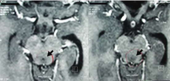
Fig 2. Contrast-enhanced axial T1-weighted image with edema around the cerebral aqueduct (arrows).
1. Takayanagui OM, Jardim E. Clinical aspects of neurocysticercosis: analysis of 500 cases. Arq Neuropsiquiatr 1983;41:50-63.
2. Sá DS, Teive HAG, Troiano AR, Werneck LC. Parkinsonism associated with neurocysticercosis. Parkinsonism Relat Disord 2005;11:69-72. 3. Verma A, Berger JR, Bowen BC, Sanchez-Ramos J. Reversible
parkinsonian syndrome complicating cysticercus midbrain encephalitis. Mov Disord 1995;10:215-219.
4. Curran T, Lang AE. Parkinsonism syndromes associated with hydrocephalus: case report, a review of the literature, and pathophysiological hypotheses. Mov Disord 1994;9: 508-520.
5. Garcia HH, Evans CAW, Nash TE, et al. Current consensus guidelines for treatment of neurocysticercosis. Clin Microbiol Rev 2002;15: 747-756.