Influences of Rich in Saturated and Unsaturated Fatty Acids Diets in
Rat Myocardium
Matheus Fécchio Pinotti, Maeli Dal-Pai Silva, Mário Mateus Sugizaki, Yeda Santana Diniz Novelli, Lea Sílvia Sant’ana,
Flávio Ferrari Aragon, Carlos Roberto Padovani, Ethel Lourenzi Barbosa Novelli, Antonio Carlos Cicogna
Faculdade Médica da Faculdade de Medicina de Botucatu – UNESP – Botucatu, SP - Brazil
Summary
Objectives: To study the influence of saturated (SFA) and unsaturated fatty acid (UFA) rich diets on mechanical function, morphology and oxidative stress in rat myocardium.
Methods: Male, 60-day-old Wistar rats were fed a control (n=8), a SFA (n=8), or a UFA-rich diet (n=8) for sixty days. Mechanical function was studied in isolated left ventricle papillary muscle under isometric and isotonic contractions, in basal conditions (1.25mM calcium chloride) and after 5.2mM calcium chloride and beta-adrenergic stimuli with 1.0µM isoproterenol. Left ventricle fragments were used to study oxidative stress and morphology under light and electron microscopy.
Results: SFA and UFA-rich diets did not change myocardium mechanical function. Both diets caused oxidative stress, with high lipid hydroperoxide and low superoxide-dismutase concentrations. UFA rich diet decreased catalase expression and SFA rich diet decreased the amount of myocardial glutathione-peroxidase. Both diets promoted light ultrastructural injuries such as lipid deposits and cell membrane injuries.
Conclusion: Results suggest that SFA and UFA rich diets do not alter isolated muscle mechanical function, but promote light myocardial morphological injuries and oxidative stress.
Key words: Diet; fatty acids; myocardium; rats; oxidative stress.
Mailing Address: Matheus Fécchio Pinotti •
Rua Dr. José Adriano Marrey Jr., 796 – ap. 101 - 18603-610 – Botucatu, SP E-mail: mpinotti@uol.com.br
Manuscript received May 09, 2006; revised manuscript received May 09, 2006; accepted June 22, 2006.
Introduction
Recent studies have reported the benefical influences of lifestyle and dietary changes, such as consuming less saturated fatty acids (SFA) and trans fatty acids, and increasing consumption of fish oil, rich in unsaturated fatty acids (UFA), on morbidity and mortality of patients with coronary artery disease1,2. While SFA rich foods are atherogenic and reduce
coronary reserve1,3,4, UFAs have an anti-atherogenic effect1,5
and protect the heart against the adverse effects of acute phase myocardial infarction5,6.
Studies on UFA have shown beneficial results in the cardiovascular system1,5,6, experiments have shown that diets
rich in UFA and SFA cause structural and metabolic damage in the myocardium6-10. While both diets cause disorganized
sarcolemma, lipid deposits9,10, and myocardial oxidative
stress6,10, the UFA rich diet also promotes necrosis7 and
abnormal muscle striation8.
Although literature shows that UFA rich diets cause morphometabolic alterations6-10, information is contradictory
with respect to their effects on myocardium mechanical function7,9,11-15. Although previous studies have shown
that diet caused depressed cardiac function depression in
isolated rabbit12 and rat 13 papillary muscle, recent data from
our laboratory15 have shown that, in agreement with other
authors7,9,11,14, UFA diet does not alter functional performance.
On the other hand, there are few reports on the relationship between SFA rich diets and cardiac function. Pepe and McLennan (2002) showed that animal origin SFA causes ventricular dysfunction in isolated rat heart, seen only after important increase in preload and the myocardial ischemia-reperfusion process. These authors demonstrate that this functional alteration is a consequence of the heart’s incapacity to increase coronary flow and adequately utilize the oxygen needed to produce energy4.
The objective of this work was to evaluate the influence of SFA and UFA supplemented diets on isolated papillary muscle mechanical function, complemented by analysis of oxidative stress and morphology in rat myocardium.
Methods
Animal model and experimental protocol - Sixty-day-old male Wistar rats from the Experimental Laboratory Biotério, Department of Internal Medicine, Botucatu School of Medicine, (FMB-UNESP), were fed a control (C, n=8), an SFA (SF, n=8) or a UFA rich diet (UF, n=8) and water ad libitum. LabinaLabinaTM commercial rat chow (Purina, Paulínia,
and Lmax was defined as muscle length at peak AT); this was allowed to stabilize for an additional 15 minutes while performing physiologically sequenced contractions. Isometric contraction parameters were determined,
including active tension (AT, g/mm2, defined as peak
isometric tension minus resting tension), resting tension (RT, g/mm2), time to peak tension (TPT, ms), peak isometric
tension development rate (+dT/dt, g/mm2/s), maximum
tension decline rate (-dT/dt, g/mm2/s), and time from peak
tension to 50% relaxation (RT50, ms).
After isometric contraction, muscles were analyzed in isotonic contraction against a weight sufficient to keep the resting muscle at Lmax. The isotonic parameters were maximum shortening velocity (-dL/dT, ML/s) and maximum relaxation velocity (+dL/dT, ML/s). The stiffness constant (km) was calculated from: log(Sm) = B + kmEm; Sm is the stiffness calculated from resting tension in different muscle lengths normalized to cross-sectional area; Em is the natural muscle strain (Em = 1n[Lm/L0.1] where Lm is the instant muscle length and L0.1 is the length with 0.1g/mm2stress). Analyses were
performed under basal conditions ([Ca2+] = 1.25mM), with
[Ca2+] = 5.2mM, and with isoproterenol 10-6 M.
The parameters used to characterize papillary muscle were length (mm), weight (mg), and cross-sectional area (CSA, mm2). L
max in vitro was measured with a Gartner
cathetometer. The muscle portion between both spring clips was cut and weighed after drying in filter paper. CSA was calculated as the relationship between weight and Lmax, considering that papillary muscle is cylindrical and its specific weight is constant.
To compare the mechanical function between different muscle lengths, isometric and isotonic parameters were normalized to CSA and Lmax.
Biochemical study - Five animals from each group were used for the biochemical study. The heart was removedThe heart was removed and cardiac adipose tissue was discarded. LV samples of 200mg were weighed and homogenized in 5ml of cold phosphate buffer (0.1M, pH7.4) containing 1mM ethylenediaminetetraacetic acid (EDTA). Tissue homogenates were prepared in a motor-driven Teflon glass Potter Elvehjem tissue homogenizer (1min. 100rpm) immersed in ice water. The homogenate was centrifuged at 10,000 rpm for 15 min and supernatant was used to determine: Total protein (TP), lipid hydroperoxide (LH), total antioxidant status (TAS), glutathione peroxidase (GSH-Px), superoxide dismutase (SOD), and catalase (CAT).
LH was measured through hydroperoxide-mediated Fe2+
oxidation under acid conditions. Samples were added to reaction mixtures containing 100µM xylenol orange, 250µM FeSO4, 25µM H2SO4, and 4mM butylated hydroxytoluene (BHT) in 90% (v/v) methanol. The mixtures were incubated for 30 min at room temperature prior to measurement at 560nm.
GSH-Px was assayed using 0.15M, pH7.0 phosphate buffer containing 5mM EDTA, 0.1ml 0.0084M NADPH,
0.005ml GSSG-reductase (Sigma), 0.01ml 1.125M NaN3
(sodium azide), and 0.1ml 0.15M GSH [31]. Superoxide dismutase activity was determined by the ability of the fat (37.45% UFA and 62.55% SFA). Diet preparation is
described below.
Rats were maintained on this dietary regimen for 60 days and then sacrificed. All animals were housed in individual cages in a room controlled at 23oC and with a 12h light:dark
cycle. All animals were weighed once a week. Initial body weight (IW), final body weight (FW), left ventricle weight (LV), right ventricle weight (RV), and LV/FW and RV/FW ratios were measured in all rats used in the functional study. Animal food consumption was determined once a week. All experiments and procedures were performed in accordance with the Guide for the Care and Use of Laboratory Animals published by the U.S. National Institute of Health and were approved by the ethics committee of Botucatu School of Medicine, UNESP, São Paulo, Brazil.
Diet preparation - The UFA and SFA rich diets were prepared with commercial rat chow. Thus all diets providedThus all diets provided sufficient vitamins, minerals, essential amino acids, and lipids. Corn oil was the source of UFA and coconut oil the source of SFA. The UFA-rich diet was prepared by mixing 220ml of corn oil and 30ml of coconut oil with 1,000g of rat chow; the SFA-rich diet was prepared by mixing 110ml of corn oil and 140ml of coconut oil with 1,000g of rat chow. The mixture was then passed through a pelleting machine. Pellets were dried in a circulating air oven for 24 hours at 70oC. After drying, both diets were stored at 6oC for use
during the experiment.
Both diets were analyzed in the Agricultural and Livestock Products Technology Laboratory of the School of Agronomy Sciences (FCA-UNESP). Fat was extracted with petroleum ether in a Soxhlet extractor16. Lipids were extracted as
per Folch et al17: fatty acid composition was determined
after esterification with methanol sulphuric acid and the resulting fatty acid methyl esters [26] were analyzed by a chromatograph CG 17 A, using a 35m DB-Wax column and flame ionization detector.
Functional study - Cardiac intrinsic contractile performance was evaluated by studying isolated LV papillary muscle by the technique used in our lab. Briefly, at the timeBriefly, at the time of study, rats were anesthetized (with sodium pentobarbital, 50mg/kg, IP) and killed; their hearts were quickly removed and placed in oxygenated Krebs-Henseleit solution at 28oC. The LV anterior or posterior papillary muscles were
dissected free, mounted between 2 spring clips, and placed vertically in a chamber containing Krebs-Henseleit solution at 28oC and oxygenated with a mixture of 95% O
2 and 5%
CO2 (pH7.38). Muscles were stimulated at 12 contractions/
min with a voltage 10% above threshold. The spring clip on the upper end of the muscle was attached to a low inertia DC pen motor (model G100-PD; General Scanning), and the lower clip to a load cell (model DSC-3; Kistler-Morse). A digital computer with an analogue-to-digital interface allowed preparation tension or length to be controlled. Tension and length data were sampled at 1kHz and stored on disk for later analysis.
enzyme to inhibit reduction of nitro blue tetrazolium (NBT; Sigma). NBT reduction rate in the absence of tissue was used as a reference. One unit of SOD was defined as the amount of protein needed to decrease the reference rate to 50% maximum inhibition. All data were expressed in SOD units per mg protein. Catalase activity was determined with pH7.0 phosphate buffer at 240nm.
Enzyme activities were determined using a microplate reader (Bio-tech Instruments INC USA). Spectrophotometric determinations were performed in a Pharmacia Biotech spectrophotometer (974213, Cambridge, England). All reagents were from Sigma (Sigma. St. Louis, MO, USA). The extinction coefficient for NADH at 340nm was 6.22µmol-1cm-1
and for DTNB at 412nm was 13.6mmol-1cm-1.
Morphological study - Five animals from each group were used for histological study. After anaesthesia with sodium pentobarbital (50mg/kg, IP), hearts were excised and ventricles separated. The LV free wall was cut into
fragments and chilled in n-hexane at –70oC cooled in
liquid nitrogen. Serial transverse 8Mm sections were
cut in a cryostat at –20oC and stained with hematoxylin
and eosin. For ultrastructural study, small pieces of the LV papillary muscle were fixed in Karnowsky’s fixative (0.12M phosphate, pH7.2) for 1-2h and postfixed in 1% osmium tetroxide in 0.1M phosphate buffer for 2h. After dehydratation in a graded ethanol series, samples were embedded in epoxy resin. Ultrathin sections were cut from selected areas with a diamond knife, double-stained with uranyl acetate and lead citrate, and examined in a Philips EM 301 electron microscope. LV myocyte cross sectional area was measured using a compound microscope attached to a computerized imUFAng analysis system (Image-Pro Plus 3.0, Media Cybernetics, Silver Spring, Md.).
Statistical analysis - Morphological, basal functional, and serum lipid data were evaluated by analysis of variance complemented by the Tukey test. Functional data with increased calcium and isoproterenol concentrations were
submitted to analysis of variance for repeated samples in three independent groups. Significance level was 5% (p<0.05).
Results
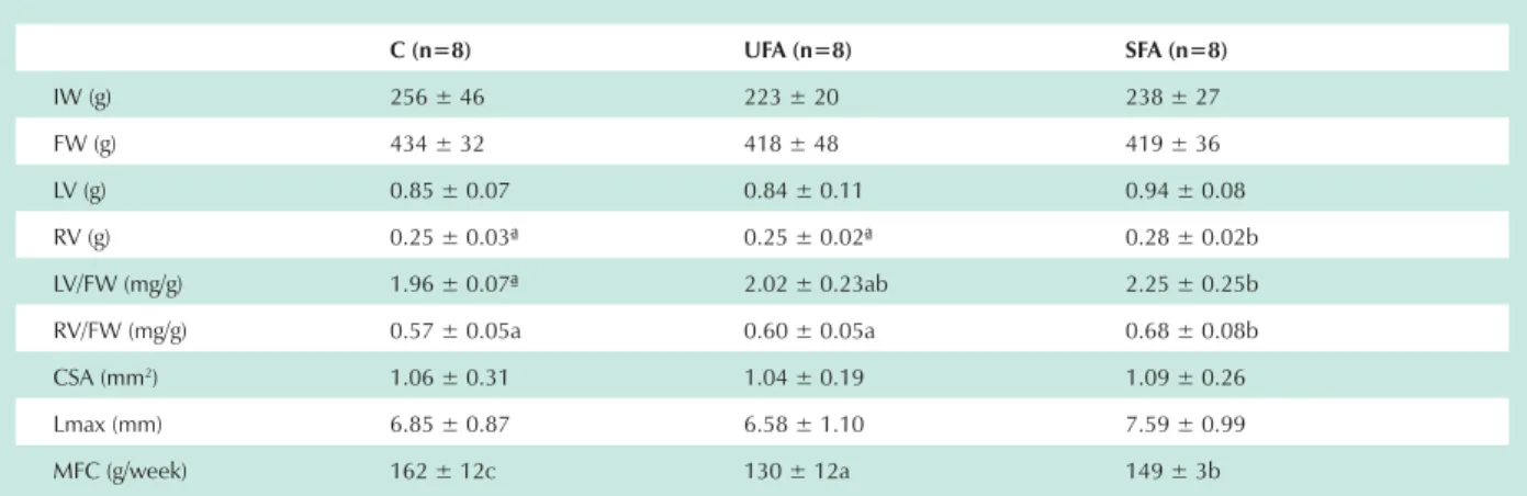
Table 1 shows the general characteristics of animals used in this experiment. Animals submitted to the SFA diet had higher VD than controls and UFA diet animals. Mean food ingestion was different between groups, higher in controls and lower in UF rats.
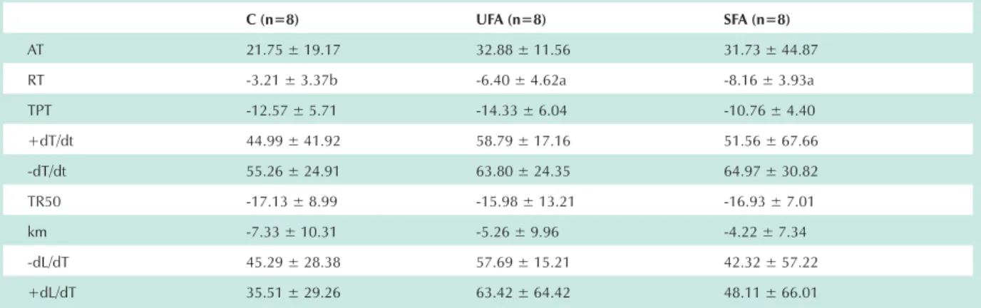
Table 2 shows data from the isolated papillary muscle functional study at basal condition in 1.25mM concentration extra-cellular calcium. There were no statistical differences between the three groups. Table 3 shows mechanical results with increased extracellular calcium concentration. The variation in resting tension (RT) was less in animals on supplemented diets than controls. The other parameters were not statistically different between groups. When stimulated with isoproterenol, the stiffness constant was less in SF than the other groups. There were no statistical differences for other analyzed variables (Tab. 4).
Biochemical analysis of the myocardium showed that SF and UF animals had increased lipid hydroperoxide and reduced total antioxidants and superoxide-dismutase than controls. Total antioxidant substances in UF were less than in SF. The UFA diet promoted reduced catalase activity and the SFA diet reduced glutathione peroxidase in relation to the other two groups. Soluble protein from the myocardium was similar in all three groups (Tab. 5).
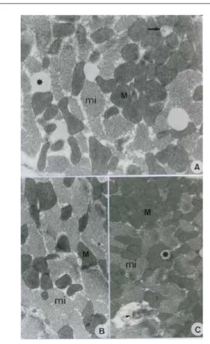
Figures 1 and 2 show ultrastructural analysis of papillary muscle from animals submitted to UFA and SFA rich diets, respectively. There are discrete alterations in both groups with small fat deposits between myocytes and focal lesions in the plasmatic membrane and mitochondrial crystals. Optical microscopy did not show any morphological alterations.
Table 1 - General experimental groups’ characteristics
C (n=8) UFA (n=8) SFA (n=8)
IW (g) 256 ± 46 223 ± 20 238 ± 27
FW (g) 434 ± 32 418 ± 48 419 ± 36
LV (g) 0.85 ± 0.07 0.84 ± 0.11 0.94 ± 0.08
RV (g) 0.25 ± 0.03ª 0.25 ± 0.02ª 0.28 ± 0.02b
LV/FW (mg/g) 1.96 ± 0.07ª 2.02 ± 0.23ab 2.25 ± 0.25b
RV/FW (mg/g) 0.57 ± 0.05a 0.60 ± 0.05a 0.68 ± 0.08b
CSA (mm2) 1.06 ± 0.31 1.04 ± 0.19 1.09 ± 0.26
Lmax (mm) 6.85 ± 0.87 6.58 ± 1.10 7.59 ± 0.99
MFC (g/week) 162 ± 12c 130 ± 12a 149 ± 3b
Discussion
The objective of this study was to evaluate the effects of SFA and UFA rich diets on isolated LV papillary muscle mechanical function, oxidative stress, myocardium morphology. Isolated muscle preparations allow measurement of the myocardium’s capacity to develop tension and shortening, independent of pre and after-load alterations, and changes in cardiac frequency which could modify in vivo cardiac performance. Inotropic stimulation made it possible to see contraction and relaxation phases that could not be seen under basal conditions, and helped in understanding the mechanisms involved in cardiac function changes.
This work showed that animals fed on UFA or SFA diets had the same body weights as controls. The lower food
consumption in hypercaloric diet rats than controls equalled out caloric intake between the three groups, which resulted in similar final body weights. These data agree with previous experiments using UFA or SFA rich diets8-10,13-15. Analysis of
relationships between ventricle weights and rat final body weights showed that animals submitted to SFA diets had biventricular hypertrophy compared to controls, and RV hypertrophy compared to UF animals.
In this study, myocardial morphological changes, with small fat deposits between myocytes, focal lesions in the plasmatic membrane, and mitochondrial crystals were only found at the ultrastructural level. The changes seen in the UFA diet were less relevant than in previous studies which showed necrosis7
and disorganization myocyte contraction apparatus8. The
mechanism which produced myocardial lesions by increasing Table 2 - Functional parameters in basal situation
C (n=8) UFA (n=8) SFA (n=8)
AT (g/mm2) 5.39 ± 1.27 4.58 ± 0.86 4.12 ± 1.02
RT (g/mm2) 1.52 ± 0.59 1.58 ± 0.25 1.28 ± 0.32
TPT (ms) 159 ± 14 162 ± 12 147 ± 12
+dT/dt (g/mm2/s) 59 ± 16 50 ± 11 49 ± 13
-dT/dt (g/mm2/s) 18 ± 6 16 ± 3 14 ± 3
TR50 (ms) 218 ± 33 213 ± 23 198 ± 21
km 19 ± 3 19 ± 2 19 ± 3
-dL/dT (ML/s) 1.24 ± 0.21 1.06 ± 0.20 1.22 ± 0.25
+dL/dT (ML/s) 2.02 ± 0.38 1.78 ± 0.58 2.22 ± 0.66
Means ± SD; n - number of animals; C - control group; UF - group treated with a diet rich in unsaturated fatty acids; SF - group treated with a diet rich in saturated fatty acids; AT - peak developed tension; RT - resting tension; TPT - time to peak tension; +dT/dt - maximum tension development rate ; -dT/dt - maximum tension decline rate; RT50 - time from peak tension to 50% relaxation; km - stiffness constant; -dL/dT - maximum shortening velocity; +dL/dT - maximum relaxation velocity. There were no statistical differences between groups (P<0.05).
Table 3 - Effects of extracellular calcium increase on functional parameters
C (n=8) UFA (n=8) SFA (n=8)
AT 21.75 ± 19.17 32.88 ± 11.56 31.73 ± 44.87
RT -3.21 ± 3.37b -6.40 ± 4.62a -8.16 ± 3.93a
TPT -12.57 ± 5.71 -14.33 ± 6.04 -10.76 ± 4.40
+dT/dt 44.99 ± 41.92 58.79 ± 17.16 51.56 ± 67.66
-dT/dt 55.26 ± 24.91 63.80 ± 24.35 64.97 ± 30.82
TR50 -17.13 ± 8.99 -15.98 ± 13.21 -16.93 ± 7.01
km -7.33 ± 10.31 -5.26 ± 9.96 -4.22 ± 7.34
-dL/dT 45.29 ± 28.38 57.69 ± 15.21 42.32 ± 57.22
+dL/dT 35.51 ± 29.26 63.42 ± 64.42 48.11 ± 66.01
Table 5 - Myocardial oxidative stress
C (n=5) UFA (n=5) SFA (n=5)
TP (g%) 21 ± 3 24 ± 1 24 ± 1
LH (nmol/ g of tissue) 158 ± 5a 176 ± 5b 187 ± 14b
TAS (g%) 35 ± 6c 15 ± 2a 24 ± 4b
SOD (U/mg of protein) 27 ± 5b 18 ± 2a 17 ± 2a
GSH-Px (U/mg of tissue) 22 ± 2b 22 ± 1b 16 ± 2a
CAT (kat.f) 9 ± 1b 7 ± 1a 9 ± 1b
Means ± SD; n - number of animals; C - control group; F - group treated with a diet rich in unsaturated fatty acids; TP - total protein; LH - lipid hydroperoxide; TAS - total antioxidant status; SOD - superoxide-dismutase; GSH-Px - glutathione peroxidase; CAT - catalase. Data with statistical differences are indicated with different lowercase letters (P<0.05).
fatty acid intake is unknown7. It is believed to be from the
oxidative stress caused by this type of diet10,18,19. Most ingested
lipids are incorporated in membrane phospholipids, making them more susceptible to attack from free radicals. This mainly occurs with UFA which has unstable molecular bonds in its structure and is therefore more susceptible to oxidative stress10,18,19. This hypothesis is supported by our experiment,
where oxidative stress and discrete ultrastructural lesions were caused by UFA and SFA rich diets.
Although prior works have associated oxidative stress and cardiac dysfunction6,20-22, this work found no alterations in
mechanical function under basal conditions. Results, such as the isolated changes in tension and relaxation with increased extracellular calcium concentration and muscle stiffness constant with isoproterenol in the SF group, allow us to conclude that the diets did not provoke significant myocardial functional changes. These findings are in agreement with studies that show that UFA diets do not modify myocardial
Table 4 - Effects of isoproterenol on functional parameters
C (n=8) UFA (n=8) SFA (n=8)
AT 12.67 ± 10.38 15.82 ± 7.54 15.46 ± 18.14
RT 0.00 ± 2.53 0.86 ± 2.14 0.45 ± 4.13
TPT -25.02 ± 7.58 -22.43 ± 5.49 -22.28 ± 7.59
+dT/dt 46.53 ± 28.01 55.41 ± 21.28 46.45 ± 33.82
-dT/dt 69.68 ± 26.70 62.21 ± 26.09 67.79 ± 41.59
TR50 -34.82 ± 8.04 -33.11 ± 7.15 -33.45 ± 11.59
km 9.08 ± 7.34b 8.44 ± 4.76b 3.53 ± 2.20a
-dL/dT 38.32 ± 21.15 43.57 ± 20.91 40.47 ± 26.95
+dL/dT 67.89 ± 16.89 62.93 ± 12.26 65.89 ± 50.20
Median ± semi-range of values in %; n: number of animals; C: control group; UF: group treated with a diet rich in unsaturated fatty acids; SF: group treated with a diet rich in saturated fatty acids; AT: peak developed tension; RT: resting tension; TPT: time to peak tension; +dT/dt: maximum tension development rate; -dT/dt: maximum tension decline rate; RT50: time from peak tension to 50% relaxation; km: stiffness constant; -dL/dT: maximum shortening velocity; +dL/dT: maximum relaxation velocity. Data with statistical differences are indicated with different lowercase letters (P<0.05).
mechanical function7,9,11,14,15, and disagree with other authors
who report depressed myocardial function with UFA12,13 and
SFA diets4.
In conclusion, this work shows that diets rich in saturated or unsaturated fatty acids cause oxidative stress and discrete ultrastructural myocardial lesions, and do not compromise myocardial mechanical function.
Acknowledgments
Our sincere thanks to J.C. Georgette, M. Bruno and C.E. Knaggs for their technical assistance and to FAPESP (02/02704-0) for believing in our work.
Potential Conflict of Interest
References
1. Hu FB, Willet WC. Optimal diets for prevention of coronary heart disease. JAMA. 2002; 288: 2569-78.
2. Iestra JA, Kromhout D, van der Schouw YT, Grobbee DE, Boshuizen HC, van Staveren WA. Effect size of lifestyle and dietary changes on all-cause mortality in coronary artery disease patients: a systematic review. Circulation. 2005; 112: 924-34.
3. Nielsen LB, Leth-Espensen P, Nordestgaard BG, Foged E, Kjeldsen K, Stender S. Replacement of dietary saturated fat with monounsaturated fat: effect on atherogenesis in cholestesterol-fed rabbits clamped at the same plasma cholesterol level. Br J Nutr. 1995; 74: 509-21.
4. Pepe S, McLennan PL. Cardiac membrane fatty acid composition modulates myocardial oxygen consumption and postischemic recovery of contractile function. Circulation. 2002; 105: 2303-8.
5. Mori TA, Beilin LJ. Long-chain omega 3 fatty acids, blood lipids and cardiovascular risk reduction. Curr Opin Lipidol. 2001;12:11-7.
6. Nageswari K, Banerjee R, Menon VP. Effect of saturated, W-3 and W-6 polyunsaturated fatty acids on myocardial infarction. J Nutr Biochem. 1999; 10: 338-44.
7. Lamers JMJ, Hartog JM, Verdouw PD, Hulsmann WC. Dietary fatty acids and myocardial function. Basic Res Cardiol. 1987; 82: 209-21.
8. Sylvén C, Glavind J. Peroxide formation, vitamin E and myocardial damage
in the rat. Int J Vitam Nutr Res. 1977; 47: 9-16.
9. Charnock JS, McLennan PL, Abeywardena MY, Dryden WF. Diet and cardiac arrhythmia: effects of lipids on age-related changes in myocardial function in the rat. Ann Nutr Metab. 1985; 29: 306-18.
10. Diniz YS, Cicogna AC, Padovani CR, Santana LS, Faine LA, Novelli EL. Diets rich in saturated and polyunsaturated fatty acids: metabolic shifting and cardiac health. Nutrition. 2004; 20: 230-4.
11. de Wildt DJ, Speijers GJ. Influence of dietary rapeseed oil and erucic-acid upon myocardial performance and hemodynamics in rats. Toxicol Appl Pharmacol. 1984; 74: 99-108.
12. Peterson DW, Griffith DW, Napolitano CA. Decreased myocardial contractility in papillary muscles from atherosclerotic rabbits. Circ Res. 1979; 45: 338-46.
13. Chemla D, Javouhey-Donzel A, Suard I, Maupoil V, Lecarpentier Y, Pourny JC, et al. Influence of dietary polyunsaturated fatty acids on contractility, lusitropy and compliance of isolated rat myocardium. J Mol Cell Cardiol. 1995; 27: 1745-55.
14. Kako KJ, Vasdev SC. Effects of a high-fat erucic acid on the lipid metabolism and contractility of the rat heart. Biochem Med. 1979; 22: 76-87.
15. Pinotti MF, Silva MDP, Sugizaki MM, Diniz YS, Sant’ana LS, Aragon FF, et al. Effects of unsaturated fatty acids on myocardial performance, metabolism
Fig. 1 – Striated cardiac muscle. Unsaturated fatty acid group. A, B, & C: Myofibrils (mi). Normal mitochondria (M). Mitochondria with crystal loss in A (arrow). Fat globules in A & C (*). Myelin figure in C (arrowhead). A: 4600X; B: 3400X; C: 2650X.
and morphology. Braz J Med Biol Res. 2006; 39: 305-12.
16. Association of Official Analytical Chemists. Official methods of analysis. 14th ed. Washington, DC: Association of Official Agricultural Chemists; 1984.
17. Folch J, Lee M, Sloane Stanley GH. A simple method for isolation and purification of total lipids from animal tissue. J Biol Chem. 1957; 226: 497-509.
18. Moreira NX, Curi R, Padovese R, Mancini-Filho J. Incorporation of dietary trans-monounsaturated fatty acids into tissues of Walker 256 tumor-bearing rats. Braz J Med Biol Res. 2001; 34: 501-8.
19. Weirik EV, Berg H, Weststrate JA, Hof KH, Graaf C. Consumption of reduced
fat products: effects on parameters of antioxidant capacity. Eur J Clin Nutr. 1996; 50: 214-9.
20. Keith M, Geranmayegan A, Sole MJ, Kurian R, Robinson A, Omran AS, et al. Increased oxidative stress in patients with congestive heart failure. J Am Coll Cardiol. 1998; 31: 1352-5.
21. Collucci WS. Molecular and cellular mechanisms of myocardial failure. Am J Cardiol. 1999; 80: 15L-25L.