Address to: Dr. Antonio Luiz Pinho Ribeiro. Rua Campanha R nº 98/101, Carmo, 30310-770 Belo Horizonte, MG, Brasil.
Phone: 55 31 3409-9437
e-mail: [email protected]
Received 10 January 2014
Accepted 17 November 2014
Ventricular arrhythmias in Chagas disease
Marco Paulo Tomaz Barbosa
[1],[2], Andre Assis Lopes do Carmo
[1],[2],
Manoel Otávio da Costa Rocha
[1]and Antonio Luiz Pinho Ribeiro
[1],[2][1]. Departamento de Clínica Médica, Faculdade de Medicina, Universidade Federal de Minas Gerais, Belo Horizonte, MG. [2]. Serviço de Cardiologia e Cirurgia Cardiovascular, Hospital das Clínicas, Universidade Federal de Minas Gerais, Belo Horizonte, MG.
ABSTRACT
Sudden death is one of the most characteristic phenomena of Chagas disease, and approximately one-third of infected patients develop life-threatening heart disease, including malignant ventricular arrhythmias. Fibrotic lesions secondary to chronic cardiomyopathy produce arrhythmogenic substrates that lead to the appearance and maintenance of ventricular arrhythmias. The objective of this study is to discuss the main clinical and epidemiological aspects of ventricular arrhythmias in Chagas
disease, the specifi c workups and treatments for these abnormalities, and the breakthroughs needed to determine a more effective approach to these arrhythmias. A literature review was performed via a search of the PubMed database from 1965 to May 31, 2014 for studies of patients with Chagas disease. Clinical management of patients with chronic Chagas disease begins with proper clinical stratifi cation and the identifi cation of individuals at a higher risk of sudden cardiac death. Once a patient develops
malignant ventricular arrhythmia, the therapeutic approach aims to prevent the recurrence of arrhythmias and sudden cardiac
death by the use of implantable cardioverter defi brillators, antiarrhythmic drugs, or both. In select cases, invasive ablation
of the reentrant circuit causing tachycardia may be useful. Ventricular arrhythmias are important manifestations of Chagas
cardiomyopathy. This review highlights the absence of high-quality evidence regarding the treatment of ventricular arrhythmias in Chagas disease. Recognizing high-risk patients who require specifi c therapies, especially invasive procedures such as the implantation of cardioverter defi brillators and ablative approaches, is a major challenge in clinical practice.
Keywords: Chagas disease. Arrhythmia. Sudden death. Pacemaker. Implantable cardioverter.
INTRODUCTION
Chagas disease is a major medical and social problem in
Latin America and affects 8-10 million people. It has substantial
impacts on the economy and on morbidity and mortality(1),(2).
More recently, with increased migration, it has also become a problem for developed countries, which now have hundreds of thousands of patients with this disease3. The clinical course of
the disease is extremely variable, and although many individuals remain asymptomatic for long periods, approximately one-third of infected patients develop life-threatening heart disease,
including malignant ventricular arrhythmias(3),(4).
Ventricular arrhythmias associated with Chagas heart
disease (ChD) have high rates of morbidity and mortality(3). In
this narrative literature review, we discuss the main clinical and
epidemiological aspects of ventricular arrhythmias in ChD, as
well as specifi c workups and treatment. Finally, we discuss the breakthroughs needed to develop a more effective approach to
these arrhythmias.
METHODOLOGY
A narrative review of the literature was performed. The PubMed database was searched for articles published from 1965 to May 31, 2014 to identify studies of patients with
Chagas disease.
The PubMed search included the search fi lter in humans and
the following keywords: [Chagas cardiomyopathy (MeSH) or
Chagas disease (MeSH)] and [Arrhythmias, cardiac (MeSH) or Pacemakers (MeSH)] or Defi brillators, implantable (MeSH) or Amiodarone (MeSH Terms) or Death, sudden, cardiac
(MeSH Terms) or Heart arrest (MeSH Terms) or Tachycardia,
ventricular (MeSH Terms) or Ventricular fi brillation (MeSH
Terms)]. We also used the related articles search strategy for
each relevant article. An additional search was conducted of the Virtual Health Library (VHL) (http://www.bireme.br/ php/index.php) using the LILACS database with Boolean
combinations of descriptors. Reference texts read in their
entirety were used as further sources of articles. The results were
ordered alphabetically by author to identify redundant studies that examined the same patients; institutions, dates, and study
designs were also examined to identify redundancies. PATHOGENESIS AND PATHOPHYSIOLOGY
The main arrhythmogenic substrates in ChD are necrotic and
fi brotic lesions caused by infl ammation of the myocardium(5).
The lesions are also associated with blood fl ow impairment
that regulate blood perfusion of the injured myocardium(6).
The lesions caused by infl ammatory processes damage the intercellular junctions that are associated with changes in
electrical potential and compromise stimulus conduction
between cells. These changes cause electrical uncoupling that results in the slow conduction of stimuli and unidirectional block. This process, coupled with the fi brotic areas, forms
the reentrant circuit that causes ventricular arrhythmias(7),(8).
It is postulated that cardiac dysautonomia, a typical fi nding
in Chagas disease, may be related to the pathogenesis of and
risk associated with ventricular arrhythmias in ChD(9),(10). In
experimental models, denervation in the severe acute phase
is followed by partial or total reinnervation, either through
axonal sprouting from intact vagal neurons or by sympathetic
terminations of axonal regrowth(11). The coexistence of denervated
and hyperinnervated areas in the diseased myocardium could result in increased electrophysiological heterogeneity during sympathetic activation and may lead to ventricular arrhythmia
and sudden cardiac death(12). Despite the well-established
association between impaired autonomic cardiac modulation
and sudden death in other clinical settings(13), this association
in ChD remains hypothetical. However, an association between
sympathetic innervation defects (as detected by iodine-123 meta-iodobenzylguanidine scintigraphic studies) and sustained
ventricular tachycardia (VT) has been demonstrated(14), and
autonomic-driven abnormal heart rate dynamics have been
shown to precede ventricular tachycardia in ChD patients(14).
DETECTING SEVERITY AND PROGNOSIS
Resting electrocardiogram and signal-averaged ECG
An electrocardiogram (ECG) is important for both the diagnosis and prognosis of ChD, although the sensitivity of
ECG for detecting myocardial injury is low. The absence of
changes in an ECG is not a reliable indicator of the absence of
cardiac involvement(15), despite several cohort studies showing
that patients with a normal ECG have an excellent prognosis after 5-10 years of follow-up and rarely develop severe global
left ventricular dysfunction(2),(16),(17). However, the onset of new
electrocardiographic alterations can help to identify patients
with substantially decreased (≥5%) left ventricular ejection
fraction (LVEF)(18).
Atrioventricular and intraventricular conduction disorders are common manifestations of ChD and are usually related to
ventricular systolic dysfunction and ventricular arrhythmias(19).
Right bundle branch block (RBBB) is the most common electrocardiographic abnormality observed in ChD. It is typically associated with left anterior hemiblock and ventricular premature beats (VPBs). Left bundle branch block (LBBB) is less common and is associated with a worse prognosis(20). RBBB
is found in 13-35% of patients with heart disease. LBBB is ten
times less common than RBBB(16),(21),(22). The QRS duration is
directly related to the left ventricular size and inversely related
to the LVEF(22). The duration of the fi ltered QRS, obtained by
means of a signal-averaged ECG, is an independent predictor of death in ChD(23).
Dynamic ECG recording (Holter monitoring)
Holter monitoring is important in the diagnosis and prognosis of ChD. VPBs are common in ChD: approximately
15-55% of individuals with positive serology for Chagas present with VPBs(21). VPBs can also be observed in approximately
10% of infected individuals with no evidence of structural
heart disease(24). The presence of numerous, polymorphic, and
complex extrasystoles is associated with more severe heart
disease(16). The occurrence of VPBs with multiple morphologies
is a relatively common fi nding that has been attributed to extensive myocardial damage and correlates with the presence
of late potentials observed using signal-averaged ECG(25).
When Chagas patients with abnormal ECGs at rest and
during heart failure are studied by dynamic electrocardiography,
virtually all of them (99%) present with VPBs, and 87% have
multiform VPBs or repetitive forms, such as non-sustained
ventricular tachycardia (NSVT)26.
Benign versus malignant arrhythmias
The presence of NSVT during electrocardiographic monitoring is a prognostic factor for overall mortality and
sudden cardiac death in patients with ventricular systolic
dysfunction(21),(26-28). Complex ventricular arrhythmias, such as
frequent or polymorphic VPBs, pairs, NSVT and sustained VT,
are often found in Chagas cardiomyopathy and translate into a
high risk of sudden cardiac death(8),(29),(30). In ChD patients with
depressed ventricular function on echocardiography, complex ventricular arrhythmias are independent predictors of fatal outcomes(23),(31).
Exercise test
Chagas cardiomyopathy patients can safely take exercise
stress tests. The test can detect exercise-induced arrhythmias
and determine a patient’s New York Heart Association functional class and the type and amount of work the patient can perform.
The presence of VT during the exercise test is a predictor of
sudden death in patients with Chagas cardiomyopathy and
ventricular arrhythmias detected using Holter monitoring(32).
However, the clinical usefulness of the exercise test is questionable. The feasibility of selecting patients at risk of
sudden death for a more aggressive treatment based on a VT-induced exercise test has not been assessed. Chronotropic incompetence and an abnormal blood-pressure response can
impair the exercise capacity of patients with ChD(3),(34).
TREATMENT
Antiarrhythmic drugs
Amiodarone is widely used as an antiarrhythmic agent in patients with ChD. Haedo et al.(35) and Rosenbaum et al.(36)
showed that amiodarone is the most effective antiarrhythmic drug in Chagas cardiomyopathy and is well tolerated.
Thyroid dysfunction and dermatological abnormalities are not uncommon, but severe pulmonary toxicity is rare. Because of
its toxicity, amiodarone should not be used in patients with
require specifi c treatment. Patients with complex ventricular extrasystoles or NSVT with no symptoms or significant ventricular dysfunction also do not require antiarrhythmic therapy. There are, however, controversies surrounding the
use of antiarrhythmic therapy in complex ventricular ectopy
in patients with reduced LVEF. Studies of patients with dilated
cardiomyopathies due to causes other than ChD found no
reduction in mortality with the use of amiodarone. A
meta-analysis of all of the randomized trials that have assessed the use of amiodarone versus a placebo for the prevention of
sudden death has recently been published(37). Amiodarone was
shown to decrease the incidence of sudden death [7.1 vs. 9.7%, odds ratio (OR) 0.71 (0.61-0.84), p=0.01] and cardiovascular death [14.0 vs. 16.3%, OR (0.71-0.94), p=0.004], but it failed to alter mortality due to all causes and was associated with a fi vefold increase in the risk of thyroid and pulmonary toxicity. These fi ndings did not differ between patients with ischemic cardiomyopathy and those with non-ischemic cardiomyopathy.
Although this meta-analysis included only a small number
of patients with ChD, amiodarone represents a viable alternative for patients with ventricular systolic dysfunction and ventricular arrhythmia who are not eligible for, or do not have access to, an implantable cardioverter defi brillator (ICD).
Frequent use of antiarrhythmic drugs is also important in the attempt to reduce the numbers of shocks triggered by implanted defi brillators. There is evidence that the use of antiarrhythmic drugs may decrease the number of shocks(30),(38),(39). In other
cardiomyopathies, the combined use of amiodarone and
beta-blockers reduced the number of therapies that were
needed(30),(38),(39). Decreasing the number of shocks triggered by
the defi brillator is an important treatment goal for a variety of reasons. A high burden of shocks can contribute to mortality
by causing myocardial necrosis and promoting or exacerbating ventricular dysfunction(40),(41).
Antiarrhythmic drugs and left ventricular dysfunction
The most frequent ventricular arrhythmias in patients with
Chagas disease are ventricular, isolated and repetitive ectopic
beats. Antiarrhythmic treatment is not required in asymptomatic patients with preserved ventricular function. In symptomatic patients without ventricular dysfunction, antiarrhythmic
treatment can be individualized. When ventricular ectopy and
NSVT are present in patients with left ventricular dysfunction,
amiodarone is a safe drug to use. Despite an absence of evidence that amiodarone changes the prognosis of these patients, its
effects on the long-term reduction in the power density of the arrhythmias and on the control of symptoms is well known.
Electrophysiologically guided therapy
Electrophysiologically (EP) guided antiarrhythmic drug
therapy has been evaluated in patients with ChD and with
other cardiomyopathies, based on the hypothesis that the
non-inducibility of VT with antiarrhythmic drug use can predict low rates of the recurrence of arrhythmia and, in some cases, preclude ICD implantation. This strategy was evaluated in the
the multicenter unsustained tachycardia trial (42) in patients with
ischemic cardiomyopathy and sustained VT-inducible EP; the
study found that the benefi ts of ICD implantation exceed those of EP-guided therapy and that patients with implanted ICDs
fared better than control patients.
According to these studies, which primarily evaluated patients with ischemic heart disease, best practices should include ICD implantation, irrespective of the therapy adopted to prevent the recurrence of VT or ventricular fi brillation (VF).
Nevertheless, prevention (or reduction of VT burden) is very
important in the management of patients with structural heart disease and in particular in chagasic patients, who experience a high frequency of VT/VF recurrence. Leite et al. evaluated the effi cacy of antiarrhythmic drug therapy (amiodarone or sotalol) in patients with Chagas cardiomyopathy and observed decreased survival in patients in whom unstable and inducible
VT persisted after drug therapy(43). Note that in this study, ICD
implantation was not evaluated; therefore, it is not possible to draw conclusions regarding the indications for its use. Therefore,
EP-guided strategies are restricted to patients unable to
undergo ICD implantation or catheter ablation, which is described below.
Implantable cardioverter defi brillator
Episodes of malignant ventricular arrhythmias are more
frequent in patients with ChD than in patients with other heart
diseases(44).
The mechanisms most frequently involved in sudden death
in ChD are malignant ventricular arrhythmia, VT degenerating
into VF and VF not preceded by VT(8).
The ICD allows early treatment of life-threatening
tachyarrhythmias, and this early treatment has become the main therapeutic strategy for preventing sudden death in
patients after myocardial infarction and in patients with dilated cardiomyopathy. The effi cacy and safety of treatment with ICDs among patients with ChD has been evaluated in previous observational studies; however, no large randomized controlled trial has examined the effi cacy and safety of treatment with ICDs in ChD. Thus, the treatment of ventricular arrhythmias in chagasic patients using ICD implantation is essentially
empirical, based on extrapolated recommendations for heart disease of other etiologies.
The American College of Cardiology (ACC), American Heart Association (AHA) and Heart Rhythm Society (HRS) 2008 Guidelines for Device-Based Therapy of Cardiac Rhythm
Abnormalities recommended ICD implantation in ChD patients
for primary and secondary prevention of sudden cardiac death(45).
The guidelines for ICD implantation defi ned by the Brazilian
Ministry of Health(46) recommend that ICDs be implanted in the
following situations: I) Resuscitation from cardiac arrest due to
documented sustained VT or VF due to a non-reversible cause,
A few prospective, observational studies and prospective,
non-randomized studies have examined a limited number of
Chagas disease patients. Studies of ICD implantation in patients with ChD have aimed to evaluate its effi cacy and safety and
to identify predictors of appropriate therapy(47),(48). In addition,
several studies have compared the results of ICD implantation in ChD patients with the results of ICD implantation in
the non-chagasic population to identify similarities and
differences between these populations(44),(49-52). All of these
studies investigated patients for whom ICD was indicated for
the prevention of arrhythmias secondary to ChD. Studies by Muratore et al.(50),(51) and Fonseca et al.(53) found no difference
between the populations with and without Chagas disease in the numbers of arrhythmia episodes, shocks, and deaths. In
contrast, the studies by Martinelli-Filho et al.(44), Rabinovich
et al.(51), Moreira(49), Cardinalli-Neto et al.(54), Cardinalli-Neto
et al.(55), Barbosa et al.(52) and the ICD registry for patients with
Chagas disease in Latin America, published in 2009(56), found
differences between the chagasic and non-chagasic populations.
ChD groups had higher numbers of ventricular arrhythmias, a higher percentage of patients receiving appropriate therapy and
increased numbers of shocks overall. The groups did not differ
in the outcomes for mortality and the number of inappropriate
shocks. Mortality predictors for Chagas disease patients with ICDs were the following: the number of shocks per patient(56),
age ≥ 65 years57 and LVEF ≤ 30%(57). Martinelli et al.(58)
investigated 116 consecutive ChD patients with ICDs implanted for the secondary prevention of arrhythmias; 50% of the cohort received appropriate shock therapy. A class III New York Heart Association score and LVEF were independent predictors of worse prognosis, while low cumulative right ventricular pacing was associated with better survival.
The use of ICDs appears to provide effective protection for Chagas patients and constitutes a safe procedure with low frequencies of inappropriate therapy and complications, despite having been assessed in only a few prospective and retrospective
observational studies that examined limited numbers of patients.
A recent study by Gali et al.(59) compared the outcomes of ChD
patients with life-threatening ventricular arrhythmias who were treated either with ICD implantation plus amiodarone or with amiodarone alone. Therapy with ICD plus amiodarone resulted in a 72% reduced risk of all-cause mortality (p=0.007) and a 95% reduced risk of sudden death (p=0.006) compared with amiodarone-only therapy. The survival benefi t associated with ICD was greatest in patients with LVEF < 40% (p=0.01) and was not signifi cant in those with LVEF ≥ 40% (p=0.15). Appropriate ICD therapies occurred in 72% of patients, and the rates of interventions were similar across patients with LVEF < 40% and those with LVEF ≥ 40%. The majority of ICD-treated
patients received appropriate therapies regardless of LV systolic
function. Four fi ndings of the present study are of particular importance. First, ICD therapy plus amiodarone was superior in reducing all-cause mortality in ChD patients with sustained ventricular arrhythmias compared with amiodarone therapy alone. Second, the benefi ts of ICD therapy resulted from a signifi cant decrease in the risk of sudden death. Third, patients with LVEF < 40% derived the most survival benefi t from ICD.
Finally, ChD patients presenting with sustained ventricular arrhythmias are at high risk for the recurrence of arrhythmias,
irrespective of the LV systolic function.
No information on the effectiveness of ICD implantation for primary prevention in Chagas patients without malignant
ventricular arrhythmia is available, and no additional criteria, such as the presence of moderate or severe systolic ventricular dysfunction, have been examined.
A clinical trial addressing this issue has been designed(60). The ACC/AHA/HRS 2008 Guidelines for Device-Based Therapy of Cardiac Rhythm Abnormalities recommends the implantation of
ICD for primary prevention based solely on the presence of ChD45.
Although benefi cial to some patients, this policy would subject all patients to the risk of inappropriate shock, pro-arrhythmia,
and perioperative and postoperative complications.
ICD implantation should be indicated only when it is cost-effective and should be restricted to patients who can benefi t
from the treatment. Although high cost and clinical uncertainties
limit the routine use of ICD in the primary prevention of
sudden cardiac death in most Latin American countries, current evidence supports its use in secondary prevention in patients
with Chagas disease.
Ablation therapy
In recent decades, ICD implantation has become the main therapy for sustained VT in patients with structural heart disease. ICDs treat VTs by delivering shocks or by providing
anti-tachycardia pacing, effectively preventing arrhythmic sudden
death. Nevertheless, the ICD-delivered shocks are painful and can substantially decrease the quality of life(40),(61), a characteristic
that calls attention to the importance of therapies that prevent VT recurrence. Because of the toxicity of antiarrhythmic drugs and their failure in preventing recurrences, ablation (surgical or catheter-based) is, for many patients, the only option to treat recurrent VT. Moreover, VT recurrence prevention decreases the
burden of ICD therapy delivered and can improve the quality of life. One study revealed that ablation of all morphologies of
VT induced by ventricular stimulation decreased heart failure-induced mortality(62).
The electrophysiological mechanism of VT in Chagas disease is reentry(63), and the main goal of ablation is to identify critical isthmuses that maintain tachycardia and to destroy small portions of the myocardium, thereby preventing the electrical
impulses that perpetuate arrhythmias (Figure 1).
The most common site of origin of VT in Chagas
cardiomyopathy is the left ventricle basal inferolateral wall(64),(65)
For patients with large areas of fi brosis, several morphologies of sustained VT can be induced, which require extensive mapping and ablation. In approximately 30% of sustained VT episodes,
the reentrant circuits are also located on the epicardial surface. A non-surgical treatment approach became possible after the initial description by Sosa et al. of the percutaneous subxiphoid access(66).
In patients with unstable VT, the traditional mapping
FIGURE 1 - Epicardial ablation of ventricular tachycardia, with arrhythmia interruption 2.5 s after radiofrequency stimulation was initiated.
I II III V1 V6
Scp
Sc3
SCD
EPI
EPI
RF ON 2,5 s
VD
10 mm/mV 50mm/s
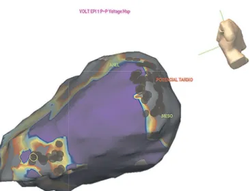
FIGURE 2 - Epicardial electroanatomical mapping of a patient with Chagas cardiomyopathy, identifying extensive scarring in the basolateral and apical walls of the left ventricle (gray area). The use of radiofrequency interrupted the VT (red dots). VT: ventricular tachycardia.
mapping (Figure 2) enables a detailed map of the scar in sinus
rhythm, allowing substrate modifi cation, even in patients with
unstable arrhythmias.
Ablation is recommended for patients with VT secondary to structural heart disease in the following situations(67):
I) For the control of symptomatic, sustained monomorphic VT, including VT terminated by ICD, which recurs despite antiarrhythmic drug therapy or when antiarrhythmic drugs are not tolerated or desirable; II) For the control of incessant,
sustained monomorphic VT or a VT storm that is not due to
a transient reversible cause; III) For the control of bundle branch reentrant or interfascicular VTs; IV) For the control of
recurrent, sustained polymorphic VT or VF that is refractory
to antiarrhythmic therapy and when there is a suspected trigger
that can be targeted for ablation.
CONCLUSIONS
Ventricular arrhythmias are important manifestations of
Chagas cardiomyopathy and are associated with increased disease severity and a high risk of death. Recognizing high-risk patients who require specifi c therapies and invasive procedures, such as ICD implantation and ablative approaches, is a major
challenge in clinical practice.
FUTURE PROPOSITIONS
The development of a strategy to accurately predict which patients are at a high risk of presenting with malignant cardiac arrhythmia would allow effective preventive actions and a
more rational use of available funds for the treatment of Chagas cardiomyopathy. Studies are also urgently needed to provide a
comparison of the two treatment modalities (amiodarone versus ICD) in the primary prevention of total mortality and sudden death in ChD patients with low left ventricular ejection fraction.
Manoel Otávio da Costa Rocha and Antônio Luiz
Pinho Ribeiro are recipients of research scholarships from
CNPq (Conselho Nacional de Desenvolvimento Científi co e Tecnológico) e FAPEMIG [Fundação de Amparo à Pesquisa de Minas Gerais (Programa Pesquisador Mineiro)].
The authors declare that there is no confl ict of interest. CONFLICT OF INTEREST
FINANCIAL SUPPORT
REFERENCES
1. Bardy GH, Lee KL, Mark DB, Poole JE, Packer DL, Boineau R, et al. Amiodarone or an implantable cardioverter‚ defi brillator for
congestive heart failure. N Engl J Med 2005; 352:225-237.
2. Ribeiro AL, Rocha MO. Forma indeterminada da doença de Chagas:
considerações acerca do diagnóstico e do prognóstico. Rev Soc Bras Med Trop 1998; 31:301-314.
3. Ribeiro AL, Nunes MP, Teixeira MM, Rocha MO. Diagnosis and
management of Chagas disease and cardiomyopathy. Nat Rev
Cardiol 2012; 9:576-589.
4. Ribeiro RA, Stella SF, Zimerman LI, Pimentel M, Rohde LE, Polanczyk
CA. Custo-efetividade de cardiodesfi briladores implantáveis no Brasil nos setores público e privado. Arq Bras Cardiol 2010; 95: 577-586.
5. Milei J, Pesce R, Valero E, Muratore C, Beigelman R, Ferrans VJ. Electrophysiologic-structural correlations in chagasic aneurysms
causing malignant arrhythmias. Int J Cardiol 1991; 32:65-73. 6. Rossi MA, Tanowitz HB, Malvestio LM, Celes MR, Campos EC,
Blefari V, et al. Coronary microvascular disease in chronic Chagas
cardiomyopathy including an overview on history, pathology, and
other proposed pathogenic mechanisms. PLoS Negl Trop Dis 2010;
7. Feldman AM, McNamara D. Myocarditis. N Engl J Med 2000; 343:1388-1398.
8. Carvalho AC, TLanowitz HB, Wittner M, Dermietzel R, Roy C, Hertzberg E, et al. Gap junction distribution is altered between cardiac myocytes infected with Trypanosoma cruzi. Circ Res 1992; 70:733-742.
9. Junqueira Jr LF. Insights into the clinical and functional signifi cance
of cardiac autonomic dysfunction in Chagas disease. Rev Soc Bras Med Trop 2012; 45:243-252.
10. Machado CR, Gomez MV, Machado AB. Changes in choline acetyltransferase activity of rat tissues during Chagas' disease.
Braz J Med Biol Res 1987; 20:697-702.
11. Machado CR, Machado AB, Chiari CA. Recovery from heart norepinephrine depletion in experimental Chagas' disease. Am J Trop Med Hyg 1978; 27:20-24.
12. Chen LS, Zhou S, Fishbein MC, Chen PS. New perspectives on the
role of autonomic nervous system in the genesis of arrhythmias. J Cardiovasc Electrophysiol 2007; 18:123-127.
13. La Rovere MT, Pinna GD, Maestri R, Mortara A, Capomolla S,
Febo O, et al. Short-term heart rate variability strongly predicts
sudden cardiac death in chronic heart failure patients. Circulation
2003; 107:565-570.
14. Miranda CH, Figueiredo AB, Maciel BC, Marin-Neto JA, Simões
MV. Sustained Ventricular Tachycardia Is Associated with Regional Myocardial Sympathetic Denervation Assessed with 123I-Metaiodobenzylguanidine in Chronic Chagas Cardiomyopathy.
J Nucl Cardiol 2011; 52:504-510.
15. Dias E, Laranja FS, Nobrega G. Doença de Chagas. Mem Inst Oswaldo Cruz 1945; 42:530-545.
16. Maguire JH, Hoff R, Sherlock I, Guimarães AC, Sleigh AC, Ramos
NB, et al. Cardiac morbidity and mortality due to Chagas' disease: prospective electrocardiographic study of a Brazilian community, Circulation 1987; 75:1140-1145.
17. Dias JC. The indeterminate form of human chronic Chagas' disease:
a clinical epidemiological review. Rev Soc Bras Med Trop 1989; 22:147-156.
18. Nascimento BR. The prognostic signifi cance of electrocardiographic
changes in Chagas disease. J Electrocardiol 2012; 45:43-48. 19. Chagas C, Villela E. Cardiac form of American Trypanosomiasis.
Mem Inst Oswaldo Cruz 2012; 14:5-91.
20. Dias JC, Kloetzel K. The prognostic value of the electrocardiographic
features of chronic Chagas' disease. Rev Inst Med Trop Sao Paulo 1968; 10:158-162.
21. Garzon SA, Lorga AM, Nicolau JC. Electrocardiography in Chagas' heart disease, Sao Paulo Med J 1995; 113:802-813.
22. Ribeiro AL, Rocha MO, Barros MV, Rodrigues AR, Machado FS. A narrow QRS does not predict a normal left ventricular function in
Chagas' disease. Pacing Clin Electrophysiol 2000; 23:2014-2017. 23. Ribeiro AL, Cavalvanti PS, Lombardi F, Nunes Mdo C, Barros MV,
Rocha MO. Prognostic value of signal-averaged electrocardiogram
in Chagas disease, J Cardiovasc Electrophysiol 2008; 19:502-509.
24. Elizari M. Arrhythmias Associated with Chagas' Disease. Cardiac Electrophysiology Review 1997; 1:270-273.
25. Muratore CA, Baranchuk A. Current and emerging therapeutic
options for the treatment of chronic chagasic cardiomyopathy.
Vasc Health Risk Manag 2010; 6:593-601.
26. Carrasco HA, Guerrero L, Parada H, Molina C, Vegas E, Chuecos R.
Ventricular arrhythmias and left ventricular myocardial function in
chronic chagasic patients. Int J Cardiol 1990; 28:35-41.
27. de Sousa MR, Morillo CA, Rabelo FT, Nogueira Filho AM, Ribeiro AL. Non-sustained ventricular tachycardia as a predictor of
sudden cardiac death in patients with left ventricular dysfunction:
A meta-analysis. Eur J Heart Fail 2008; 10:1007-1014.
28. Marin Neto JA, Simões MV, Sarabanda AV. Chagas' heart disease.
Arq Bras Cardiol 1999; 72:247-280.
29. Rocha MO, Ribeiro AL, Teixeira MM. Clinical management of
chronic Chagas cardiomyopathy. Front Biosci 2003; 8:44-54. 30. Rassi Jr A, Rassi A, Little WC, Xavier SS, Rassi SG, Rassi AG,
et al. Development and validation of a risk score for predicting death in Chagas' heart disease. N Engl J Med 2006; 355:799-808.
31. Guerrero L, Carrasco H, Parada H, Molina C, Chuecos R. Ventricular
mechanics and cardiac arrhythmias in patients with chagasic
and primary dilated cardiomyopathy. Echo-electrocardiographic
follow-up. Arq Bras Cardiol 1991; 56:465-469.
32. de Paola AA, Gomes JA, Terzian AB, Miyamoto MH, Martinez Fo EE. Ventricular tachycardia during exercise testing as a predictor of
sudden death in patients with chronic chagasic cardiomyopathy and
ventricular arrhythmias. Br Heart J 1995; 74:293-295.
33. Rocha AL. Chronotropic incompetence and abnormal autonomic modulation in ambulatory Chagas disease patients. Ann Noninvasive
Electrocardiol 2006; 11:3-11.
34. Rocha AL. Índice cronotrópico-metabólico na doença de Chagas.
Rev Soc Bras Med Trop 2005; 38:373-376.
35. Haedo AH, Chiale PA, Bandieri JD, Lázzari JO, Elizari MV, Rosenbaum MB. Comparative antiarrhythmic effi cacy of verapamil,
17-monochloracetylajmaline, mexiletine and amiodarone in patients
with severe chagasic myocarditis: relation with the underlying arrhythmogenic mechanisms. J Am Coll Cardiol 1986; 7:1114-1120. 36. Rosenbaum M, Posse R, Sgammini H, Núñez Burgos J, Chiale PA, Pastori JD, et al. Comparative multicenter clinical study of fl ecainide
and amiodarone in the treatment of ventricular arrhythmias
associated with chronic Chagas cardiopathy. Arch Inst Cardiol Mex
1987; 57:325-330.
37. Piccini JP, Berger JS, O'Connor CM. Amiodarone for the prevention
of sudden cardiac death: a meta-analysis of randomized controlled trials. Eur Heart J 2009; 30:1245-1253.
38. Satomi K, Kurita T, Takatsuki S, Yokoyama Y, Chinushi M, Tsuboi N, et al. Amiodarone therapy in patients implanted with cardioverter-defi brillator for life-threatening ventricular arrhythmias. Circulation 2006; 70:977-984.
39. Rassi Júnior A, Gabriel Rassi A, Gabriel Rassi S, Rassi Júnior L, Rassi A. Ventricular arrhythmia in Chagas disease. Diagnostic,
prognostic, and therapeutic features. Arq Bras Cardiol 1995;
65:377-387.
40. Connolly SJ, Dorian P, Roberts RS, Gent M, Bailin S, Fain ES, et al.
Comparison of {beta}-blockers, amiodarone plus {beta}-blockers, or sotalol for prevention of shocks from implantable cardioverter defi brillators: The OPTIC study: A randomized Trial. JAMA 2006; 295:165-171.
41. Poole JE, Johnson GW, Hellkamp AS, Anderson J, Callans DJ, Raitt MH, et al. prognostic importance of defi brillator shocks in patients with heart failure, N Engl J Med 2008; 359:1009-1017.
42. Buxton AE, Lee KL, Fisher JD, Josephson ME, Prystowsky EN, Hafl ey G. A randomized study of the prevention of sudden death in patients with coronary artery disease. N Engl J Med 1999; 341:
1882-1890.
43. Leite LR, Fenelon G, Simoes A Jr, Silva GG, Friedman PA, de Paola AA. Clinical usefulness of electrophysiologic testing in patients
with ventricular tachycardia and chronic chagasic cardiomyopathy treated with amiodarone or sotalol. J Cardiovasc Electrophysiol 2003; 14:567-573.
44. Martinelli Filho M, De Siqueira SF, Moreira H, Fagundes A, Pedrosa A, Nishioka SD, et al. Probability of occurrence of
life-threatening ventricular arrhythmias in Chagas' disease versus
45. Epstein AE, Di Marco JP, Ellenbogen KA, Estes NA 3rd, Freedman RA, Gettes LS, et al. ACC/AHA/HRS 2008 Guidelines for Device-Based Therapy of Cardiac Rhythm Abnormalities: A Report of the American College of Cardiology/American Heart Association
Task Force on Practice Guidelines (Writing Committee to Revise the ACC/AHA/NASPE 2002 Guideline Update for Implantation of Cardiac Pacemakers and Antiarrhythmia Devices): Developed
in Collaboration With the American Association for Thoracic Surgery and Society of Thoracic Surgeons. Circulation 2008; 117:350-408.
46. Ministério da Saúde. Secretaria de Atenção à Saúde. Portaria 152 de 08 de março de 2007. Diário Ofi cial 8-3-2007. Brasília:
Ministério da Saúde; 2007.
47. Cardinalli-Neto A, Greco OT, Bestetti RB. Automatic implantable cardioverter-defi brillators in Chagas' heart disease patients with
malignant ventricular arrhythmias. Pacing Clin Electrophysiol
2006; 29:467-470.
48. Cardinalli-Neto A, Bestetti RB, Cordeiro JA, Rodrigues VC.
Predictors of all-cause mortality for patients with chronic Chagas' heart disease receiving implantable cardioverter defi brillator therapy. J Cardiovasc Electrophysiol 2007; 18:1236-1240.
49. Moreira HB. Probabilidade de ocorrência de morte súbita cardíaca
na cardiomiopatia chagásica e não chagásica. Reblampa 2003; 16:55-58.
50. Muratore C, Rabinovich R, Iglesias R, González M, Darú V, Liprandi AS. Implantable cardioverter defi brillators in patients with Chagas' disease: are they different from patients with coronary
disease? Pacing Clin Electrophysiol 1997; 20:194-197.
51. Rabinovich R, Muratore C, Iglesias R, Gonzalez M, Darú V, Valentino M, et al. Time to fi rst shock in implantable cardioverter defi brillator (ICD) patients with Chagas cardiomyopathy. Pacing
Clin Electrophysiol 1999; 22:202-205.
52. Barbosa MP, da Costa Rocha MO, de Oliveira AB, Lombardi F, Ribeiro AL. Effi cacy and safety of implantable cardioverter-defi brillators in patients with Chagas disease, Europace 2013; 15:957-962.
53. da Fonseca SM, Belo LG, Carvalho H, Araújo N, Munhoz C,
Siqueira L, et al. Clinical follow-up of patients with implantable cardioverter-defi brillator. Arq Bras Cardiol 2007; 88:8-16.
54. Cardinalli-Neto A, Greco OT, Bestetti RB. Automatic implantable cardioverter-defi brillators in Chagas' heart disease patients with
malignant ventricular arrhythmias. Pacing Clin Electrophysiol
2006; 29:467-470.
55. Cardinalli-Neto A, Bestetti RB, Cordeiro JA, Rodrigues VC.
Predictors of all-cause mortality for patients with chronic Chagas' heart disease receiving implantable cardioverter defi brillator Therapy. J Cardiovasc Electrophysiol 2007; 18:1236-1240. 56. Muratore CA, Batista Sa LA, Chiale PA, Eloy R, Tentori MC,
Escudero J, et al. Implantable cardioverter defi brillators and Chagas' disease: results of the ICD Registry Latin America. Europace 2009; 11:164-168.
57. di Toro D, Muratore C, Aguinaga L, Batista L, Malan A, Greco
O, et al. Predictors of All-cause 1-year mortality in implantable cardioverter defi brillator patients with chronic Chagas’ Cardiomyopathy. Pacing Clin Electrophysiol 2011; 34:1063-1069. 58. Martinelli M, de Siqueira SF, Sternick EB, Rassi Jr A, Costa R,
Ramires JA, et al. Long-Term Follow-Up of Implantable Cardioverter-Defi brillator for Secondary Prevention in Chagas'
Heart Disease. Am J Cardiol 2012; 110:1040-1045.
59. Gali WL, Sarabanda AV, Baggio JM, Ferreira LG, Gomes GG,
Marin-Neto JA, et al. Implantable cardioverter-defi brillators for treatment of sustained ventricular arrhythmias in patients with Chagas' heart disease: comparison with a control group treated with amiodarone alone. Europace 2014; 6: 674-680.
60. Martinelli M, Rassi Jr A, Marin-Neto JA, de Paola AA, Berwanger O, Scanavacca MI, et al. Chronic use of amiodarone against implantable cardioverter-defi brillator therapy for primary prevention of death in patients with Chagas cardiomyopathy Study: Rationale and design of a randomized clinical trial. Am Heart J 2013; 166:976-982. 61. Schron EB, Exner DV, Yao Q, Jenkins LS, Steinberg JS, Cook JR,
et al. Quality of Life in the Antiarrhythmics Versus Implantable Defi brillators Trial: Impact of Therapy and Infl uence of Adverse Symptoms and Defi brillator Shocks. Circulation 2002; 105:589-594. 62. Della Bella P, De Ponti R, Uriarte JA, Tondo C, Klersy C,
Carbucicchio C, et al. Catheter ablation and antiarrhythmic drugs for haemodynamically tolerated post-infarction ventricular tachycardia. Long-term outcome in relation to acute electrophysiological
fi ndings. Eur Heart J 2002; 23:414-424.
63. de Paola AA, Horowitz LN, Miyamoto MH, Pinheiro R, Ferreira DF,
Terzian AB, et al. Angiographic and electrophysiologic substrates of ventricular tachycardia in chronic Chagasic myocarditis.
Am J Cardiol 1990; 65:360-363.
64. Sosa E, Scanavacca M, D'Avila A, Bellotti G, Pilleggi F. Radiofrequency catheter ablation of ventricular tachycardia guided
by nonsurgical epicardial mapping in chronic Chagasic heart disease. Pacing Clin Electrophysiol 1999; 22:128-130.
65. Sarabanda AV, Sosa E, Simões MV, Figueiredo GL, Pintya AO,
Marin-Neto JA. Ventricular tachycardia in Chagas' disease: a comparison of clinical, angiographic, electrophysiologic and myocardial perfusion
disturbances between patients presenting with either sustained or nonsustained forms. Int J Cardiol 2005; 102:9-19.
66. Sosa E, Scanavacca M, d'Avila A, Pilleggi F. A new technique to
perform epicardial mapping in the electrophysiology laboratory,
J Cardiovasc Electrophysiol 1996; 7:531-536.
67. Aliot EM, Stevenson WG, Almendral-Garrote JM, Bogun F, Calkins CH, Delacretaz E, et al. EHRA/HRS Expert Consensus
on catheter ablation of ventricular arrhythmias: Developed in a
partnership with the European Heart Rhythm Association (EHRA),
a Registered Branch of the European Society of Cardiology (ESC),
and the Heart Rhythm Society (HRS); in collaboration with the
American College of Cardiology (ACC) and the American Heart