Cop
yright
© ABE&M t
odos os dir
eit
os r
eser
vados
.
46,XX DSD and Antley-Bixler syndrome
due to novel mutations in the
cytochrome P450 oxidoreductase gene
DDS 46,XX e síndrome de Antley-Bixler causada por novas mutações no gene da enzima P450 oxidorredutase
Guilherme Guaragna-Filho1,2, Carla Cristina Telles de Sousa Castro1,2,
Rodrigo Ribeiro De Carvalho3, Fernanda Borchers Coeli3, Lúcio
Fábio Caldas Ferraz3, Reginaldo José Petroli3, Maricilda Palandi
De Mello3, Letícia Esposito Sewaybricker1,2, Soia Helena Valente
Lemos-Marini1,Lilia Freire Rodrigues D'Souza-Li1, Márcio Lopes
Miranda4, Andréa Trevas Maciel-Guerra2,5, Gil Guerra-Junior1,2
SUMMARY
Deiciency of the enzyme P450 oxidoreductase is a rare form of congenital adrenal hyperplasia with characteristics of combined and partial impairments in steroidogenic enzyme activities, as P450 oxidoreductase transfers electrons to CYP21A2, CYP17A1, and CYP19A1. It results in disorders of sex development and skeletal malformations similar to Antley-Bixley syndrome. We report the case of a 9-year-old girl who was born with virilized genitalia (Prader stage V), absence of palpable gonads, 46,XX karyotype, and hypergonadotropic hypogonadism. During the irst year of life, ovarian cyst, par-tial adrenal insuficiency, and osteoarticular changes, such as mild craniosynostosis, carpal and tarsal synostosis, and limited forearm pronosupination were observed. Her mother presented severe virilization during pregnancy. The molecular analysis of P450 oxidoreductase gene revealed compound heterozygosis for the nonsense p.Arg223*, and the novel missense p.Met408Lys, inherited from the father and the mother, respec-tively. Arq Bras Endocrinol Metab. 2012;56(8):578-85
SUMÁRIO
A deiciência da enzima P450 oxidorredutase é uma forma rara de hiperplasia congênita da adre-nal com características de inibição combinada e parcial de enzimas esteroidogênicas, pois a enzima P450 oxidorredutase participa da transferência de elétrons para as enzimas CYP21A2, CY-P17A1 e CYP19A1. Essa deiciência causa um distúrbio do desenvolvimento do sexo e alterações esqueléticas semelhantes às da síndrome de Antley-Bixley. Relatamos o caso de uma menina, atualmente com 9 anos de idade, que apresentava ao nascimento genitais virilizados (Prader 5) sem gônadas palpáveis, com cariótipo 46,XX e hipogonadismo hipergonadotróico. No primeiro ano de vida, foram observados cisto ovariano, insuiciência adrenal parcial e alterações osteoar-ticulares como leve craniossinostose, sinostose carpal e tarsal e limitação de pronossupinação dos membros superiores. Sua mãe apresentou intensa virilização durante a gestação. O estudo molecular do gene P450 oxidorredutase revelou a heterozigose composta das mutações nonsen-se p.Arg223* e da misnonsen-sennonsen-se nova p.Met408Lys, herdadas do pai e da mãe, respectivamente. Arq Bras Endocrinol Metab. 2012;56(8):578-85
1 Departamento de Pediatria,
Unidade de Endocrinologia Pediátrica, Faculdade de Ciências Médicas, Universidade Estadual de Campinas (FCM-Unicamp), Campinas, SP, Brazil
2 Grupo Interdisciplinar de Estudos
da Determinação e Diferenciação do Sexo (GIEDDS), FCM-Unicamp, Campinas, SP, Brazil
3 Centro de Biologia Molecular e
Engenharia Genética (CBMEG), Unicamp, Campinas, SP, Brazil
4 Departamento de Cirurgia,
Disciplina de Cirurgia Pediátrica, FCM-Unicamp, Campinas, SP, Brazil
5 Departamento de Genética
Médica, FCM-Unicamp, Campinas, SP, Brazil
Correspondence to:
Gil Guerra-Junior Departamento de Pediatria, FCM-Unicamp
13083-100 – Campinas, SP, Brazil [email protected]
Received on July/31/2012 Accepted on Oct/29/2012
INTRODUCTION
C
ytochrome P450 monooxygenases are enzymesthat catalyze the oxidation of several organic compounds. Two biochemical classes of P450 en-zymes have been distinguished in the human genome.
Cop
yright
© ABE&M t
odos os dir
eit
os r
eser
vados
.
turn, type II enzymes are expressed in the endoplas-mic reticulum and receive electrons from NADPH via a single P450 lavoprotein. Cytochrome P450 oxido-reductase (OMIM *124015; POR) is the only elec-tron donor lavoprotein for the cytochrome P450 type II enzyme complex, including the steroidogenic
17α-hydroxylase/17,20-lyase (CYP17A1),
21-hy-droxylase (CYP21A2), and aromatase (CYP19A1) (1). POR is an 82-kDa membrane-associated protein
en-coded by the POR gene, which is formed by 15 exons
spanning 32 kb on the 7q11.2 chromosomal band (2). Association with the allosteric cytochrome b5 can en-hance POR activity, as demonstrated by 17,20-lyase reactions and some hepatic P450 enzymes (3).
POR deiciency (OMIM #613571; PORD) is a rare and complex form of congenital adrenal hyper-plasia that results from partial and combined impair-ment of steroidogenic enzymes. This autosomal re-cessive disorder was described for the irst time in 2004, when mutations in POR encoding gene were identiied (4,5). PORD has a wide spectrum of clini-cal signs and symptoms, including partial and com-bined enzymatic adrenal dysfunction associated with disorders of sex development (DSD) in 46,XX and 46,XY individuals, and skeletal abnormalities of Antley-Bixler syndrome (OMIN #201750) (6). Maternal hyperandrogenism and virilization during pregnancy can also be observed. Skeletal malforma-tions observed in many, but not all, patients with PORD are considered to be due to impairment of enzyme activities involved in sterol synthesis, such
as 14α-lanosterol demethylase (CYP51A1) and
squalene epoxidase, disruption of retinoic acid me-tabolism catalyzed by CYP26 isozymes (6,7), and hepatic drug metabolism (8).
To date, the public and private Human Gene Mutation Databases report, respectively, 57 and 79 mutations in the
POR gene (9). Most of them are missense, followed by
nonsense, splicing, and a couple of deletions. Depending
on the type of mutation and its location within the POR
gene, it may reduce CYP17A1, CYP21A2, CYP19A1 ac-tivities differently. Therefore, POR variants must be stud-ied separately for each potential P450 target enzyme (10). Here, we report a 46,XX patient with PORD and manifestations of the Antley-Bixler syndrome in whom
POR gene analysis revealed compound
heterozygo-sis for the nonsense p.Arg223* and the novel mis-sense p.Met408Lys, inherited from the father and the mother, respectively.
CASE REPORT
A girl, who was the third child of a healthy mother, was born at term by vaginal delivery. At birth, she weighed 2,830 g and her height was 48 cm, with head circum-ference of 32.5 cm. Due to the virilized genital ap-pearance, she was assigned as a male. The mother, who denied the use of drugs during gestation, re-ported severe virilization with hirsutism, clitoro-megaly, and voice deepening (Figure 1A). All these signs regressed after delivery. She also reported two previous uneventful gestations: a normal girl from her irst marriage and a healthy boy.
At the irst appointment in our service, the pa-tient was 8 months old with male assignment. General physical examination indicated virilized genitalia with Prader stage V and 2.3-cm phallus, without palpable gonads; balanic urethral opening and complete fusion
Figure 1. Mother´s (A) and child’s (B) external genitalia 8 months after delivery.
A
Cop
yright
© ABE&M t
odos os dir
eit
os r
eser
vados
.
of labioscrotal folds, which were not pigmented, were also observed (Figure 1B). No other dysmorphic signs or physical alterations were observed. The mother re-ported that the phallus length had diminished since birth. Initial cytogenetic investigation revealed a 46,XX
karyotype in 30 metaphases analyzed. A 1.1-cm3 uterus
and gonads with typical ovarian shape were observed upon pelvic ultrasonography, with an anechoic homo-geneous image in the left adnexial region, suggestive of a simple ovarian cyst.
The moderate increase of 17-OH progesterone le vel and low androgen (DHEA, androstenedione and tes-tosterone) levels did not conirm either 21-hydroxylase
or 11b-hydroxylase deiciencies (Table 1). Therefore,
the irst hypothesis considered for the diagnosis was the CYP19 deiciency, based on the severe virilization of
the mother during pregnancy. However, the CYP19
gene analysis did not conirm such diagnosis. Although the etiology of the clinical signs remained undeined, when the patient was 18 months old, it was decided to reassign the sex from male to female, with the agree-ment of the family, and feminizing introitoplasty was performed. Gonadal biopsy conirmed the presence of two normal ovaries.
Almost one year after the irst appointment,
muta-tions in the POR gene sequence were investigated. The
c.667C>T and c.1223T>A nucleotide changes located in exon 6 and in exon 10, respectively, were identiied (Figure 2). The c.667C>T nucleotide change affects codon 223, causing a CGA>TGA substitution that forms a premature stop codon; whereas the c.1223T>A nucleotide change leads to a change in residue 408 from a methionine (ATG) to a lysine (AAG).
Sequen-cing of the POR gene of her parents indicated that the
p.Arg223* and p.Met408Lys mutations were inher-ited from the father and the mother, respectively. The biological importance of p.Met408Lys change in the structure of the enzyme was investigated by modeling the mutant enzyme and comparing it to its wild type.
Laboratory follow-up from 8 to 105 months is de-tailed in table 1. She had never presented any abnormal-ities related to neurological and growth development until the age of 64 months, when X-ray examinations demonstrated discrete reduction in cephalic diameters,
Table 1. Hormonal proile of the patient with POR deiciency during the irst 105 months of life
Age (months)
8 18 48 67 105
Basal Post ACTH Post hCG
FSH (IU/L)*1 23.3 10.4 1.3 - - 1.2 1.3
LH (IU/L)*2 13.5 0.5 < 0.1 - - < 0.1 < 0.1
ACTH (pg/mL)*3 20.3 26.8 20.9 - - 35.3 42.9
Cortisol (µg/dL)#4 9.0 8.8 7.8 14.3 - 9.1 8.9
17-OH progesterone (ng/mL)#5 11.6 2.9 3.8 13.3 - 6.6 5.0
DHEA (ng/mL)#6 0.6 0.5 1.0 1.1 - 1.1 1.4
Androstenedione (ng/mL)*7 0.2 0.1 0.1 0.5 - 0.1 0.2
Testosterone (ng/mL)*8 < 0.02 < 0.02 <0.02 - 0.09 < 0.02 < 0.02
* Electrochemiluminescence assay; # radioimmunoassay; normal range (pre-pubertal girls): 1 < 0.1 - 3.8; 2 < 0.1 - 1.4; 3 < 46; 4 5.0 - 22.0; 5 0.07 - 1.5; 6 < 2.5; 7 0.05 - 0.5; 8 < 0.2.
Figure 2. Partial electropherograms of POR gene sequencing of a patient with POR deiciency. A) The c.667C>T nucleotide change in exon 6 causes the CGA>TGA substitution at codon 223; an arginine is replaced by a stop codon, representing the null mutation p.Arg223*. B) The c.1223T>A nucleotide change located in exon 10 causes the ATG>AAG substitution in codon 408, leading to the missense p.Met408Lys. The p.Arg223* and p.Met408Lys mutations were inherited from the father and the mother, respectively.
A
B
Codon 223
Cop
yright
© ABE&M t
odos os dir
eit
os r
eser
vados
.
“hammered silver” appearance usually associated to craniosynostosis (Figure 3A), and carpal and tarsal syn-ostoses (Figures 3B and 3C). When she was 7 years old, she complained of limited movement of the arms; radiological investigation was normal, despite limited pronosupination. The score of osteoarticular anomalies
was 5, according to Krone and cols. (11).
She had never required hydrocortisone replacement therapy, except before and after the two additional sur-gical procedures that she had been submitted to: vagi-nal dilation at 5 years of age due to vagivagi-nal stenosis and endometritis, and clitoroplasty at the age of 6. She is now 9 years old, her weight is 31 kg (z = 0.32), height 133 cm (z = -0.02, within her target height), and head circumference 51 cm (slightly below the mean) (Figure 4A); her bone age is compatible with chronological age, she has normal female genitalia with no signs of puberty (Figure 4B), and she takes no medications.
Figure 3. Radiographs of the skull (A), left hand (B) and left foot (C) of the patient with POR deiciency, showing a shortened diameter of the head and hammered-silver appearance (A), carpal synostosis (B), and tarsal synostosis (C).
A
A
B
B C
Cop
yright
© ABE&M t
odos os dir
eit
os r
eser
vados
.
MATERIALS AND METHODS
Samples of peripheral blood leukocytes were obtained and genomic DNA extraction was performed by standard
techniques (12). The 10 exons of CYP19A1 gene and the
15 exons of the POR gene, as well as exon-intron
junc-tions were ampliied by PCR in different fragments. Be-fore sequencing, each PCR fragment was puriied using
the Wizard® SV Gel and PCR clean-up system (Promega,
Madison, WI, USA). Further direct sequencing using ABI PRISM Big Dye Terminator v3.1 Cycle Sequencing Kit (ABI PRISM/PE Biosystems, Foster City, CA, USA) was carried out in separated reactions using sense and antisense primers. The sequences were obtained in an automatic sequencer ABI PRISM 3130 DNA Analyzer (ABI PRISM/PE Biosystems). Free software Chromas Pro v.1.5 and CLC Sequence Viewer v.6.6.2 were used to analyze and compare sequences with the published
CYP19A1 and POR sequences at Ensembl database
(ENSG00000137869 and ENSG00000127948, re-spectively, www.ensembl.org). The structure used in modeling analyses for p.Met408Lys mutation was the crystallographically-resolved POR structure PDB ID: 3QE2 (13). Molecular modeling was performed using MODELLER web server software. Modelled images were examined and edited using a free version of
Py-MOL® and internal contacts were investigated using the
Millennium STING (CNPTIA-Embrapa, Brazil).
DISCUSSION
A case of PORD caused by the compound heterozy-gosis for p.Arg223* and p.Met408Lys is reported. The p.Arg223* creates a premature stop codon and may probably activate nonsense mRNA decay before translation. This mutation was described recently in a patient who carried p.Arg223* in compound hetero-zygosis with p.Arg287Pro, which is considered the most common POR mutation in Caucasians (14). The p.Met408Lys is described here for the irst time. It is a non-conservative substitution, since it replaces the neutral nonpolar hydrophobic methionine residue by the basic polar hydrophilic lysine residue at codon 408. Residue 408 is located at the boundary of lavin adenine dinucleotide (FAD) domain and a hinge re-gion that links FAD domain to lavin mononucleotide (FMN) domain (Figure 5A). The alignment of mod-eled wild-type and mutated POR structures did not show differences in protein folding. However, diver-gences in two disordered regions can be observed in the
hinges. In the wild-type protein, the Met408 residue is in contact with Leu404 and Leu418 (Figure 5B). The methionine-to-lysine substitution creates novel internal contacts with neighboring residues, such as L389 and W422 (Figure 5C). Those four residues, in turn, estab-lish additional contacts with other residues and with the p.Met408Lys mutation (Figure 5D-K). If such modii-cations cause disturbances in enzymatic activity should be further investigated. However some possibilities can be considered based on data in the literature.
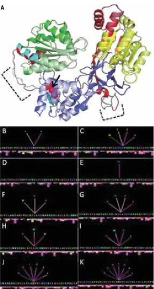
Figure 5. Alignment of structures of wild-type and p.Met408Lys proteins
(A); FMN domain is denoted in green (dark green = wild-type; light green = mutant); FAD domain is denoted in blue (dark blue = wild-type; light blue = mutant); and NADPH domain is denoted in yellowish tones (yellow = wild-type; greenish-yellow = mutant); red and cyan represent the hinge regions of the wild-type and mutant proteins, respectively; black arrow points to the normal Met408 (purple) and mutated Lys408 (red) residues; dashed lines delimit hinge divergences. Internal contacts for residues 408, 389, 404, 418, and 422 are shown in the left (B, D, F, H, J) and in the right (C, E, G,
I, K) for the wild-type and mutant proteins, respectively. The color of lines and bars below each amino acid represents different chemical interactions: hydrophobic (pink), hydrogen bond (rose), aromatic stacking (grey).
Cop
yright
© ABE&M t
odos os dir
eit
os r
eser
vados
.
Hamdane and cols. (15) demonstrated that the hinge
region facilitates electron transfer from FAD to FMN by properly aligning and orienting the two lavin domains. They conclude that electron low from FAD to FMN and from FMN to CYPs may be regulated by adjusting the distance and orientation of the two lavin cofactors. Furthermore, their report provided evidence that POR enzyme may undergo conformational changes for proper electron transfer to its redox partners. As p.Met408Lys mutation causes disturbances in the conformational ar-rangement of the hinge and establishes novel local inter-actions, it may alter enzymatic activity by modifying the conformation of the hinge region, so that the lexibility necessary to promote electron transfer is impaired (16).
The reported patient did not present neonatal signs of skeletal malformations; however, during childhood, she developed a few osteoarticular abnormalities, such as carpal and tarsal synostosis, reduced skull diameter and “hammered-silver” appearance (suggestive of cra-niosynostosis), and limited forearm pronosupination, indicating that periodic clinical and image evaluations were necessary (6,10,11,17,18). Considering that p.Arg223* is a null allele in the compound heterozy-gosis with p.Met408Lys, it can be inferred that the residual activity of p.Met408Lys missense carrying al-lele is deining the patient’s phenotype. Different from the most studied p.Ala287Pro and p.Arg457His muta-tions, which have been frequently observed in Japanese patients and are related to severe inborn skeletal mal-formations (6,10,18), p.Met408Lys seems to be more deleterious to POR activity related to the androgenic pathway than to skeletal development, since the patient presented severe virilizition and no signs of skeletal mal-formations at birth. Krone and cols. (11) proposed a score for the evaluation of osteoarticular manifestations in PORD patients. According to this methodology, total malformation score (TMS) can vary from 0 to 16. Fol-lowing this score method, our patient presented TMS of 5, which refers to a moderate grade malformations.
PORD may lead to genital ambiguity in both sexes: boys may present undervirilization due to inadequate testosterone production resulting from CYP17A1 im-pairment, and genital ambiguity in girls may be the re-sult of more complex mechanisms, mainly those from defective CYP19A1 activity with excessive androgen production and activation of the alternative SRD5A1 and AKR1C pathways, as well as direct conversion of 17OH-progesterone to dihydrotestosterone (DHT) (2,11,19,20). Probably, only CYP19A1 was affected in
this case, leading severe virilization of both mother and child with low testosterone and high 17OH-progester-one levels.
The literature is still controversial regarding the need for daily glucocorticoid replacement in PORD. However, most authors agree with drug administra-tion only in case of moderate to severe stress, especially for those patients who do not present adequate corti-sol response to ACTH stimulus, as was the case of our patient (11,18,21-25). Therefore, glucocorticoid was only administrated upon stress situations. Mineralocor-ticoid reposition is not necessary, although deicient POR activity may affect CYP21A2 catalyzed reactions (11,24).
PORD deiciency differs from other congenital ad-renal hyperplasias because it affects multiple enzymes, leading them to partial activities and resulting in a vari-able hormonal proile. Upon CYP21A2 activity, there are moderately elevated basal and ACTH-stimulated 17OH-progesterone levels. However, dihydroepian-drosterone (DHEA), DHEA-sulphate and androstene-dione levels are generally low or normal due to the low 17,20-lyase activity of CYP17A1. This hormonal pro-ile, which is usually observed in patients with PORD, including this patient, indicates that the 21-hydrox-ylation activity by CYP21A2 is as diminished as the 17-hydroxylation activity by CYP17A1 (4,6,18,25).
Puberty is another important aspect to be consid-ered; however, knowledge regarding its evolution and the hormonal proile of patients with PORD is not to-tally available yet. It seems that male patients spontane-ously begin and progress to puberty more easily than female patients, although both sexes present different levels of hypergonadotropic hypogonadism in all re-ported cases (4,14,17,18,24).
Cop
yright
© ABE&M t
odos os dir
eit
os r
eser
vados
.
catalyzes the conversion from lanosterol to meiosis-acti-vating sterols. Follicular luid meiosis-actimeiosis-acti-vating sterols have been shown to be crucial to resume oocyte meiosis at puberty, and to support oocyte maturation (31). It is suggested in the literature that hormonal replacement could ameliorate the presence of polycystic ovaries (14,18). Although our patient still have not shown any pubertal signs, she presented elevated gonadotropin levels and a suggestive image of an ovarian cyst upon ultrasonography in the irst months of life.
Concluding, we present a case report on a girl with PORD who is compound heterozygous for the nonsense p.Arg223* and the novel missense p.Met408Lys mutations associated with severe ma-ternal virilization, 46,XX DSD, partial glucocorti-coid insuficiency, hypergonadotropic hypogonadism, and ovarian cysts during the irst year of life, as well as late onset features of Antley-Bixler syndrome.
Disclosure: no potential conlict of interest relevant to this article was reported.
REFERENCES
1. Miller WL. Minireview: regulation of steroidogenesis by electron transfer. Endocrinology. 2005;146:2544-50.
2. Scott RR, Gomes LG, Huang N, Van Vliet G, Miller WL. Apparent manifesting heterozygosity in P450 oxidoreductase deiciency and its effect on coexisting 21-hydroxylase deiciency. J Clin En-docrinol Metab. 2007;92:2318-22.
3. Auchus RJ, Lee TC, Miller WL. Cytochrome b5 augments the 17,20-lyase activity of human P450c17 without direct electron transfer. J Biol Chem. 1998;273:3158-65.
4. Flück CE, Tajima T, Pandey AV, Arlt W, Okuhara K, Verge CF, et al. Mutant P450 oxidoreductase causes disordered steroido-genesis with and without Antley-Bixler syndrome. Nat Gen-et. 2004;36:228-30.
5. Arlt W, Walker EA, Draper N, Ivison HE, Ride JP, Hammer F, et al. Congenital adrenal hyperplasia caused by mutant P450 oxidore-ductase and human androgen synthesis: analytical study. Lancet. 2004;363:2128-35.
6. Scott RR, Miller WL. Genetic and clinical features of P450 oxidore-ductase deiciency. Horm Res. 2008;69:266-75.
7. Polusani SR, Kar R, Riquelme MA, Masters BS, Panda SP. Regu-lation of gap junction function and connexin 43 expression by cytochrome P450 oxidoreductase (CYPOR). Biochem Biophys Res Commun. 2011;411:490-5.
8. Tomalik-Scharte D, Maiter D, Kirchheiner J, Ivison HE, Fuhr U, Arlt W. Impaired hepatic drug and steroid metabolism in congenital adrenal hyperplasia due to P450 oxidoreductase deiciency. Eur J Endocrinol. 2010;163:919-24.
9. Human Gene Mutation Database. Available at: http://www.hgmd. cf.ac.uk/ac/index.php. Accessed on: Nov 17, 2012.
10. Flück CE, Pandey AV. Clinical and biochemical consequences of P450 oxidoreductase deiciency. Endocr Dev. 2011;20:63-79. 11. Krone N, Reisch N, Idkowiak J, Dhir V, Ivison HE, Hughes BA, et al.
Genotype-phenotype analysis in congenital adrenal hyperplasia
due to P450 oxidoreductase deiciency. J Clin Endocrinol Metab. 2012;97:E257-67.
12. Sambrook J, Fristsch EF, Maniatis TE. Molecular Cloning: a labora-tory manual. Cold Spring Harbor, NY: Cold Spring Harbor Labora-tory Press; 1989.
13. Xia C, Panda SP, Marohnic CC, Martásek P, Masters BS, Kim JJ. Structural basis for human NADPH-cytochrome P450 oxidoreduc-tase deiciency. Proc Natl Acad Sci U S A. 2011;108:13486-91. 14. Idkowiak J, O’Riordan S, Reisch N, Malunowicz EM, Collins F,
Kerstens MN, et al. Pubertal presentation in seven patients with congenital adrenal hyperplasia due to P450 oxidoreductase dei-ciency. J Clin Endocrinol Metab. 2011;96(3):E453-62.
15. Hamdane D, Xia C, Im SC, Zhang H, Kim JJ, Waskell L. Structure and function of an NADPH-cytochrome P450 oxidoreductase in an open conformation capable of reducing cytochrome P450. J Biol Chem. 2009;284(17):11374-84.
16. Xia C, Hamdane D, Shen AL, Choi V, Kasper CB, Pearl NM, et al. Conformational changes of NADPH-cytochrome P450 oxidore-ductase are essential for catalysis and cofactor binding. J Biol Chem. 2011;286:16246-60.
17. Herkert JC, Blaauwwiekel EE, Hoek A, Veenstra-Knol HE, Kema IP, Arlt W, et al. A rare cause of congenital adrenal hyperpla-sia: Antley-Bixler syndrome due to POR deiciency. Neth J Med. 2011;69:281-3.
18. Fukami M, Hasegawa T, Horikawa R, Ohashi T, Nishimura G, Hom-ma K, et al. Cytochrome P450 oxidoreductase deiciency in three patients initially regarded as having 21-hydroxylase deiciency and/or aromatase deiciency: diagnostic value of urine steroid hormone analysis. Pediatr Res. 2006;59:276-80.
19. Flück CE, Miller WL. P450 oxidoreductase deiciency: a new form of congenital adrenal hyperplasia. Curr Opin Pedi-atr. 2006;18:435-41.
20. Homma K, Hasegawa T, Nagai T, Adachi M, Horikawa R, Fuji-wara I, et al. Urine steroid hormone proile analysis in cyto-chrome P450 oxidoreductase deiciency: implication for the back-door pathway to dihydrotestosterone. J Clin Endocrinol Metab. 2006;91:2643-9.
21. But WM, Lo IF, Shek CC, Tse WY, Lam ST. Ambiguous genitalia, impaired steroidogenesis, and Antley-Bixler syndrome in a patient with P450 oxidoreductase deiciency. Hong Kong Med J. 2010;16:59-62.
22. Iijima S, Ohishi A, Ohzeki T. Cytochrome P450 oxidoreduc-tase deiciency with Antley-Bixler syndrome: steroidogenic capacities. J Pediatr Endocrinol Metab. 2009;22:469-75. 23. Ko JM, Cheon CK, Kim GH, Yoo HW. A case of Antley-Bixler
syndrome caused by compound heterozygous mutations of the cytochrome P450 oxidoreductase gene. Eur J Pe-diatr. 2009;168:877-80.
24. Sahakitrungruang T, Huang N, Tee MK, Agrawal V, Russell WE, Crock P, et al. Clinical, genetic, and enzymatic characterization of P450 oxidoreductase deiciency in four patients. J Clin En-docrinol Metab. 2009;94:4992-5000.
25. Adachi M, Tachibana K, Asakura Y, Yamamoto T, Hanaki K, Oka A. Compound heterozygous mutations of cytochrome P450 oxidoreductase gene (POR) in two patients with Antley-Bixler syndrome. Am J Med Genet A. 2004;128:333-9.
26. New MI. Nonclassical congenital adrenal hyperplasia and the polycystic ovarian syndrome. Ann NY Acad Sci. 1993;687:193-205. 27. Rosa S, Duff C, Meyer M, Lang-Muritano M, Balercia G, Boscaro M, et al. P450c17 deiciency: clinical and molecular characteriza-tion of six patients. J Clin Endocrinol Metab. 2007;92:1000-7. 28. Shima M, Tanae A, Miki K, Katsumata N, Matsumoto S, Nakajima
Cop
yright
© ABE&M t
odos os dir
eit
os r
eser
vados
.
29. Belgorosky A, Pepe C, Marino R, Guercio G, Saraco N, Vaiani E, et al. Hypothalamic-pituitary-ovarian axis during infancy, early and late prepuberty in an aromatase-deicient girl who is a compound heterozygote for two new point mutations of the CYP19 gene. J Clin Endocrinol Metab. 2003;88:5127-31.
30. Fukami M, Horikawa R, Nagai T, Tanaka T, Naiki Y, Sato N, et al. Cy-tochrome P450 oxidoreductase gene mutations and Antley-Bixler
syndrome with abnormal genitalia and/or impaired steroidogen-esis: molecular and clinical studies in 10 patients. J Clin Endocri-nol Metab. 2005;90:414-26.