Fisioter. Mov., Curitiba, v. 30, n. 3, p. 453-461, Jul./Sep. 2017 Licenciado sob uma Licença Creative Commons DOI: http://dx.doi.org/10.1590/1980-5918.030.003.AO03
Relationship between head posture and lumbar curve
in a sitting position: a biomechanical study
Relação entre a postura da cabeça e a
curvatura lombar na posição sentada:
um estudo biomecânico
Rozilene Maria Cota Aroeira, Renata Maria Moreira Moraes Furlan,
Antônio Eustáquio de Melo Pertence, Estevam Barbosa de Las Casas, Marcelo Greco*
Escola de Engenharia de Estruturas da Universidade Federal de Minas Gerais (UFMG), Belo Horizonte, MG, Brazil
[R] Abstract
Introduction: The sitting position routinely used for a wide variety of tasks increases the potential of devel-oping forward head posture, which can seriously compromise the health of different systems in the human body. Objective: A static equilibrium analysis was conducted, comparing the position of the head with the lumbar curve in three different sitting positions. Methods: The approximate force and flexion moment of the head extensor muscles in static equilibrium was calculated in each of the following positions: (A) with-out a backrest; (B) using a backrest with a 100° tilt angle; (C) using a 100° tilted backrest associated with a cylindrical lumbar support cushion at the level of the L3 vertebra. Results: The C7-tragus angles were 43°, 50° and 52°; Frankfort horizontal plane (FH) angles were 5°, 9° and 9°; force of the head extensor muscles was 53.0N, 59.7N and 43.5N and flexion moments were 2.60Nm, 2.05Nm and 1.78Nm, in positions A, B and C, respectively. Conclusion: The results revealed that the sitting position using a 100° tilted backrest and lumbar support with the smallest L3-tragus horizontal distance required less effort by the head and neck extensor muscles to retain the head in equilibrium. This study demonstrated the need to preserve the
454
physiology of the lumbar spine, characterized by the position of the L3 vertebra, in order to ensure good head position.
Keywords: Posture. Spine. Biomechanics. Photogrammetry. Employee Health
Resumo
Introdução: A postura do indivíduo sentado, utilizada rotineiramente na execução de grande variedade de ta-refas, constitui-se num potencial aumentado para o desenvolvimento da postura de projeção da cabeça, a qual pode ocasionar sérios comprometimentos à saúde de vários sistemas no corpo humano. Objetivo: Um estudo do equilíbrio estático foi realizado, relacionando a posição da cabeça com a curvatura da coluna lombar em três diferentes posturas do indivíduo sentado. Métodos: Foi realizado o cálculo do valor aproximado de força e
momento fletor dos músculos extensores da cabeça na manutenção do equilíbrio estático em cada uma das se -guintes posturas: (A) sem uso de encosto para as costas; (B) com uso de encosto de 100° de inclinação; (C) com uso de encosto de 100° associado a um suporte lombar cilíndrico em nível da vértebra L3. Resultados: Os ân-gulos tragus-C7 foram 43°, 50° e 52°; os ânân-gulos de Frankfort foram 5°, 9° e 9°; as forças musculares extensoras
da cabeça foram 53,0N, 59,7N e 43,5N e os momentos fletores foram 2,60N.m, 2,05N.m e 1,78N.m, nas posturas
A, B e C, respectivamente. Conclusão: Os resultados revelaram que a postura sentada com encosto inclinado a 100° e calço lombar, onde houve a menor distância horizontal tragus-L3, apresentou menor esforço para a musculatura cervical extensora na manutenção do equilíbrio da cabeça. Este estudo demonstrou a necessidade
da preservação do eixo lombar fisiológico, caracterizado pelo posicionamento da vértebra L3, para garantir o
bom posicionamento da cabeça.
Palavras-chave: Postura. Coluna vertebral. Biomecânica. Fotogrametria. Saúde do Trabalhador.
Introduction
The physiology and sound functioning of the hu-man body are closely related to body posture, which affects and governs everything from breathing to hormone production (1). Correct posture is con-sidered an important indicator of musculoskeletal health, with disorders of this system primarily caused by mechanical stress (2, 3). The sitting position rou-tinely used for a wide variety of tasks increases the potential of developing forward head posture, con-sidered abnormal and frequently observed in medical practice (4). According to Marques et al. (5), sitting for more than four hours may pose a risk to the mus-culoskeletal system.
Forward head posture has been associated with chronic musculoskeletal and functional disorders in the craniofacial region, neck and shoulders (6, 7, 8, 9). The annual prevalence of neck pain varies in dif-ferent countries, from 27.1% to 47.8% (10). However, prevalence can increase significantly as a result of specific work-related tasks, such as in dentistry and among professionals who use visual display units in
their work routines (11, 12, 13). Gazzola et al. (14) found a high prevalence (98.6%) of musculoskeletal disorders in 71 young dentists, with the most affect-ed regions being the cervical spine (77.5%), lumbar spine (73.3%) and shoulders (69%). In a study by Kang et al. (15), a group of individuals who remained seated at a computer for six or more hours a day for over ten years exhibited forward head posture, a forward shift in the body’s center of gravity, and re-duced balance and postural control when compared to a control group. In a literature review, Côté et al. (10) reported that neck pain is a significant health problem among employees. Every year, at least 5% of the population is expected to develop frequent or persistent neck disorders and there is evidence that head posture is a risk factor associated with these conditions. Zandi et al. (16) highly recommend head posture assessment for patients with head and neck pain for the purpose of diagnosis, planning, treatment strategy and monitoring.
455 of the head and neck, weight and height. Photographs were taken using a Sony Cyber-Shot 4.1 megapixel camera mounted on a tripod, a chair without a back-rest and one with a 100° tilted backback-rest, as well as a Mckenzie® lumbar support cushion. In order to en
-sure the images could be compared, the camera and tripod were positioned at right angles to the volun-teer at a focal length of 2.0m, respecting the 1.2 to 2.4m interval recommended by Ricieri (24) and using minimum zoom.
Computerized Photogrammetry
After photograph collection the images were submitted to angular kinematics via computerized photogrammetry, using Corel Draw 13® software.
Angles were measured after identifying the center of anatomical markers on the computer screen using the zoom feature, which was standardized for the entire photographic analysis process in order to pre-vent measurement errors. The C7-tragus angle was measured according to the protocol recommended by Braun & Amundson (17), analyzing the position of the head in relation to the body. This cervical angle is highly reliable in assessing forward head posture (25). Deviation of the thoracic and lumbar segments related to the L3 vertebra was also assessed. The ver-tical direction of the gaze was analyzed by measur-ing the angle of the Frankfort plane, which passes through the tragus and the outer edge of the eye socket. The angle is positive when the lower edge of the eye socket is higher than the tragus (26).
Photo Interpretation
This involves interpreting photogrammetric measurements in order to determine their meaning within the field of knowledge related to the object of this study.
Mathematical Analysis of the Data
Head and neck measurements
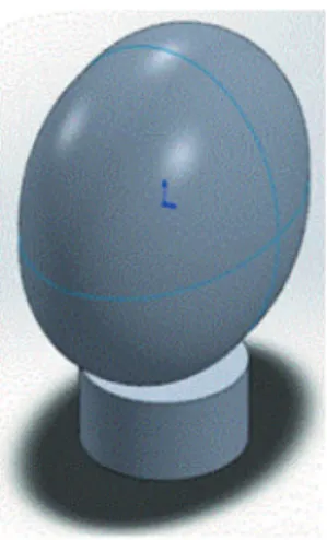
First, the dimensions of the volunteer’s head were measured: e = 15cm, f = 20cm and g = 22cm, where e, f and g are the axes of the ellipse that is an approximate and a horizontal line through the spinous process
of the C7 vertebra (17). Studies have shown that this angle, measured in adults, is between 49 and 55° (18, 19, 20). The head-neck ratio should be as -sessed along the sagittal plane. According to Nordin & Frankel (21), the ratio is evaluated when the subject is in a relaxed standing position, where the line of gravity passes immediately in front of the outer ear and descends ventrally to the lumbar spine. This line moves further forward in a relaxed sitting position with no back support. In this condition, the center of mass (CM) moves toward the ischial tuberosity, the pelvis is tilted and lumbar curvature is neutral or inverted. This movement creates a longer moment arm for the force exerted by the weight of the upper portion of the body. When sitting upright the CM co-incides with the ischial tuberosity, the pelvis is in a neutral position and the moment arm is shorter, but still slightly longer than that observed in a relaxed standing position (22).
The aim of this study was to: i. compare the posi-tion of the head to lumbar curvature in three different sitting positions, using posture analysis by computer-ized photogrammetry; ii. measure the approximate force and flexion moment of the head extensor mus -cles in the three positions at static equilibrium.
Methods
Photograph collection and angle measurement by computerized photogrammetry
An 18-year-old female volunteer with a weight of 53kg and height of 1.65m was photographed from the left sagittal plane after markers were placed on anatomical reference points. A 13 mm-wide circular white marker was placed on the left tragus (at the en-trance to the external auditory canal) and anatomical tracking markers measuring 45 mm by 18.79 Ø were positioned on the spinous processes of vertebrae C7 to L5 (23). The volunteer was photographed in three different sitting positions: (A) without a backrest; (B) using a backrest with a 100° tilt angle; (C) using a 100° tilted backrest associated with a cylindrical lumbar support cushion at the level of the L3 verte-bra, after approval by the Research Ethics Committee (COEP/UFMG under protocol number ETIC 579/07.
456
The CG of the head and neck assembly of the vol-unteer was 11.56cm from the apex of the head in the Y direction; 11cm from the rearmost point of the head in the Z direction and 7.5cm from either ear in the x direction.
Results
Measurement by computerized photogrammetry
Sitting position without a backrest
The C7-tragus angle with the horizontal line, mea-sured by computerized photogrammetry, was 43° and the Frankfort plane angle was 5°. In this position, posterior projection of L3 and forward head posture were observed, prompting an increase in the distance between the tragus and the spinous process of L3 (Tr-L3), whose value on the virtual scale was 15.53mm. As a result, changes occurred in the physiological curvature of the spine, such as: correction of cervical lordosis, increased thoracic kyphosis, and reversal of lordosis, as shown in Figure 2A.
Sitting position in a chair with a 100° tilted backrest
The forward head angle was 50° and the angle of the Frankfort plan was 9°. In this position there was less posterior projection of L3 compared to the thoracic segment and a significant reduction in the horizontal Tr-L3 distance, measured at 9.05mm (Figure 2B).
Sitting position in a chair with a 100° tilted backrest and lumbar support at L3
The forward head angle was 52° and the angle of the Frankfort plan was 9°. This position exhibited the smallest horizontal Tr-L3 distance (8.41mm) of all three postures. In addition, alignment of the posterior thoracic curve and hip was observed (Figure 2C). representation of the head, with f representing the
vertical axis. Neck dimensions and head-neck length were: neck perimeter s = 31cm and distance from the base of the neck to the top of the head H = 25cm, respectively. The following simplifying assumptions were made: the geometric center (centroid) is equal to the center of gravity (CG); ii- the head-neck assem-bly used in the calculation obtained by simplification of the lateral areas of the ellipse and cylinder (27). Based on these measurements, the areas of the el-lipse (Equation 1) and rectangle (Equation 2) were calculated, for subsequent determination of the CG (Equation 3). In other words, it is assumed that the geometric center was in approximately the same position as the center of gravity. This simplifying as-sumption is valid due to the approximate symmetry of the head-neck assembly, as shown in Figure 1.
Figure 1 - Head-neck assembly.
The CG of the head-neck assembly was calculated as follows:
Calculation of the area of the ellipse:
Aellipse = π · f · g (1) Calculation of the lateral area of the cylinder: Acylinder = 2 · r · h (2)
Where r and h are, respectively, the radius of the cylinder and the height of the rectangle, which are an approximate representation of the neck.
Calculation of the CG of the assembly:
y =
∑
2 i=1yi · Ai
457
After conversion into real values, the Tr-L3 distances were: (A) Tr-L3 = 18.88 cm; (B) Tr-L3 = 11.0 cm and (C) Tr-L3 = 10.23 cm.
Calculation of the force and lexion moment of head extensor muscles
The head extensor muscles and C7-T1 joint were identified by palpation of surface anatomy. The lines representing the force vectors were drawn based on these references. The mass of the head was considered to be 8% of body weight (53kg), that is, approximately 4kg (28). The magnitude of the resulting muscle force exerted by the head extensor muscles was denominated FM and the reaction force on the C7-T1 joint, FJ (Figure 3).
Figure 2 - Sitting position of the volunteer: (A) in a chair with no backrest, C7-tragus angle of 43°, Frankfort plane 5° and Tr-L3 distance of 15.53 mm ;(B) with a 100° tilted backrest, C7-tragus angle of 50°, Frankfort plane 9° and Tr-L3 distance of 9.05 mm and (C) with a 100° tilted backrest and lumbar support cushion at the level of vertebra L3, C7-tragus angle of 52°, Frankfort plane 9° and Tr-L3 distance of 8.41 mm.
Figure 3 - FM represents the magnitude of the resulting muscle force exerted by the head extensor muscles, θ depicts the angle between the FM vector and the horizontal line, FJ the reaction force on the C7-T1, β the angle between the FJ vector and the horizontal line and W the weight of the head. Adapted from Nordin & Frankel (1989).
15,53 mm
(A) sem encosto (B) com encosto e sem calço (B) com calço em L3
458
The angles between FM and FJ and the horizon-tal line were obtained by computerized photogram-metry, as shown in Figure 4. Based on equilibrium equations, since the relative weights FM and FJ are competing forces, the force required by the head ex-tensor muscles to support the head in the different positions studied was calculated (Equations 4 to 7).
|FMX| = FM · cos(θ) (4)
|FMY| = FM · sen(θ) (5)
|FJX| = FJ · cos(β) (6)
|FJY| = FJ · sen(β) (7)
The static equilibrium equations using CG as refer-ence provided equations (8) and (9):
|FM| = 39 · 24
tan(β) · cos(θ) – sen(θ) (8)
|FJ| = cos(θ)
cos(β) |FM| (9)
The flexion moment was calculated using linear interpolation to obtain the real distance of the C7-y axis measurement, expressed in millimeters in the measurement software (Figure 4).
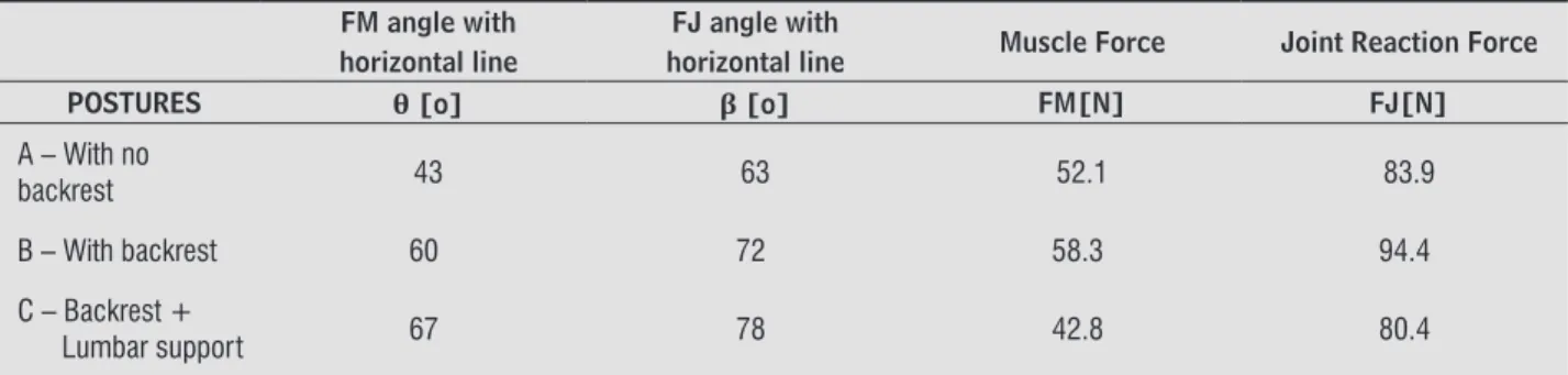
Figure 4 - Sitting positions: (A) in a chair with no backrest, angle of the head extensor muscles with the horizontal line 43°, angle of the reaction of the C7-T1 joint with the horizontal line 63°, C7-y axis distance 5.32 mm; (B) with a 100° tilted backrest, angle of the head extensor muscles with the horizontal line 60°, angle of the reaction of the C7-T1 joint with the horizontal line 72°, C7-y axis distance 4.21mm; (C) with a 100° tilted backrest and lumbar support cushion at L3, angle of the head extensor muscles with the horizontal line 67°, angle of the reaction of the C7-T1 joint with the horizontal line 78°, C7-y axis distance 3.66mm.
The values obtained for muscle and joint reaction forces are shown in Table 1. The values calculated for the flexion moment M(C7-T1) were 2.27Nm, 2.56Nm and 1.73Nm in positions A, B and C, respectively.
Table 1 - Force values obtained
FM angle with horizontal line
FJ angle with
horizontal line Muscle Force Joint Reaction Force
POSTURES θ [o] β [o] FM[N] FJ[N]
A – With no
backrest 43 63 52.1 83.9
B – With backrest 60 72 58.3 94.4
C – Backrest +
Lumbar support 67 78 42.8 80.4
459 moment. By contrast, Nordin and Frankel (21) found low electrical activity in all the positions despite the increased load in the different head positions. This suggests that the flexion moment is balanced by pas -sive connective tissue structures such as capsules and ligaments.
Thus, studies show that the load on the neck is related to the trunk and the position of the head. The load moment is balanced by muscle force and the traction of passive connective tissue. The moment arm and corresponding muscle force are 50% higher at a forward head angle of 30° when compared to values obtained with zero tilt (2). Load on the C7-T1 segment is 3 to 4 times greater with full head flexion. Vassavada et al. (30) evaluated the three-dimensional moment arm during maximal voluntary contraction of the neck muscles in humans. The authors calcu-lated the maximum moment arm, generated under strain, at different points along the cervical spine of men and women. Magnitudes quantified were those related to the moment arms for extension, flexion, lat -eral tilt and axial rotation of the head. They concluded that the maximum extension moment of the head in men was 52±11Nm and 21±12Nm in women, and that the magnitude of the flexion moment decreased linearly with the vertical distance of the lower cer-vical spine toward the mastoid process. According to the authors, it is unclear how the size, sex and geometry of individuals affects the ability of neck muscles to generate momentum. Additionally, the authors measured the forward head angle in rela-tion to C7-tragus and the Frankfort plane in 11 men and women considered to have good posture. The forward head posture (C7-tragus) measured for men and women was 50±4 degrees, with a mean Frankfort plane of 8±5 degrees for both sexes. These data cor -roborate the findings of the present study, where forward head posture in a sitting position using a backrest and combined with lumbar support was 50° and 52°, respectively. The Frankfort plane measure-ment for both postures was 9°.
Nordin and Frankel (21) reported that the head flexion moments generated around the C7-T1 seg -ment for static posture at slight and maximum flex -ion were 3.7Nm (3.0-6.2) and 4.3Nm (3.7-6.5), re -spectively. In a neutral neck position with the head straight, the flexion moment was 0.9Nm, indicating a substantial increase in the load on C7-T1 as the head moved toward the front of the neck. The au-thors did not specify the angles measured for what Discussion
The present study demonstrated the relationship between the position of vertebra L3 and the head when sitting. The results indicated an increase in forward head posture and neck muscle strain in the presence of a posterior tilt of vertebra L3, character-ized by a reduction or even reversal of lordosis.
Head and neck pain is frequently associated with incorrect sitting posture. The sitting position is gen-erally influenced by several factors, including the design of the chair, its ergonomic adaptation to the individual and the task to be performed. When sitting without a backrest, the pelvis tilts backwards and lumbar curvature is reduced or reversed, converting lordosis into kyphosis. Pressure on the intervertebral disc in this position (no backrest), measured at the level of L3, was 40% greater than that recorded with the subject standing (2). In the erect sitting position, the forward tilt of the pelvis preserves the concavity of lordosis, promoting a reduction in the load on this vertebral segment (21). However, this erect posture without a backrest puts excess strain on the muscles, making it unsuitable for performing tasks over pro-longed periods. As such, the chair should allow for postural adjustments in order to reduce pressure on the intervertebral disc.
Nordin and Frankel (21) studied the influence of sitting position in a chair with a 90° and 110° tilted backrest, with and without lumbar support. The authors found that sitting with a 110° tilted back-rest reduces compression in the spinal discs when compared to the 90° backrest. Moreover, the authors concluded that combining lumbar support with the tilted backrest further reduces intradiscal pressure. In the present study, use of a tilted backrest and lum -bar support cushion favored the physiological curva-ture of the vertebra, in addition to reducing forward head posture.
460
studies using groups of individuals with different occupational activities are suggested in order to im-prove the quantification protocol of musculoskeletal exertion related to head and trunk posture.
Acknowledgments
The authors are grateful for the financial support provided by the National Council of Scientific and Technological Development (CNPq), Coordination for the Improvement of Higher Education Personnel (CAPES) and Research Support Foundation of the State of Minas Gerais (FAPEMIG).
References
1. Lennon J, Shealy NC, Cady RK, Matta W, Cox R, Simpson WF. Postural and respiratory modulation of autonom-ic function, pain, and health. Am J Pain Management. 1994;4(1):36-9.
2. Nordin M, Andersson GBJ, Pope MH. Musculoskeletal Disorders in the Workplace, principles and practice. St. Louis: Mosby; 1997.
3. McEvoy MP, Grimmer K. Reliability of upright posture measurements in primary school children. BMC Mus-culoskelet Disord. 2005;6(1):35-42.
4. Bricot B. Posturologia. 2nd ed. São Paulo: Ícone; 2001.
5. Marques NR, Hallal CZ, Gonçalves M. Características ergonômicas e clínicas da postura sentada: uma re -visão. Fisioter Pesqui. 2010;17(3):270-6.
6. Nejati P, Lotfian S, Moezy A, Nejati M. The study of correlation between forward head posture and neck pain in Iranian office workers. Int J Occup Med Envir Health. 2015;28(2):295-303.
7. Yip CHT, Chiu TTW, Poon ATK. The relationship be-tween head posture and severity and disability of pa-tients with neck pain. Man Ther. 2008;13(2):148-54. 8. Fernandez-de-las-Peñas C, Alonso-Blanco C, Cuadrado ML, Pareja JA. Forward Head Posture and Neck Mo -bility in Chronic Tension-Type Headache. A Blinded, Controlled Study. Cephalalgia. 2006;26(3):314-9.
9. Svensson HF, Svensson OK. The influence of the view-ing angle on neck-load durview-ing work with video display units. J Rehabil Med. 2001;33(3):133-6.
they referred to as slight and maximum flexion. In the present study, flexion moments of 2.27Nm, 2.56Nm and 1.73Nm were obtained for head angles of 43°, 60° and 67°, respectively; where 43° represented the posture with the greatest head flexion. For extensor muscle force, a value of 52.1N was recorded for an angle of 43°in the sitting position without a backrest. In research by Ozkaya & Nordin (31), head extensor muscle force of 50 N was observed for an angle of 30°.
The Tr-L3 horizontal distance in this study was directly related to forward head posture. The larg-est head inclination was measured in a sitting posi-tion with no backrest for a Tr-L3 horizontal distance of 18.88 cm, while the smallest forward inclination was observed for a Tr-L3 distance of 10.23 cm when the subject was seated with a backrest and lumbar support cushion. There was a 46% decrease in the Tr-L3 horizontal distance and 9° decline in forward head posture when sitting with a 100° tilted backrest combined with lumbar support when compared to sitting without backrest.
Ruivo et al. (32) studied adolescents in a stand-ing position with worrisome results, where 68% of participants exhibited forward head posture and 58% had rounded shoulders. Thus, biomechani -cal studies demonstrate the need for an extensive postural education project at the elementary school level. Moreover, the present study contributes to the development of ergonomic measures and increased awareness among employees whose work activities require them to sit. The use of chairs with a backrest, associated with a lumbar support cushion, proved to be an important additional preventive measure for the health of workers required to remain seated for prolonged periods.
Conclusion
461
23. Aroeira RMC, Leal JS, Pertence AEM. New Meth-od of Scoliosis Assessment: Preliminary Results Using Computerized Photogrammetry. Spine. 2011;36(19):1584-91.
24. Ricieri DV. Biofotogrametria - Análise Cinemática Angular dos Movimentos. 2nd ed. Curitiba (Brazil): Inspirar; 2005.
25. Shumway-Cook A, Woollacott MH. Motor control: theory and practical applications. 2nd ed. Maryland: Williams & Wilkins; 2001.
26. Bjerin AA. Comparison between the Frankfort hori -zontal and sella turcica-nasion as reference planes in cephalometric analysis. Acta Odontol Scond. 1957;15(1):1-12.
27. Melo SIL, Santos SG. Antropometria em Biomecânica: Características, Princípios e Modelos Antropomé -tricos. Rev Bras Cineantropom Desempenho Hum. 2000;2(1):97-105.
28. Rodacki ALFR. Análise dos fatores antropométricos em biomecânica [cited 2011 Dec 15]. Available from: http://tinyurl.com/jzygb55.
29. Carneiro JP, O`Sullivan P, Burnet A, Barach A, O’Neil D, Tveit O, et al.The influence of different sitting posture on head/neck posture and muscle activity. Man Ther. 2010;15(1):54-60.
30. Vassavada AN, Li S, Delp SL. Three-Dimensional Iso -metric Strength of Neck Muscles in Humans. Spine J. 2001;26(17):1904-9.
31. Ozkaya N, Nordin M. Fundamental of Biomechan-ics – equilibrium, motion and deformation. New York: Springer Science; 1998.
32. Ruivo RM, Pezarat-Correia P, Carita AI. Cervical and shoulder postural assessment of adolescents between 15 and 17 years old and Association with upper quad-rant pain. Braz J Phys Ther. 2014;18(4):364-71.
Received in 10/07/2015
Recebido em 07/10/2015
Approved in 08/18/2016
Aprovado em 18/08/2016
10. Côté P, Velde G, Cassidy D, Carrol L, Hogg-Johnson S, Hom LW, et al. The Burden and Determinants of Neck Pain in Workers. Eur Spine J. 2008;17(Suppl 1):60-74.
11. Raine S, Twoey L. Posture of the head, shoulders and thoracic spine in comfortable erect standing. Aust J Physioter. 1994;40(1):25-32.
12. Alexopoulos EC, Stathi IC, Charizani F. Prevalence of musculoskeletal disorders in dentists. BMC Musculo-skelet Disord. 2004;5:16.
13. Michalak-Turcotte C. Controlling dental hygiene work related musculoskeletal disorders: the ergonomic process. J Dent Hyg. 2000;74(1):41-8.
14. Gazzola F, Sartor N, Ávila SN. Prevalência de desordens musculoesqueléticas em odontologistas de Caxias do Sul. Ciência & Saúde. 2008;1(2):50-6.
15. Kang JH, Park RY, Lee SJ, Kim JY, Yoon SR, Jung KI. The effect of the forward head posture on postural balance in long time computer based worker. Ann Rehabil Med. 2012;36(1):98-104.
16. Zandi S, Rajabi R, Alizadeh MH. Validity intratester and intertester reliability of a noninvasive quantita-tive forward posture assessment method. J Research Sport Rehabilitation. 2015;2(4):51-6.
17. Braum BL, Amundson LR. Quantitative assessment of head and shoulder posture. Arch Phys Med Rehabil. 1989;70(4):322-9.
18. Dalton MB. The effect of age on cervical posture in a normal female population. In: Jones HM, Jones MA, Milde MR, editors. Proceeding of the Sixth Biennial Conference of the Manipulative Therapists Associa-tion of Australia. Adelaide, 1989. p. 34-44.
19. Braum BL. Postural differences between asymptom-atic men and women and craniofacial pain patients. Arch Phys Med Rehabil. 1991;72(9):653-6.
20. Watson DH, Trott PH. Cervical headache: an investiga-tion of natural head posture and upper cervical flexor performance. Cephalagia. 1993;13(4):272-84.
21. Nordin M, Frankel V. Basic Biomechanics of the Mus-culoskeletal System. 2nd ed. Malvern (PA): Lea & Fe-biger; 1989.