ORIGINAL
RES
EAR
CH
Correspondence address: Juliana Ferreira Sauer – Rua Cipotânea, 51 – Cidade Universitária – CEP: 05360-000 – São Paulo (SP), Brasil – E-mail: [email protected] Presentation: Jun. 2013 – Accepted for publication: May 2014 – Financing source: São Paulo Research Foundation (FAPESP) and Coordenação de Aperfeiçoamento de Pessoal de Nível Superior (CAPES) – Conflict of interests: nothing to declare – Approval’s report at the Ethics Committee No. 0959/08.
ABSTRACT | To assess the articular range of motion in children with congenital visual impairment. Were evalu-ated 75 children between five and twelve years old, 49 with normal visual acuity and 26 visually impaired. Two evaluators performed the measure of active and pas-sive range of motion by goniometry of shoulder and hip in all axes of motion. All examiners made a test of correlation between data obtained, to determine the in-ter-rater reliability, using the intraclass correlation coef-ficient (ICC). In individuals with visual impairments was statistically significant difference, with higher range of motion in individuals with visual impairments to medial and lateral rotation of shoulder and hip lateral rotation. Were found: high correlation in the inter-rater reliability (ICC>0.70) for 9 (22.5%) groups of articular range of mo-tion, moderate correlation (0.7>ICC>0.5) for 25 (62.5%) groups and low correlation to 6 (15%) groups ranges of motion. Sampled children with congenital visual impair-ment showed greater joint mobility for rotational range of motion of the shoulder and hip than children with normal visual acuity, although they have also shown lower values for articular range of motion in abduction and extension in shoulders.
Keywords | Child; Visually Impaired Persons; Range of Motion, Articular.
Children with visual impairments may have
altered joint movement: an observational
case-control study
Crianças com deiciência visual podem ter a amplitude de movimento
articular alterada: um estudo observacional tipo caso-controle
Niños con deiciencia visual pueden tener la amplitud de movimiento
articular alterada: un estudio observacional tipo caso-control
Silvia Maria Amado João1, Michelle de Pádua2, Ulisses Tirollo Taddei3, Yuri Carvalho Mendes3,
Juliana Ferreira Sauer2
Study carried out at the Musculoskeletal Assessment Laboratory of the Physical Therapy, Speech Language Therapy and Occupational Therapy Department from the Medical School of Universidade de São Paulo (USP ) – São Paulo (SP), Brazil.
1Professor and PhD in the Physical Therapy Course of the Medical School at USP − São Paulo (SP), Brasil.
2Physical Therapist; Master’s degree by the Post-graduate Program of Rehabilitating Sciences from the Medical School of USP – São
Paulo (SP), Brazil.
3Physical Therapist graduated by USP − São Paulo (SP), Brazil.
RESUMO | Avaliou-se a amplitude de movimento articular em crianças com deficiências visuais congênitas. Foram avaliadas 75 crianças entre 5 e 12 anos de idade, sendo 49 com acuidade visual normal e 26 portadoras de deficiência visual. Dois avalia-dores realizaram a medida da amplitude de movimento arti-cular ativa e passiva, pela goniometria do ombro e quadril, em todos os eixos de movimento. Todos os avaliadores realizaram um teste de correlação entre os dados obtidos, para determi-nação da confiabilidade interavaliador, por meio do coeficiente de correlação intraclasse (ICC). Nos indivíduos com deficiên-cia visual houve diferença estatisticamente significativa, com maiores valores de amplitude de movimento para rotação me-dial e lateral de ombro e rotação lateral de quadril. Foram en-contradas: alta correlação na confiabilidade interexaminador (ICC>0,70) para 9 (22,5%) grupos de amplitude de movimento articular, correlação moderada (0,7>ICC>0,5) para 25 (62,5%) grupos e baixa correlação para 6 (15%) grupos de amplitudes de movimento. As crianças amostradas com deficiência visual congênita apresentaram maior mobilidade articular para as amplitudes de movimento rotacional do ombro e quadril que as crianças sem comprometimentos visuais, embora tenham apresentado também menores valores para amplitude de mo-vimento articular de abdução e extensão de ombros.
INTRODUCTION
Vision plays an essential role in the development of the body, being a primary source of stimuli that enable the direct relationship between the individual and the
ex-ternal environment1,2. his relationship is represented
by the ability to move and explore the environment, thus determining the acquisition of essential experienc-es that enable the global development and the
adapta-tion to the environment3.
Several studies have been trying to ind relation-ships between the incidence of visual impairment and
anthropometric changes in the afected population4-7,
which are often motivated by the stereotyped posture that is present in some of these individuals or by the mannerisms that can be a result of poor motor develop-ment during childhood, usually associated with
imper-fect or absent visual stimuli8,9.
Other studies try to correlate such observations with
the static posture10,11, or to correlate posture to
func-tional tasks12 and gait13. Bouchard e Tétreault2 found
that children with visual impairment presented with poorer and weaker motor skills and, in order for their movements to be functional, they would tend to change the way they performed several tasks. his study also described the decreased range of motion involving ro-tations, however, with no experimental conirmation, which could be related to the poor repertoire acquired during motor development.
Changes in the joint range of motion may cause alterations in the development of the musculoskeletal system of the children; especially when considering the transition period to adolescence, in which severe
physi-cal and structural changes happen14,15. Besides, it should
be considered that children have higher values of range of motion in comparison to adults, and they can also present with benign hypermobility in childhood, which
afecs 5 to 30% in the populations16.
According to the Brazilian Institute of Geography and Statistics (IBGE), in 2000, 14% of the Brazilians had some type of visual impairment, and, out of these, 57% had a permanent diiculty to see; nowadays, it is
the most incident visual impairment in Brazil17.
he objective of this study was to compare the joint range of movement in children aged 5 to 12 years old with blindness and congenital low vision with asymp-tomatic controls.
he hypothesis is that children with visual impair-ment during developimpair-ment will present with lower val-ues of joint range of movement.
METHODOLOGY
Male and female children, aged from 5 to 12 years
old, attending the institutions Escola Júlio Mesquita,
Instituição Padre Chico, Associação Brasileira de Assistência ao Deiciente Visual and patients assisted by the Sector of Sub-Normal Vision of Hospital das Clínicas, in São Paulo. Children who presented with neuromuscular, musculoskeletal and cardiorespiratory
diseases were excluded18.
All of the participants were contacted by educa-tional institutions that were interested to participate in the study, or families were contacted by telephone. An assessment report was ofered to each one of the participants after the study was concluded.
RESUMEN | Se evaluó la amplitud de movimiento articular en ni-ños con deficiencias visuales congénitas. Se evaluaron a 75 nini-ños con edades entre 5 y 12 años, siendo 49 con acuidad visual normal y 26 con deficiencia visual. Dos evaluadores realizaron la medición de la amplitud de movimiento articular activa y pasiva mediante goniometría del hombro y de la cadera, en todos los ejes de mo-vimiento. Todos los evaluadores realizaron un test de correlación entre los datos obtenidos para la determinación de la fiabilidad interevaluador, por medio del coeficiente de correlación intracla-se (ICC). En los individuos con deficiencia visual hubo diferencia estadísticamente significativa, con mayores valores de amplitud de movimiento para la rotación medial y lateral del hombro y rotación
lateral de la cadera. Fueron encontradas: alta correlación de fiabili-dad interevaluadores (ICC>0,70) para 9 (22,5%) grupos de ampli-tud de movimiento articular, correlación moderada (0,7>ICC>0,5) para 25 (62,5%) grupos y baja correlación para 6 (15%) grupos de amplitudes de movimiento. Los niños incluidos en la muestra con deficiencia visual congénita presentaron mayor movilidad articular para las amplitudes de movimiento de rotación del hombro y de la cadera que los niños sin deficiencias visuales, aunque también presentaron valores más bajos para la amplitud de movimiento ar-ticular de abducción y extensión de hombros.
The participation of the children depended on the parents or parties in charge signing the Informed Consent Form. The study was approved by the Research Ethics Committee of Hospital das
Clínicas, in the Medical School of Universidade de
São Paulo (CAPPesq).
Participants formed two groups: the control group (CG), composed of individuals with no visual altera-tions, and the experimental group (EG), composed of children with visual impairment caused by congenital, infectious, genetic or parasitic disorders, which would lead the visual capacity to be higher than 70% with the best correction, that is, low vision.
he dysfunctions included in the study for the EG approach the disorders that afect the sampled
popula-tion the most8, being retinal or refraction problems,
ab-normality in crystalline lens or oculomotor pathologies. In the CG, there were 49 children with mean age
of 8.8±1.5 years old, being 25 male and 24 female
par-ticipants, with mean weight of 30.5±11.5 kg, mean
height of 1.3±0.2 m, mean body mass index (BMI) of
17.5±4.7 kg/m2; in the EG, there were 26 children with
mean age of 8.4±2.4 years old, 12 male and 14 female
participants, with mean weight of 31.5±8.7 kg, mean
height of 1.3±0.1 m, mean BMI of 17.2±3.1 kg/m2,
with visual impairment.
Goniometry was conducted by a universal goniom-etry by Carci®, according to the methodology described
by Marques19 for lexion, extension, abduction, lateral
rotation and medial rotation movements in shoulder and hip joints, actively and passively, with participants wearing bathing suits (speed or bikini).
Measurements were taken by two diferent examiners, in order to ensure the reliability of the values. Inter-rater reliability was calculated by the intraclass correlation coeicient (ICC). Measurements were taken on two diferent days, so that the result from the irst measurement would not inluence the next one.
Statistics
he software Statistica 8.0 was used, and the Shapiro-Wilk test was conducted to verify data normality.
Afterwards, the Student’s t-test was performed for
in-dependent variables, in order to assess the diferences in mean values of joint motion between the CG and the
EG, with α=0.01.
By using the ICC, measurements from both evalua-tors were compared to determine inter-rater reliability.
ICC values of 0.00 to 0.25 were classiied as little or no reliability; low reliability comprised values from 0.26 to 0.49; moderate reliability, from 0.50 to 0.69; high reli-ability, from 0.70 to 0.89; and very high relireli-ability, for
values that were higher than 0.9020-22.
RESULTS
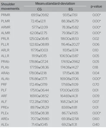
Values concerning the range of movement of pas-sive and active medial shoulder rotation are shown in Table 1. here was a signiicant diference (p<0.01) between the EG and the CG, and range values are higher in the EG.
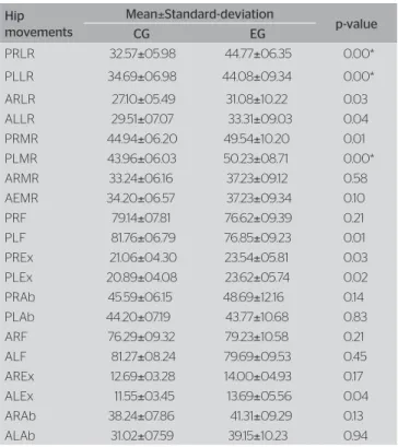
For the hip range of motion, movements that presented statistically signiicant diferences in the Student’s t- test were the passive lateral rotations and the passive left hip medial rotation, as observed in Table 2.
In inter-rater reliability, the ICC found: high reli-ability in 9 groups (22.5%), moderate relireli-ability in 25 groups (62.5%) and low reliability in 6 groups (15%).
Table 1. Mean, standard-deviation and comparison between groups for move-ments of medial rotation, lateral rotation, abduction, flection and extension of the shoulder, passively and actively, for the right and left upper limb
Shoulder movements
Mean±standard-deviation
p-value
CG EG
PRMR 69.51±09.82 93.15±17.61 0.00*
PLMR 72.45±12.11 88.38±15.79 0.00*
ARMR 57.71±12.09 78.38±15.82 0.00*
ALMR 62.08±12.75 79.38±17.25 0.00*
PRLR 125.02±08.45 118.00±18.53 0.02
PLLR 122.82±08.89 116.46±20.27 0.06
ARLR 117.76±10.03 113.15±13.74 0.10
ALLR 111.91±10.15 106.50±13.87 0.05
PRAb 178.86±07.24 174.92±09.62 0.05
PLAb 177.59±08.36 174.08±14.27 0.18
ARAb 178.08±12.18 171.15±16.38 0.04
ALAb 176.86±07.71 169.08±17.06 0.00*
PRF 172.49±07.19 171.69±11.09 0.70
PLF 175.10±06.44 170.00±10.55 0.01
ARF 169.10±08.52 164.69±14.31 0.09
ALF 172.28±07.80 168.23±11.34 0.07
PREx 89.79±08.29 83.69±11.81 0.01
PLEx 93.55±08.38 86.77±11.65 0.00*
AREx 70.73±09.80 69.38±12.58 0.60
ALEx 71.43±10.45 69.23±11.31 0.40
DISCUSSION
he objective of this study was to observe possible changes in the joint range of motion among children with visual impairment. Data show that children with visual impairment presented increased hip and shoul-der range of motion, when compared to children at the same age group with no visual impairment.
According to Decree n. 5,296, from December 2nd,
2004, the person characterized with blindness is the one with visual acuity similar to or lower than 0.05 in the best eye, with the best optical correction; low vision consists of visual acuity between 0.3 and 0.05 in the best eye, also with the best optical correction, and in cases when the sum of the visual ield measurement in both eyes is equal to or lower than 60º, or the simul-taneous occurrence of any of the previous conditions. It is possible that the use of rotational movements during childhood for visual impaired children is higher due to the need to discover the environment around
them in an alternative way23, therefore, they are more
developed during maturation.
Gaunet et al.24 reported that early blindness afects
both exploratory patterns and the performance of indi-viduals, by interacting with objects disposed in a space, when compared to blind-folded individuals.
As previously described, it was expected that indi-viduals with imperfect or absent visual acuity had dif-ferences concerning functional repertoire in their motor development, which would result in possible biome-chanical changes. Results difered from the expected as to the form of these diferences, since the hypothesis was that individuals with such impairment would ob-tain lower values of range of motion, which is exactly the opposite of what was found.
Another efect that would possibly lead to the ob-tained results would be sampling-related problems, once most individuals in the EG were institutionalized and participated in numberless extracurricular tasks, such as physical activities, musical education, arts, among others. Children in the CG were not stimulated nor did they have the access ofered by the school to the same activities.
In order to verify this last possibility, the precision of comparisons between groups was decreased between groups. he expectation was that, in case the airmative was true, higher values of range of motion would be higher in the EG than in the CG.
For non-rotational shoulder movements, signii-cantly diferent values were obtained between groups
for passive and active movements of abduction, lexion, and extension; the CG presented the highest values of range of motion.
hese indings may be explained by the presuppo-sition that, during the period of motor learning, these children experiment stimuli to reach lower levels in com-parison to a situation in which they would not have vi-sual impairment. Another possibility would be the delay to acquire cephalic stabilization, even if not completely, thus interfering in the modulation of relexes in
child-hood and making it diicult to stabilize the cinguli25,
therefore afecting the use of limbs. hese indings are also in contrast with the hypothesis that institutions would be a source of error in the inferences of this study.
he intra-rater correlation index demonstrated that, for most measurements, correlation was moderate.
CONCLUSION
his study shows that children with congenital low vi-sion have higher ranges of motion for medial shoul-der rotations, both in the passive and the active forms of motion, besides the higher mobility in the passive
Table 2. Mean, standard-deviation and comparison between groups for mo-vements of medial rotation, lateral rotation, abduction, flection and extension of the shoulder, passively and actively, for the right and left lower limb
Hip movements
Mean±Standard-deviation
p-value
CG EG
PRLR 32.57±05.98 44.77±06.35 0.00*
PLLR 34.69±06.98 44.08±09.34 0.00*
ARLR 27.10±05.49 31.08±10.22 0.03
ALLR 29.51±07.07 33.31±09.03 0.04
PRMR 44.94±06.20 49.54±10.20 0.01
PLMR 43.96±06.03 50.23±08.71 0.00*
ARMR 33.24±06.16 37.23±09.12 0.58
AEMR 34.20±06.57 37.23±09.34 0.10
PRF 79.14±07.81 76.62±09.39 0.21
PLF 81.76±06.79 76.85±09.23 0.01
PREx 21.06±04.30 23.54±05.81 0.03
PLEx 20.89±04.08 23.62±05.74 0.02
PRAb 45.59±06.15 48.69±12.16 0.14
PLAb 44.20±07.19 43.77±10.68 0.83
ARF 76.29±09.32 79.23±10.58 0.21
ALF 81.27±08.24 79.69±09.53 0.45
AREx 12.69±03.28 14.00±04.93 0.17
ALEx 11.55±03.45 13.69±05.56 0.04
ARAb 38.24±07.86 41.31±09.29 0.13
ALAb 31.02±07.59 39.15±10.23 0.94
medial and lateral hip rotations. hese indings point out to a new focus concerning training programs for motor skills and preventive interventions among chil-dren with low vision.
REFERENCES
1. Gianini RJ, Masi Ed, Coelho EC, Oréfice FR, Moraes RA. Prevalence of low visual acuity in public school’s students from Brazil. Rev Saude Publica. 2004;38(2);201-8.
2. Bouchard D, Tetreault S. The Motor development of sighted children and children with moderate low vision aged 8-13. J Visual Impair Blin 2000;98(9):564-73.
3. Gilbert C, Foster A. Blindness in children: control priorities and research opportunities. Br J Ophthalmol. 2001;85(9):1025-27.
4. Evensen KA, Lindqvist S, Indredavik MS, Skranes J, Brubakk AM, Vik T. Do visual impairments afect risk of motor problems in preterm and term low birth weight adolescents? Eur J Paediatr Neurol. 2009;13(1):47-56.
5. Zebrowska A, Gawlik K, Zwierzchowska A. Spirometric measurements and physical eficiency in children and adolescents with hearing and visual impairments. J Physiol Pharmacol. 2007;58(Pt 2):847-57.
6. Singh R, Singh HJ. Anthropometric and physiological profiles of active blind malaysian males. J Sports Med Phys Fitness. 1993;33(4):378-82.
7. Horvat M, Nocera J, Ray C, Croce R. Comparison of isokinetic peak force and power in adults with partial and total blindness. Percept Mot Skills. 2006;103(1):231-7
8. Brito PR, Veitzman S. Causas de cegueira e baixa visão em crianças. Arq Bras Oftalmol. 2000;63(1):49-54.
9. Haddad MAO. Habilitação e reabilitação de escolares com baixa visão: aspectos médico-sociais [Tese]. São Paulo: Universidade de São Paulo; 2006. 169p.
10. Catanzariti JF, Salomez E, Bruandet JM, Thevenon A. visual deficiency and scoliosis. Spine (Phila Pa 1976). 2001;26(1):48-52.
11. Aulisa L, Bertolini C, Piantelli S, Piazzini DB. Axial deviations of the spine in blind children. Ital J Orthop Traumatol. 1986;12(1):85-92.
12. Schmid M, Nardone A, De Nunzio AM, Schmid M, Schieppati M. Equilibrium during static and dynamic tasks in blind subjects: no evidence of cross-modal plasticity. Brain. 2007;130(Pt 8):2097-107.
13. Logan D, Kiemel T, Dominici N, Cappellini G, Ivanenko Y, Lacquanit F,
et al. The many roles of vision during walking. Exp Brain Res. 2010;206(3):337-50.
14. Asher C. Variações na postura da criança. 1ª ed. São Paulo: Manole; 1976.
15. Reimer AM, Cox RF, Nijhuis-Van der Sanden MW, Boonstra FN. Improvement of fine motor skills in children with visual impairment: An explorative study. Research in Developmental Disabilities. 2011;32(5):1924-33 .
16. Murray KJ, Woo P. Benign joint hypermobility in childhood. Rheumatology. 2001;40(5):489-91.
17. Moura e Castro J, Costa O, de Freitas F. Evaluation of the aerobic capacity of blind people, by direct vo2 maximal measurement. Rev Port Cardiol. 1992;11(6):525-9.
18. Kussuki MOM, João SMA, Cunha ACP. Caracterização postural da coluna de crianças obesas de 7 a 10 anos. Fisioter Mov. 2007;20(1):77-84.
19. Marques AP. Manual de Goniometria. 2ª ed. Barueri: Manole; 2003.
20. Domholdt E. Physical Therapy Research. Philadelphia: W.B. Saunden Company; 1993.
21. Jonson SR, Gross MT. Intraexaminer Reliability, Interexaminer Reliability, and Mean Values for Nine Lower extremity Skeletal measures in healthy naval midshipmen. J Orthop Sports Phys Ther. 1997;25(4):253-63.
22. Wahlund K, List T, Dworkin SF. Temporomandibular disorders in children and adolescents: reliability of a questionnaire, clinical examination, and diagnosis. J Orofac Pain. 1998;12(1):42-51.
23. Thinus-Blanc C, Gaunet F. Representation of space in blind persons: vision as a spatial sense? Psychol Bull. 1997;121(1):20-42.
24. Gaunet F, Martinez JL, Thinus-Blanc C. Early-blind subjects’ spatial representation of manipulatory space: exploratory strategies and reaction to change. Perception. 1997;26(3):345-66.