Cystoscopy-assisted laparoscopy for bladder endometriosis:
modified light-to-light technique for bladder preservation
_______________________________________________
Rafael Mamprin Stopiglia
1, Ubirajara Ferreira
1, Daniel Gustavo Faundes
2, Carlos Alberto Petta
31 Grupo de Urologia Oncológica, Universidade de Campinas, UNICAMP, SP, Brasil; 2 Centro de Reprodução
Humana Campinas, SP, Brasil; 3 Departamento de Ginecologia, Universidade de Campinas, UNICAMP,
SP, Brasil
ABSTRACT
ARTICLE
INFO
______________________________________________________________ ______________________
Introduction: Endometriosis is a disease with causes still unclear, affecting approximately 15% of women of reproductive age, and in 1%-2% of whom it may involve the uri-nary tract. The bladder is the organ most frequently affected by endometriosis, observed around 85% of the cases. In such cases, the most effective treatment is partial cystec-tomy, especially via videolaparoscopy.
Study Objective, Design, Size and Duration: In order to identify and delimit the extent of the intravesical endometriosis lesion, to determine the resection limits, as well as to perform an optimal reconstruction of the organ aiming for its maximum preservation, we performed a cystoscopy simultaneously with the surgery, employing a modified light-to-light technique in 25 consecutive patients, from September 2006 to May 2012.
Setting: Study performed at Campinas Medical Center – Campinas – Sao Paulo – Brazil. Participants/materials, setting and methods: Patients aged 27 to 47 (average age: 33.4 years) with deep endometriosis with total bladder involvement were selected for the study. The technique used was conventional laparoscopy with a transvaginal uterine manipulator and simultaneous cystoscopy (the light-to-light technique). A partial vide-olaparoscopic cystectomy was performed with cystoscopy-assisted vesical reconstruction throughout the entire surgical time. The lesions had an average size of 2.75cm (ranging from 1.5 to 5.5cm). The average surgical time was 137.7 minutes, ranging from 110 to 180 minutes.
Main Results: Postoperative follow-up time was 32.4 months (12-78 months), with clini-cal evaluation and a control cystoscopy performed every six months. No relapse was observed during the follow-up period.
Conclusions: A cystoscopy-assisted partial laparoscopic cystectomy with a modified light-to-light technique is a method that provides adequate identification of the lesion limits, intra or extravesically. It also allows a safe reconstruction of the organ aiming for its maximum preservation.
Keywords:
Endometriosis; Urinary Bladder; Cystoscopy
Int Braz J Urol. 2017; 43: 87-94
_____________________
Submitted for publication: January 07, 2015
_____________________
Accepted after revision: July 14, 2016
_____________________ Published as Ahead of Print: September 09, 2016
INTRODUCTION
Endometriosis is a gynecologic disease with causes still unclear. It was first described in 1860, but its most accepted etiopathogeny
pos-tulating retrograde menstruation was proposed in 1921 (1, 2).
or embedding on the walls of the pelvic organs (3). It is an estrogen-dependent disorder associa-ted with chronic pelvic pain and infertility (4).
It is estimated that approximately 15% of women of reproductive age are affected by endo-metriosis (5).
In 1979, the American Fertility Society initially classified endometriosis in 4 stages of severity, but reviewed this classification in 1985 (6, 7). The present classification was introduced in 1997, whereby it stages endometriosis as superfi-cial when it affects the parietal and visceral layers of the peritoneal membrane, and deep when there is more than 5cm penetration of the walls of the organs (8).
The most common sites affected by endo-metriosis in the pelvic cavity are the torus uterinus, the posterior fornix, the uterosacral ligaments, the rectum, the vagina and the urinary tract (9). Ho-wever, it may affect other sites, such as the dia-phragm, the umbilical cord, the ileum, the lungs, the pleura, the pericardium and the brain (10, 11).
Endometriosis may cause dysmenorrhea, even at the beginning of a woman´s fertile age, dyspareunia, chronic pelvic pain, and peri-mens-trual pain (12).
Another frequent disorder is infertility, oc-curring in up to 60% of the cases.
Specifically, in the urinary tract, there is a 0.3% to 12% incidence of endometriosis; however, it is usually reported as 1%-2% (13), and the most commonly affected sites are the bladder (85%), ureter (9%), kidneys (4%), and the urethra (2%), as shown in Figure 1A below.
When the bladder is affected, 70% of wo-men present pain during urination, dysuria,
su-deep endometriosis when the bladder is affected. This surgical procedure is excisional and consists of the removal of the entire bladder wall affected by endometriosis. For this type of procedure, the bladder must ideally present good functional ca-pacity, show a single lesion and be located >5mm of the urethral meatus.
PATIENTS AND METHODS
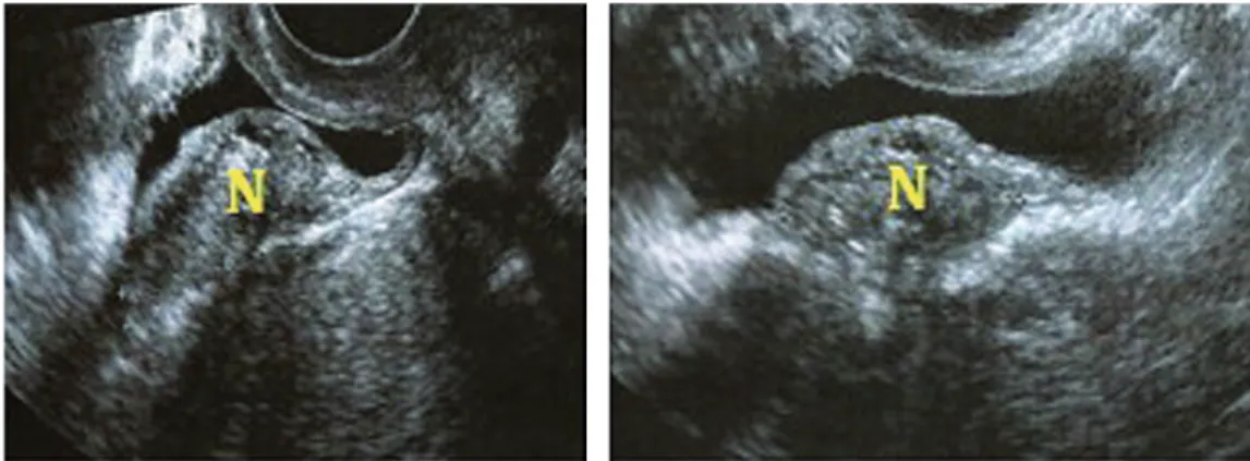
From September 2006 to May 2012, 25 pa-tients with initial diagnosis of deep endometrio-sis affecting the bladder wall were treated by the cystoscopy-assisted videolaparoscopic cystectomy with the light-to-light technique (16). (The asso-ciation of both procedures is meant to identify and delimit the extent of the intravesical endo-metriotic lesion, to determine the resection limits, as well as to perform an optimal reconstruction of the organ, aiming for its maximum preservation. The patient’s average age was 33.4 years, ranging from 27 to 47 years. After clinical assessment and a physical examination with bimanual palpation, the patients were tested for serum urea and cre-atinine levels, urine (proteinuria or microscopic hematuria) and urine culture, all of which were normal. All patients were submitted to transva-ginal ultrasound (TVUS) to diagnose the disease (Figure-1B), and to magnetic resonance (MRI) of the pelvis for surgical planning purposes (Figure--1C). The vesical lesion depicted on MRI is cha-racterized by hyper-signal on T1 and hypo-signal on T2 (17, 18).
in the umbilical scar, one 10mm trocar in the bi-sector of the imaginary line going from the ante-rior supeante-rior iliac crest to the umbilical scar on the right, and one 5mm in the exact same position on
Figure 1A - Endometriosis incidence in utrinary tract
Figure 1B - Transvaginal ultrasound with endometriotic endovesical lesion. (N)
Figure 1C - MRI of the pelvis depicting hypo-signal on T2 (lesion is highlighted).
URETHRA
0% 20% 40% 60% 80% 100% KIDNEYS
URETER
BLADDER
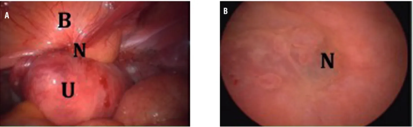
the left side, as per the representation below. A videolaparoscopy subsequently perfor-med inventory of the abdominal and pelvic cavity and identified a solid nodular lesion on the vesi-cal dome and vesico-uterine fossa, at times with significant adherence of such organs, as shown in Figure-1D (a).
The procedure above was followed by po-sitioning the transvaginal uterine manipulator and performance of the light-to-light cystoscopy technique, originally described by Seracchioli et al. The endoscopic diagnosis was confirmed by vi-sualization of tissue compatible with endometrio-sis on the vesical mucous surface. These lesions were blistered purple-blue nodules, containing en-dovesical material, as shown in Figure1D (b).
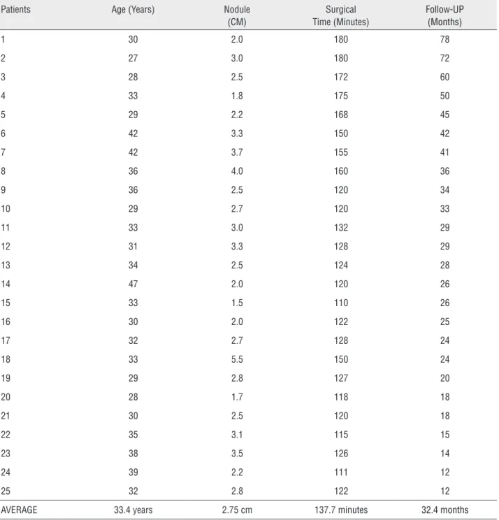
Follow-up was made every six months by means of clinical assessment and a cystoscopy, with total follow-up time of 32.4 months in ave-rage (ranging from 12 to 78 months) (Table-1).
There was no relapse of the disease in all cases. The patients presented normal vesi-cal physiology without alterations in bladder filling or emptying, evidenced by clinical asses-sment and cystoscopy.
DISCUSSION
The morpho-physiology of the vesical en-dometriosis lesions may vary according to the menstrual cycle. However, the lesions are better identified during menstruation. At cystoscopy, these lesions may appear in several colors, such as shades of red, blue, brown or even black. The urothelium is usually rarely ulcerated (19).
Biopsies for differential diagnosis with was then performed with some modifications, such
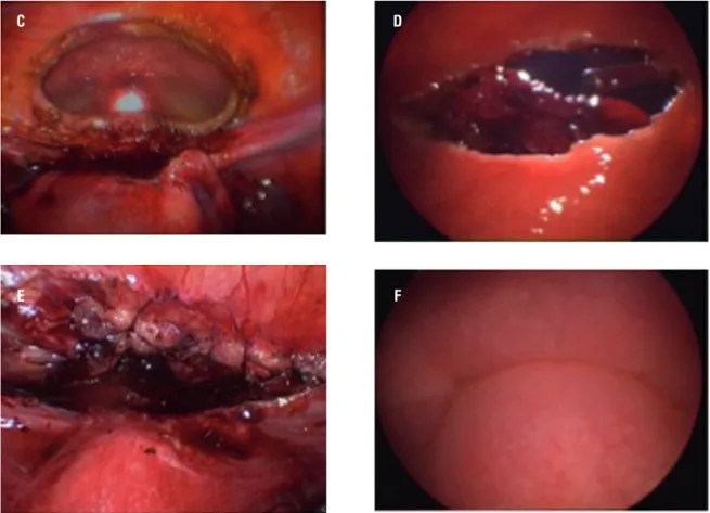
as initially not inserting urethral catheters. As the lesions affected the entire bladder wall, a partial cystectomy was performed assisted by cystosco-pic visualization throughout the procedure. Both surgeons identified and delimited the lesion, kee-ping a margin of at least 5mm of healthy tissue. Biopsies of the lower, right lateral, left lateral and superior margins were performed after exeresis of the lesion to eliminate permanence of the disease. The subsequent vesical reconstruction consisted of a one-layer suture with monofilament absorbable 3.0 thread, with continuous cystoscopy monito-ring, to ensure better visualization of the suture and final checking of the procedure, thus allowing maximum possible preservation of the healthy ve-sical tissue (Figure 1E).
All patients maintained a urethral catheter for 7 days.
Figure 1D - A - Endometriosis in laparoscopic view, B - Endometriosis in cistoscopic view.
Figure 1D, (a) (left): laparoscopic view; (B=Bladder, N=Node, U=Uterus); Figure 1D, (b) (right): cystoscopic view; (N=node)
diagnostic method, whether for superficial or deep lesions, is laparoscopy (21).
Treatment of pelvic endometriosis affec-ting the bladder may depend on several factors, such as age, symptom intensity, fertility, extent of the disease, presence in other organs and le-vel of menstrual dysfunction. As the disease ori-ginates outside the bladder (in the peritoneum), subsequently invading it, a vesical transurethral resection is usually an ineffective method (22, 23).
The disease is hormone (estrogen)-depen-dent, therefore the treatment of superficial lesions is based on hormonal blockade. The most com-monly adopted therapy for this purpose is the as-sociation of GnRH analogues, progestogens and oral contraceptives (24). This treatment aims at temporarily suppressing endometriosis, reason why it is more recommended for younger patients
Figure 1E - C e E - Endometriosis ressection and suture in laparoscopic view D e F - Endometriosis ressection and suture in cistoscopic view.
Clockwise: c = partial cystectomy, laparoscopic view; d = partial cystectomy, cystoscopic view; e = cystorraphy, laparoscopic view; f = cystorraphy, cystoscopic view. C
E
D
F
without deep endometriosis who wish to preserve their fertility. An intrauterine device (IUD) with le-vonorgestrel may also be used in these more con-servative cases, in addition to acting as an adju-vant in corrective surgeries. The IUD importance rests on the fact that is has a duration of up to 5 years and maintains fertility upon discontinuance of its use (25).
There are some options available for cases of deep vesical endometriosis, depending on the extent and site of the lesion in relation to its dis-tance from the urethral meatus.
Table 1 - Date table.
Patients Age (Years) Nodule (CM)
Surgical Time (Minutes)
Follow-UP (Months)
1 30 2.0 180 78
2 27 3.0 180 72
3 28 2.5 172 60
4 33 1.8 175 50
5 29 2.2 168 45
6 42 3.3 150 42
7 42 3.7 155 41
8 36 4.0 160 36
9 36 2.5 120 34
10 29 2.7 120 33
11 33 3.0 132 29
12 31 3.3 128 29
13 34 2.5 124 28
14 47 2.0 120 26
15 33 1.5 110 26
16 30 2.0 122 25
17 32 2.7 128 24
18 33 5.5 150 24
19 29 2.8 127 20
20 28 1.7 118 18
21 30 2.5 120 18
22 35 3.1 115 15
space, identification and dissection of the nodu-le, allowing exeresis of its total extension, and verification of the margins free of the disease.
Healthy 5mm margins of the bladder and a distance of at least 1cm of the urethral meatus should ideally be preserved (29).
The laparoscopic approach has as advan-tages less blood loss, less time of hospital stay, less use of pain killers and better aesthetic re-sults (30).
We also agree that the interaction betwe-en gynecologists and urologists is relevant for the best treatment of this disease and for the perfor-mance of successful procedures.
CONCLUSIONS
A cystoscopy-assisted partial laparoscopic cystectomy with a modified light-to-light techni-que is a method that provides adequate identifi-cation of the lesion limits, intra or extravesically. It also allows a safe reconstruction of the organ aiming at its maximum preservation.
CONFLICT OF INTEREST
None declared.
REFERENCES
1. Von Rokitansky C. Ueber uterusdrusen-neubildung in uterus and ovarilsarcomen. Z Ges Aerzte wein 1860;37:577-93 2. Sampson JA. Ovarian hematomas of endometrial
type (perforating hemorrhagic cysts of the ovary) and implantation adenomas of endometrial type. Boston Med Surg J 1922; 186: 445–73.
3. Olive DL, Pritts EA. Treatment of endometriosis. N Engl J Med. 2001;345:266-75.
4. Giudice LC, Kao LC. Endometriosis. Lancet. 2004;364:1789-99. 5. Pérez-Utrilla Pérez M, Aguilera Bazán A, Alonso Dorrego
JM, Hernández A, de Francisco MG, Martín Hernández M, et al. Urinary tract endometriosis: clinical, diagnostic, and therapeutic aspects. Urology. 2009;73:47-51.
6. [No authors] American Fertility Society. Classification of endometriosis. Fertil Steril 1979:32;633-45
7. [No authors] Revised American Fertility Society classification of endometriosis: 1985. Fertil Steril. 1985;43:351-2.
8. Koninckx PR, Martin DC. Deep endometriosis: a consequence of infiltration or retraction or possibly adenomyosis externa? Fertil Steril. 1992;58:924-8.
9. Chapron C, Fauconnier A, Vieira M, Barakat H, Dousset B, Pansini V, et al. Anatomical distribution of deeply infiltrating endometriosis: surgical implications and proposition for a classification. Hum Reprod. 2003;18:157-61.
10. Ludwig M, Bauer O, Wiedemann GJ, Diedrich K. Ureteric and pulmonar endometriosis. Arch Gynecol Obstet. 2001;265:158-61.
11. Karaman K, Pala EE, Bayol U, Akman O, Olmez M, Unluoglu S, et al. Endometriosis of the terminal ileum: a diagnostic dilemma. Case Rep Pathol. 2012;2012:742035.
12. Petta CA, Matos AM, Bahamondes L, Faúndes D. Current practice in the management of symptoms of endometriosis: a survey of Brazilian gynecologists. Rev Assoc Med Bras (1992). 2007;53:525-9.
13. Collinet P, Marcelli F, Villers A, Regis C, Lucot JP, Cosson M, et al. [Management of endometriosis of the urinary tract]. Gynecol Obstet Fertil. 2006;34:347-52.
14. Abrao MS, Dias JA Jr, Bellelis P, Podgaec S, Bautzer CR, Gromatsky C. Endometriosis of the ureter and bladder are not associated diseases. Fertil Steril. 2009;91:1662-7. 15. Kennedy S, Bergqvist A, Chapron C, D’Hooghe T, Dunselman
G, Greb R, et al. ESHRE guideline for the diagnosis and treatment of endometriosis. Hum Reprod. 2005;20:2698-704.
16. Seracchioli R, Mannini D, Colombo FM, Vianello F, Reggiani A, Venturoli S. Cystoscopy-assisted laparoscopic resection of extramucosal bladder endometriosis. J Endourol. 2002;16:663-6.
17. Balleyguier C, Chapron C, Dubuisson JB, Kinkel K, Fauconnier A, Vieira M, et al. Comparison of magnetic resonance imaging and transvaginal ultrasonography in diagnosing bladder endometriosis. J Am Assoc Gynecol Laparosc. 2002;9:15-23.
18. Manganaro L, Fierro F, Tomei A, Irimia D, Lodise P, Sergi ME, et al. Feasibility of 3.0T pelvic MR imaging in the evaluation of endometriosis. Eur J Radiol. 2012;81:1381-7.
19. Chapron C, Bourret A, Chopin N, Dousset B, Leconte M, Amsellem-Ouazana D, et al. Surgery for bladder endometriosis: long-term results and concomitant management of associated posterior deep lesions. Hum Reprod. 2010;25:884-9.
20. Bogart LM, Berry SH, Clemens JQ. Symptoms of interstitial cystitis, painful bladder syndrome and similar diseases in women: a systematic review. J Urol. 2007;177:450-6. Erratum in: J Urol. 2007;177:2402.
22. Comiter CV. Endometriosis of the urinary tract. Urol Clin North Am. 2002;29:625-35.
23. Maccagnano C, Pellucchi F, Rocchini L, Ghezzi M, Scattoni V, Montorsi F, et al. Diagnosis and treatment of bladder endometriosis: state of the art. Urol Int. 2012;89:249-58. 24. de Ziegler D, Gayet V, Aubriot FX, Fauque P, Streuli I, Wolf JP,
et al. Use of oral contraceptives in women with endometriosis before assisted reproduction treatment improves outcomes. Fertil Steril. 2010;94:2796-9.
25. Petta CA, Ferriani RA, Abrao MS, Hassan D, Rosa E Silva JC, Podgaec S, et al. Randomized clinical trial of a levonorgestrel-releasing intrauterine system and a depot GnRH analogue for the treatment of chronic pelvic pain in women with endometriosis. Hum Reprod. 2005;20:1993-8. 26. Garry R. Endometrial ablation and resection: validation of a new
surgical concept. Br J Obstet Gynaecol. 1997;104:1329-31. 27. Chapron C, Dubuisson JB. Laparoscopic management
of bladder endometriosis. Acta Obstet Gynecol Scand. 1999;78:887-90.
28. Nezhat CH, Malik S, Osias J, Nezhat F, Nezhat C. Laparoscopic management of 15 patients with infiltrating endometriosis of the bladder and a case of primary intravesical endometrioid adenosarcoma. Fertil Steril. 2002;78:872-5.
29. Chapron C, Dubuisson JB, Jacob S, Fauconnier A, Da Costa Vieira M. Laparoscopy and bladder endometriosis. Gynecol Obstet Fertil. 2000;28:232-7.
30. Nerli RB, Reddy M, Koura AC, Prabha V, Ravish IR, Amarkhed S. Cystoscopy-assisted laparoscopic partial cystectomy. J Endourol. 2008;22:83-6.
_______________________ Correspondence address: