J. bras. pneumol. vol.34 número2 en v34n2a10
Texto
Imagem
Documentos relacionados
A computed tomography (CT) scan revealed an atypical horseshoe kidney with cysts and three-dimensional spiral CT reconstruction showed the pre- sence of a single ureter.. The
Chest radiography demonstrated dextrocardia (Figure 2), and computed tomography demonstrated abnormal configuration of the calvaria, with the typical trilobed “cloverleaf”
Figure 2 - Three-dimensional reconstruction of postoperative CT scan with OsiriX software showing left endograft and femoro-femoral bypass graft patency, right iliac occlusion
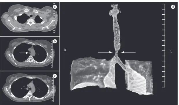
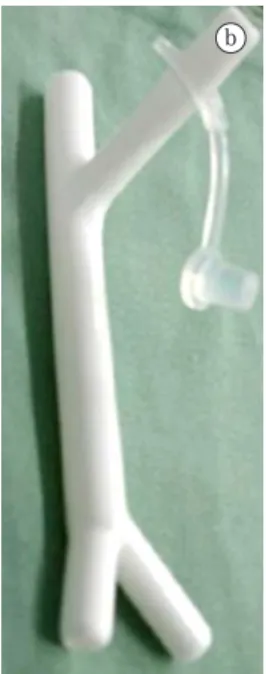
On the evening after the day of hospitalization, with esophagoscopy and computed assisted tomography of the chest scheduled for the following day, the patient presented with
After diagnostic procedures, including chest X-ray and computed tomography scan of the chest, endobronchial lesion with fat density was suspected.. The diagnostic hypothesis
Figure 2 - A) Computed tomography scan of the chest showing free bilateral pleural effusion, greater on the right and atelectasis by compression of the lower lobes, greater on
Figure 2 - Computed tomography scan of the chest showing an extensive mediastinal lesion (arrow) in contact with the right tracheal wall and causing anterior deviation
A computed tomography scan of the chest revealed a 4-cm mass with heterogeneous content and pleural extension to the level of the lingula, as well as two micronodules in
