J. bras. pneumol. vol.39 número2
Texto
Imagem
Documentos relacionados
A transesophageal echocardiogram revealed a 30x35 mm multicavitated ovoid mass in the left atrium, attached to the interatrial septum, at the level of the fossa ovalis
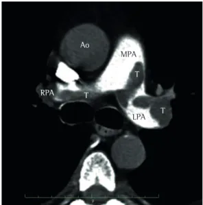
We report on the rare case of partial anomalous return of four pulmonary veins in the right atrium and superior vena cava with intact interatrial septum in a five-year-old
The echocardiogram (Figure 2) confirmed the marked mitral valve insufficiency with increase mainly of the left heart chambers and preserved ventricular function (left
Figure 4 - Echocardiogram subcostal view showing the pacemaker electrode path: it enters the right atrium, crosses the interatrial septum, goes through the left atrium and mitral
At the operation, a tumor was found in the left atrium with extensive infiltration into the atrial septum and adhesion of the anterior leaflet of mitral valve, which was
Transthoracic echocardiogram showed a dilated left atrium and a mass could be seen attached to the anterior leaflet of the mitral valve.. Transesophageal echo- cardiogram showed
Transeophageal echo- cardiography was performed to access: left atrium enlar- gement; communication or aneurysm of the interatrial septum; patent foramen ovale; spontaneous
2 - Implant sequence in plastic model documented by fluoroscopy; (A and B) distal disk set after the delivery catheter reached the left atrium; C) left atrium disk withdrawn towards
