en 0100 3984 rb 47 06 00XI
Texto
Imagem
Documentos relacionados
Finally, the recognition of imaging findings of uncommon liver lesions as well as of atypical findings of more frequent liver lesions play a relevant role in the construction of
Posteroanterior chest radiography and chest computed tomography showing bullous emphysema in the upper portions of the lungs, predominantly at right.. Diagnosis: Bullous emphysema in
In the present issue of Radiologia Brasileira there is an in- teresting study aimed at determining reference intervals for the fetal cisterna magna volume by means of
In 1949, Raul Leborgne revitalized the interest in mammography calling the attention over the need for technical qualification for patients positioning and over radiological
MDs, Residents, Department of Radiology and Imaging Diagnosis, Hospital das Clínicas – Universidade Federal de Goiás (UFG), Goiânia, GO,
In 2008, the American Cancer Society, in association with the US Multi-Society Task Force on Colorectal Cancer (representing the three major American gastroenterological societies
Therefore, despite all efforts of academic and university centers, as well as of both public and private services that promote professional training pro- grams accredited by
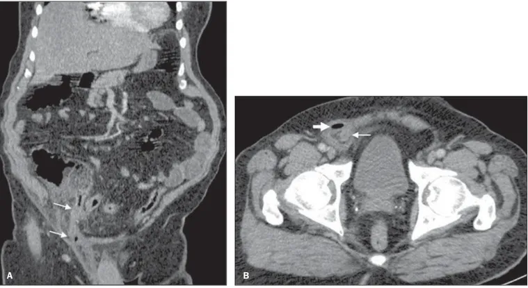
equi infection should be considered in the differential diagnosis of cavitated consolidations in AIDS patients, with a particular difficult differentiation from lesions caused
