Prevalence of self-reported
medical diagnosis of uterine
leiomyomas in a Brazilian
population: Demographic and
socioeconomic patterns in the
Pro-Saúde Study
Prevalência de diagnóstico médico
auto-relatado de miomas uterinos
em população brasileira: Padrões
demográicos e socioeconômicos no
Estudo Pró-Saúde*
Karine de Lima Sírio Boclin
Eduardo Faerstein
Institute of Social Medicine, University of the State of Rio de Janeiro – UERJ.
* This research project was funded by Capes (Coordination for the Improvement of Higher Educa-tion Personnel) through the NaEduca-tional Post-Doctoral Program in Health. Pós-Doc Capes/SUS. Pro-cess 23038009349/2010-65.
Corresponding author: Karine de Lima Sírio Boclin. Instituto de Medicina Social, Universidade do Estado do Rio de Janeiro – UERJ. Rua São Francisco Xavier, 524, Pavilhão João Lyra Filho, 6º andar / blocos E, e 6º andar, sala 6003, Maracanã, 20550-900 Rio de Janeiro, RJ, Brasil. E-mail: [email protected]
Abstract
Introduction: Uterine leiomyomas (UL) are considered the most common tumors of the female reproductive system. However, there are few epidemiological studies about this condition in Brazil. Aim: To estimate the prevalence of self-reported history of UL according to demographic and socioeco-nomic characteristics, and to markers of access to health care. Methods: We analyzed data from 1,733 university employees who participated at the baseline waves of the Pro-Saude Study (1999-2001), in relation to three outcomes: (1) self-reported medical diagnosis of UL, (2) UL with symptoms prior to diagnosis, and (3) cases with hys-terectomy due to UL. Prevalence and 95% confidence intervals (95% CI) were estima-ted in relation to strata of variables relaestima-ted to demographic (age, color/race) and so-cioeconomic characteristics (education, income) and of markers of access to health care (Pap smear, breast clinical exam and private health insurance status). Results: The prevalence of medically-diagnosed UL was 23.3% (95% CI - 21.3, 25.2), the UL with symptoms prior to diagnosis of 13.3% (95% CI - 11.7, 15.0) and hysterectomy due to UL, 8.4% (95% CI - 7.5, 10.3). Among par-ticipants younger than 45 years old, higher prevalence was observed among women with worse socioeconomic conditions and of black color/race. Among those with 45 years or more, there was higher prevalence among women with better access to health care. Conclusion: In this study population of Brazilian women, UL is a relevant health problem, and its prevalence and associated socio-demographic gradients are similar to those observed in other countries.
Resumo
Introdução: Os miomas uterinos (MU) são considerados os tumores mais comuns do sistema reprodutor feminino; no entanto, existem poucos estudos epidemiológicos sobre essa condição no Brasil. Objetivo: Estimar as prevalências de história auto--relatada de MU segundo características demográficas, socioeconômicas e de acesso a serviços de saúde. Métodos: Foram ana-lisados dados de 1.733 trabalhadoras de universidade no Rio de Janeiro, participan-tes da linha de base do Estudo Pró-Saúde (1999-2001), em relação a três desfechos: (1) diagnóstico médico de MU, (2) MU com sintomas prévios ao diagnóstico e (3) casos que realizaram histerectomia pelo tumor. As prevalências e seus intervalos de 95% de confiança (IC 95%) foram estimadas em relação a estratos de variáveis demográficas (idade, cor/raça), socioeconômicas (esco-laridade, renda) e marcadoras de acesso a serviços de saúde (teste Papanicolaou, exa-me de mama, plano de saúde). Resultados: A prevalência de diagnóstico médico de MU foi de 23,3% (IC 95% - 21,3; 25,2); a de MU com sintomas prévios ao diagnóstico, de 13,3% (IC 95% - 11,7; 15,0) e a de histerecto-mia pelo tumor, de 8,4% (IC 95% - 7,5; 10,3). Entre participantes abaixo de 45 anos de idade, foram observadas prevalências mais elevadas nos estratos de piores condições socioeconômicas e de cor/raça preta. Entre aquelas com 45 anos ou mais, foram encon-tradas maiores prevalências entre mulheres com melhor acesso a serviços de saúde. Conclusão: Entre as mulheres brasileiras investigadas, os MU constituem problema relevante de saúde, com prevalências e gradientes sociodemográficos similares aos observados em populações de outros países.
Palavras-chaves: Leiomioma. Prevalência. Saúde da mulher.
Introduction
Uterine leiomyomas (UL) are slow-gro-wing monoclonal benign neoplasms that develop in several locations in the uterus1,2. They are considered to be the most common tumors in the female reproductive system3. Studies performed in the United States have suggested that between 70% and 80% of women aged from 40 to 50 years have UL; however, almost half of these tumors are not even diagnosed, nor do they require treatment, as they do not show clinical signs or symptoms4-6.
Although rarely associated with ma-lignization or mortality2,7, UL can have a significant impact on the quality of life of women of reproductive age8. Depending on their anatomical position, number and size, these tumors can cause excessive uterine bleeding and/or a prolonged menstrual pe-riod9-11; feeling of pelvic pressure, increase in abdominal volume10,12,13; pain during sexual intercourse14 and urinary incontinence15,16. Additionally, the UL can have a negative impact on the reproductive function and it is associated with infertility and adverse gestational outcomes, such as spontaneous abortions, fetal anomalies, premature bir-ths, and a higher number of indications for Cesarean sections10,17-22.
menopause36 and those who consumed greater amounts of fruits, vegetables and fish28,35.
Whereas studies conducted in the United States indicate prevalences of up to 80%, depending on the characteristics of the sub-groups studied, European studies have revealed significantly lower prevalences of tumors43. In Germany, 10.7% of participants of a study performed with 10,241 women aged less than 65 years reported having re-ceived a diagnosis of “benign tumor in the uterus”43,44. In Italy, UL cases were detected by ultrasound in 21.4% of participants of a study conducted with 341 women aged between 30 and 60 years45. In Sweden, these tumors were also diagnosed by ultrasound in 3.3% of women aged from 25 to 32 years and in 7.8% of those aged from 33 to 40 years in a random sample comprised of 335 women46. In Brazil, there are few epidemiological data on UL. At present, only one study has been identified, which was conducted with a low-income population cared for in a health clinic of the city of São Paulo. In this study, UL was found in 23% of white women and 42% of black ones. The occurrence of hyste-rectomy for UL also varied between groups, totaling 4% among white women and 16% among black ones47.
Several reasons indicate the need for better identification of the characteristics of occurrence of this condition among Brazilian women. UL has a great negative impact on women’s health, whether due to the reduction in the quality of life of a significant number of young women of reproductive age8 or due to the increase in the number of mutilating surgeries3. Not less important are the differences between the Brazilian and American contexts, especially with regard to ethnic relations and their interfaces with the remaining demographic and socioeconomic characteristics48.
Contrasting this lack of evidence, there is growing space for reflection and social poli-cies aimed at women in general and, speci-fically, the African Brazilian population49,50. Governmental policies have sought, mainly in the last decade, “to increase, qualify and
humanize comprehensive care for women’s health in the Sistema Único de Saúde (SUS – Unified Health System) […] aiming to re-duce female morbidity and mortality […], considering ethnic peculiarities”50.
In this sense, special attention has been given to the most frequent diseases and conditions found in the black population, among which is UL49.
Thus, aiming to contribute to the know-ledge about epidemiology of UL, the present study was conducted with women partici-pating in the Pró-Saúde Study and had the purpose of estimating the prevalence of (1) cases of self-reported medical diagnosis of UL; (2) cases of self-reported medical nosis of UL, with symptoms prior to diag-nosis; and (3) cases of self-reported medical diagnosis of UL, with the performance of hysterectomy. Additionally, the prevalences of these three outcomes were identified in different demographic and socioeconomic strata and according to access to and use of health services.
Methods
The Pró-Saúde Study
The Pró-Saúde Study is a longitu-dinal investigation conducted among technical-administrative workers of a university located in the city of Rio de Janeiro, Southeastern Brazil. The social determinants of health and health beha-vior are its main thematic focus. All active workers found in the institution at the beginning of the study (1999) were invited to participate51.
Study population
of 1,733 workers were analyzed after exclu-ding those who did not provide information about the medical diagnosis of UL (n = 86); in general, the latter had poorer socioecono-mic conditions, compared to those analyzed in the present study.
Data collection and study variables
Self-administered questionnaires were applied in the workplace by trained field researchers, assisted by supervisors. Questions about the following aspects were included: socioeconomic conditions, gender, ethnic group, geographic and social mobility, experience of discrimina-tion, work-related stress, social support and network patterns, women’s health, morbidities, work accidents, work-related behavioral disorders and common mental disorders. Methods used to improve the quality of information, such as pilot stu-dies, validation of scales and test-retest reliability tests, were performed51.
Outcome
Information about the medical diagno-sis of UL was obtained from participants in 1999 through the following question: “Have you ever been informed by a physician that you had uterine leiomyoma, a benign tumor in the uterus?”. The test-retest reliability of responses was assessed in a stratified sam-ple (age and level of education) of 98 women who were not eligible for the Pró-Saúde Study (temporary workers of the same uni-versity), with an interval of two weeks, and it was considered to be excellent (kappa = 0.94 – 95%CI: 0.86; 1.00).
Additionally, participants provided information about age of diagnosis of UL, previous symptoms and performance of hysterectomy as a result of UL. Based on this information, three case definitions were developed and explored separately as ou-tcomes of interest, and the second and third definitions were a sub-set of the first one: (1) totality of cases of self-reported medical diagnosis of UL; (2) cases of self-reported
medical diagnosis of UL, with symptoms prior to diagnosis; and (3) cases of self--reported medical diagnosis of UL, with the performance of hysterectomy.
Socioeconomic and demographic variables
Age: discrete variable categorized in two ways: (1) younger than 35 years, from 35 to 44 years, from 45 to 54 years, and older than 54 years; and (2) younger than 45 years, and 45 years and older.
Color/race: in 1999, information about participants’ color/race was collected with the open question (“In your opinion, what is your color/race?”). A total of 41 distinct terms were reported to identify participants’ color/race (including men). These terms were categorized as follows: white, brown (typical Portuguese words such as parda, morena, mulata, mestiça and cabocla were used to describe mixed ethnicity), black (ty-pical Portuguese words such as negra, preta, Africana and escura were used to describe mixed ethnicity) and of Asian descent52. Participants of Asian descent were excluded from the analyses due to their small number (n = 8; - 0.5%).
Level of education: complete primary school, complete secondary school, and complete university undergraduate level or higher.
Per capita household income: variable obtained by dividing the total income of those contributing to the household ex-penses by the number of residents. It was categorized into: less than three minimum wages (MW), three to six MW, and more than six MW (One MW was R$136.00 or US$ 71.57 at the time of this study).
Variables markers of health service access and use
Pap smear test: has never had it perfor-med or had it perforperfor-med more than three years before, and had it performed less than three years before.
never had a breast examination performed or had it more than three years before, and had it performed less than three years before.
Health insurance: yes and no.
Statistical analyses
Prevalences and their 95% confidence intervals (95%CI) were estimated for the three outcomes in the entire study popu-lation in demographic, socioeconomic and health service access and use strata. Prevalences were also stratified according to age groups (younger than 45 years, 45 years and older). The cut-off point was defined after changes in the pattern of prevalences were verified close to the age of 45 years in the population studied. Pearson’s chi-square test was used to assess the heterogeneity of proportions of sub-groups. Differences with a p-value < 0.05 were considered to be statistically significant.
Data entry and consistency checking were performed with the Epi-Info software. Analyses were made in the R statistical sof-tware, version 2.6.2. The present study was approved by the Research Ethics Committee of the University of the State of Rio de Janeiro. There were no conflicts of interest.
Results
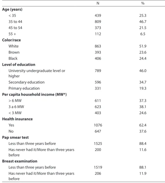
Of all 1,733 study participants, 72% were younger than 45 years, 51.9% reported they were white, 62.4% had health insurance, 88.4% had a Pap smear test performed less than three years before and 88.1% had a breast examination performed less than three years before. Slightly less than half (46%) reported they had completed their university undergraduate program and 37% had a per capita household income higher than six MW (Table 1).
The prevalence of medical diagnosis of UL was 23.3% (95%CI - 21.3; 25.3), that of UL with symptoms prior to diagnosis was 13.3% (95%CI - 11.7; 15.0), and that of hys-terectomy due to tumor was 8.4% (95%CI - 7.5; 10.3). Figure 1 shows the prevalences
of three outcomes according to age groups. There was an increase in the prevalences with the increase in age until 45 years, with a subsequent stabilization after this age (Figure 1).
Table 2 shows the prevalences of three outcomes among the sub-groups formed by the demographic, socioeconomic and health service access and use strata. With regard to the medical diagnosis of UL, apart from the increase in prevalence with age, there were also higher prevalences among black women (32.8% - p < 0.001), those who had completed primary education (33.8% -
p < 0.001) and those who had had a breast examination less than three years before (24% - p = 0.021).
This pattern of distribution of prevalen-ces in the strata of covariables was similar to other two outcomes (UL with symptoms prior to diagnosis and cases of hysterectomy due to UL). In both cases, higher prevalen-ces were also found among women with a lower per capita household income (19.7% of those with symptoms prior to diagnosis and 14.3% of cases of hysterectomy due to UL, each with p < 0.001) and among those who did not have health insurance (16.6% of those with symptoms prior to diagno-sis, with p = 0.002; and 11.3% of cases of hysterectomy due to UL, with p = 0.007). Differences were not statistically significant according to time of performance of breast examination (Table 2).
Table 3 shows the prevalences of UL by covariables, stratified by age for the three outcomes. Considering the two age groups analyzed, the patterns found were different. Black women and those who had completed primary education had significantly higher prevalences of medical diagnosis of UL in the stratum of women younger than 45 years exclusively (25.6% - p < 0.001 and 25.4% - p = 0.013, respectively). On the other hand, wo-men aged 45 years and more, who had had the Pap smear test and breast examination less than three years before, showed higher prevalences (40.2% - p = 0.023 and 40.8% - p
age groups (Table 3).
Stratifications of prevalences by age also showed different patterns for UL with symptoms prior to diagnosis. Being black and having a per capita household income lower than three MW were directly asso-ciated with the diagnosis of symptomatic UL among women younger than 45 years (13.5% - p = 0.006, and 15% - p = 0.002, res-pectively). In contrast, those aged 45 years
and more showed no statistically significant differences with regard to the characteristics studied (Table 3).
Concerning cases of hysterectomy due to UL, the same variables were associated with higher prevalences among women younger than 45 years. There were no sta-tistically significant differences with regard to the characteristics studied among women aged 45 years and more (Table 3).
Table 1 - Demographic, socioeconomic and health services access characteristics in the study population. The Pro-Saúde Study (1999-2001).
Tabela 1 - Características demográicas, socioeconômicas e de acesso a serviços de saúde da população estudada. Estudo Pró-Saúde (1999-2001).
N %
Age (years)
< 35 439 25.3
35 to 44 809 46.7
45 to 54 373 21.5
55 + 112 6.5
Color/race
White 863 51.9
Brown 393 23.6
Black 406 24.4
Level of education
University undergraduate level or higher
789 46.0
Secondary education 596 34.7
Primary education 331 19.3
Per capita household income (MW*)
> 6 MW 611 37.3
3 a 6 MW 623 38.1
< 3 MW 403 24.6
Health insurance
Yes 1076 62.4
No 647 37.6
Pap smear test
Less than three years before 1525 88.4
Has never had it/More than three years before
200 11.6
Breast examination
Less than three years before 1519 88.1
Has never had it/More than three years before
206 11.9
* One minimum wage (MW) was R$136.00 or US$ 71.57 at the time of this study.
The sum of each variable may not correspond to the total number of participants due to loss of data. * Salário-mínimo (R$136,00) .
Discussion
The prevalences of UL reported in the international literature show great varia-tion: from 3.3% in a Swedish study46 to 80% in an American study4, depending on the geographic origin and age group of the population analyzed3,4. As many cases of UL are asymptomatic, the methods used to estimate the frequency of tumors can also influence these results5. Thus, studies based on the previous diagnosis of UL can
generate underestimated prevalences, as asymptomatic cases among participants without a history of diagnosis will probably pass unnoticed, especially if they do not have adequate access to health services5. On the other hand, this underestimation decreases in studies that use ultrasound tests, for example.
The prevalences estimated in the present study are in an intermediate position, closer to the American estimates. One must take into consideration the fact that the history Figure 1 - Prevalence and Intervals Conidence of 95% (95%) of self-reported medical diagnosis of uterine leiomyoma, uterine leiomyoma symptoms prior to diagnosis and hysterectomy for uterine leiomyoma by age. The Pro-Saude Study (1999-2001).
of medical diagnosis of UL was reported by participants and that, consequently, these data are subject to underestimation, due to
the existence of asymptomatic cases. Some strategies were used to minimize this limi-tation. The first one, although indirect, was Table 2 - Prevalence (%) of self-reported medical diagnosis of uterine leiomyoma (UL), UL with symptoms prior to diagnosis and hysterectomy for UL by demographic, socioeconomic and health services access characteristics. Pro-Saude Study (1999-2001).
Tabela 2 - Prevalências (%) de diagnóstico médico auto-relatado de mioma uterino (MU), MU com sintomas prévios ao diagnóstico e histerectomia por MU segundo características demográicas, socioeconômicas e de acesso a serviços de saúde. Estudo Pró-Saúde (1999-2001).
Self-reported medical diagnosis of UL
UL with symptoms prior to diagnosis
Hysterectomy due to UL
N n (%) N n (%) N n (%)
Age (years)
< 35 439 33 (7.5) 439 20 (4.6) 423 3 (0.7)
35 to 44 809 184 (22.7) 806 92 (11.4) 745 52 (7.0)
45 to 54 373 144 (38.6) 373 90 (24.1) 330 62 (18.8)
55 + 112 42 (37.5) 112 28 (25.0) 97 24 (24.7)
p-value* <0.001 <0.001 <0.001
Color/race
White 863 167 (19.4) 863 86 (10.0) 805 43 (5.3)
Brown 393 90 (22.9) 393 55 (14.0) 363 31 (8.5)
Black 406 133 (32.8) 403 80 (19.9) 366 60 (16.4)
p-value* <0.001 <0.001 <0.001
Level of education
University undergraduate level or higher
789 167 (21.2) 789 80 (10.1) 742 45 (6.1)
Secondary education 596 118 (19.8) 594 73 (12.3) 552 43 (7.8)
Primary education 331 112 (33.8) 330 74 (22.4) 287 51 (17.8)
p-value* <0.001 <0.001 <0.001
Per capita household income (MW)
< 3 MW 611 133 (21.8) 611 61 (10.0) 579 33 (5.7)
3 to 6 MW 623 130 (20.9) 623 70 (11.2) 574 42 (7.3)
> 6 MW 403 106 (26.3) 402 79 (19.7) 364 52 (14.3)
p-value* 0.107 <0.001 <0.001
Health insurance
Yes 1076 242 (22.5) 1075 121 (11.3) 1003 72 (7.2)
No 647 157 (24.3) 645 107 (16.6) 584 66 (11.3)
p-value* 0.431 0.002 0.007
Pap smear test
Less than three years before 1525 364 (23.9) 1522 206 (13.5) 1404 121 (8.6) Has never had it/More than three
years before
200 35 (17.5) 200 21 (10.5) 183 17 (9.3)
p-value* 0.055 0.279 0.87
Breast examination
Less than three years before 1519 365 (24.0) 1517 205 (13.5) 1404 127 (9.0) Has never had it/More than three
years before
206 34 (16.5) 205 22 (10.7) 183 11 (6.0)
p-value* 0.021 0.320 0.218
the assessment of reliability of the question about the diagnosis of UL, which indicated an excellent pattern. The second one was
the exploration of the three outcomes, which, apart from enabling the assessment of the severity of UL, tested different levels
Table 3 - Prevalence (%) by age of self-reported medical diagnosis of uterine leiomyoma (UL), UL with symptoms prior to diagnosis and hysterectomy for UL by demographic, socioeconomic and health services access characteristics. Pro-Saude Study (1999-2001).
Tabela 3 - Prevalências (%) estratiicadas por idade de diagnóstico médico auto-relatado de mioma uterino (MU), MU com sintomas prévios ao diagnóstico e histerectomia por MU segundo características demográicas, socioeconômicas e de acesso a serviços de saúde. Estudo Pró-Saúde (1999-2001).
Self-reported medical diagnosis of UL
UL with symptoms prior to diagnosis
Hysterectomy due to UL
< 45 years ≥45 years < 45 years ≥45 years < 45 years ≥45 years
n (%) n (%) n (%) n (%) n (%) n (%)
Color/race
White 94 (14.1) 73 (37.2) 46 (6.9) 40 (20.4) 17 (2.7) 26 (14.8)
Brown 53 (18.9) 37 (32.7) 28 (10.0) 27 (23.9) 10 (3.7) 21 (21.9)
Black 67 (25.6) 66 (45.8) 35 (13.5) 45 (31.2) 27 (11.4) 33 (25.4)
p-value* <0.001 0.084 0.006 0.071 <0.001 0.061
Level of education
University undergraduate level or higher
115 (17.6) 52 (38.5) 53 (8.1) 27 (20.0) 26 (4.2) 19 (15.3)
Secondary education 66 (14.4) 52 (37.7) 39 (8.6) 34 (24.6) 18 (4.2) 25 (20.5)
Primary education 33 (25.4) 79 (39.3) 19 (14.7) 55 (27.4) 10 (8.7) 41 (23.8)
p-value* 0.013 0.955 0.051 0.306 0.094 0.199
Per capita household income (MW)
> 6 MW 84 (17.4) 49 (38.3) 34 (7.0) 27 (21.1) 17 (3.7) 16 (14.0)
3 to 6 MW 76 (15.8) 54 (38.3) 40 (8.3) 30 (21.3) 15 (3.4) 27 (21.1)
< 3 MW 46 (19.7) 60 (35.5) 35 (15.0) 44 (26.0) 21 (9.8) 31 (20.8)
p-value* 0.427 0.841 0.002 0.503 <0.001 0.285
Health insurance
Yes 150 (17.9) 92 (38.7) 69 (8.2) 52 (21.8) 33 (4.2) 39 (18.1)
No 66 (16.2) 91 (38.1) 43 (10.6) 64 (26.8) 21 (5.6) 45 (21.8)
p-value* 0.500 0.971 0.211 0.251 0.373 0.407
Pap smear test
Less than three years before 197 (17.7) 167 (40.2) 102(9.2) 104 (25.1) 48 (4.6) 73 (19.9)
Has never had it/more than three years before
19 (14.1) 16 (24.6) 10 (7.4) 11 (16.9) 7 (5.7) 10 (17.9)
p-value* 0.345 0.023 0.594 0.203 0.823 0.853
Breast examination
More than three years before 201 (18.0) 164 (40.8) 105 (9.4) 100 (24.9) 52 (5.0) 75 (20.9)
Has never had it/More than three years before
15 (11.8) 19 (24.1) 7 (5.6) 15 (19.0) 3 (2.5) 8 (12.3)
p-value* 0.105 0.007 0.204 0.328 0.342 0.149
of specificity of information about tumors reported. The third one was the use of varia-bles markers of access to health services in the analyses. In this sense, a great part of the study population had reasonable conditions of obtaining a medical diagnosis of tumors, although asymptomatic, as they had health insurance and had had a Pap smear test and breast examination performed less than three years before.
On the other hand, as workers excluded from the analyses, as a result of their not providing information about UL, had a poo-rer socioeconomic profile than participants, the prevalences of UL, symptomatic UL and hysterectomy due to UL were probably underestimated.
As in other studies, the prevalences of UL increased with age4-6. Epidemiological studies have found higher frequencies of tumors in women aged between 40 and 50 years, thus strengthening the hypothesis of the role of hormonal imbalance in their development. According to this hypothesis, UL tumors would result from an excess of blood estrogen and progesterone; thus, the longer one is exposed to this imbalance, the greater the chance of developing tumors2,7. Despite the cross-sectional design of this study, the results suggest that age is a possi-ble modifier of the association between UL and the remaining variables analyzed, as the pattern of distribution of prevalences varied in the demographic, socioeconomic and health service access and use sub-groups according to the age group analyzed. Significant differences were more frequent among women younger than 45 years. In this group, the greatest prevalences were found among black participants and those with poorer socioeconomic conditions.
On the other hand, women aged 45 years and more did not show significant differences in estimates, according to their demographic and socioeconomic pattern. Higher prevalences were only found among those who reported having greater access to medical diagnosis, i.e. Pap smear test and breast examination performed less than three years before.
The remaining results corroborate the findings of studies performed in the United States with women from different color/race groups. According to these studies, compa-red to white women, tumors in black women occur between two and nine times more frequently in all age groups, in addition to the higher number of tumors, more severe symptoms, younger ages when diagnosed, and higher rates of hysterectomy4,23-26,53,54. However, the causes of ethnic inequality in the occurrence of UL remain unknown and possible explanatory mechanisms have been scarcely studied in the literature.
Epidemiological studies suggest that risk factors established for these tumors, such as those associated with reproductive life and lifestyle, could only explain a small fraction of color/race inequalities4,23,26. Marshall et al, for example, found a relative risk for UL of 3.25 (95%CI 2.71; 3.88) and that of hys-terectomy due to UL of 1.82 (95%CI 1.17; 2.82) among black women, after adjusting for variables such as age, body mass index (BMI), length of time since previous preg-nancy, history of infertility, alcohol use, smoking, leisure-time physical activities, age of menarche, age of first pregnancy, use of oral contraceptives and marital status23. Faerstein et al reported that black women had a nine times greater chance of presence of UL (OR: 9.4; 95%CI: 5.7; 15.7), after adjusting for age of menarche, use of oral contraceptives, smoking, body weight, arterial hypertension, diabetes mellitus, and history of pelvic inflammatory disea-se26. Baird et al observed an odds ratio of 2.7, (95%CI: 2.3; 3.2) for black women, after adjusting for BMI and parity4.
have been analyzed30,31,34,39,42 as possible confounders of the remaining associations, thus not being the main focus of analysis.
Nonetheless, considering the few studies that approach the association between level of education and UL, some were found to lack such association4,40,55,56 and one study showed a direct association28. Two studies dealt with the influence of exposures oc-curring during the intrauterine life and childhood57,58 and found direct associations between tumors and parents’ low level of education, food insecurity, and low hou-sehold income during childhood among white women57, but not among black ones58. There is much yet to be investigated in
terms of how biological mechanisms are associated with socioeconomic and demo-graphic characteristics, influencing health outcomes such as UL and including ways of determination apart from hormonal imba-lance or health behavior. The present study showed indications that the frequency of UL is higher among black women and those with poorer socioeconomic conditions. Other epidemiological studies of a longi-tudinal analytical nature can better clarify the causal relations of interest, enabling action strategies related to this problem to be more effective and regulated by the search for greater equality in the sphere of women’s health.
References
1. Jolley S. An overview of uterine fibroids. Nurs Standard (Royal College of Nursing) (Great Britain). 2009; 24(6): 44-8.
2. Parker WH. Etiology, symptomatology, and diagnosis of uterine myomas. Fertil Steril 2007; 87(4): 725-36.
3. Reynolds A. Diagnosis and management of uterine fibroids. Radiol Technol 2007; 79(2): 157-78; 79-82.
4. Day Baird D, Dunson DB, Hill MC, Cousins D, Schectman JM. High cumulative incidence of uterine leiomyoma in black and white women: ultrasound evidence. Am J Obstet Gynecol 2003; 188(1):100-7.
5. Schwartz SM, Marshall LM, Baird DD. Epidemiologic contributions to understanding the etiology of uterine leiomyomata. Environ Health Perspec 2000 Oct; 108: 821-7.
6. Divakar H. Asymptomatic uterine fibroids. Best Pract Res Clin Obstet Gynaecol 2008; 22(4): 643-54.
7. Walker CL, Stewart EA. Uterine fibroids: the elephant in the room. Science 2005: 10; 308(5728): 1589-92.
8. Flake GP, Andersen J, Dixon D. Etiology and
pathogenesis of uterine leiomyomas: a review. Environ Health Perspect 2003; 111(8): 1037-54.
9. Wegienka G, Baird DD, Hertz-Picciotto I, Harlow SD, Hartmann KE. Uterine leiomyomata (fibroids): are bleeding symptoms more likely to be reported after diagnosis? J Clin Epidemiol 2004; 57 (3): 318-20.
10. Stovall DW. Clinical symptomatology of uterine leiomyomas. Clin Obstet Gynecol 2001; 44(2): 364-71.
11. Wegienka G, Baird DD, Hertz-Picciotto I, Harlow SD, Steege JF, Hill MC et al. Self-reported heavy bleeding associated with uterine leiomyomata. Obstet Gynecol 2003; 101(3): 431-7.
12. Taylor E, Gomel V. The uterus and fertility. Fertil Steril 2008; 89(1): 1-16.
13. Bukulmez O, Doody KJ. Clinical features of myomas. Obstet Gynecol Clin North Am 2006; 33(1): 69-84.
14. Lippman SA, Warner M, Samuels S, Olive D, Vercellini P, Eskenazi B. Uterine fibroids and gynecologic pain symptoms in a population-based study. Fertil Steril 2003; 80(6): 1488-94.
15. Waetjen LE, Dwyer PL. Estrogen therapy and urinary incontinence: what is the evidence and what do we tell our patients? Int Urogynecol J Pelvic Floor Dysfunct 2006; 17(5): 541-5.
16. Waetjen LE, Liao S, Johnson WO, Sampselle CM, Sternfield B, Harlow SD et al. Factors associated with prevalent and incident urinary incontinence in a cohort of midlife women: a longitudinal analysis of data: study of women’s health across the nation. Am J Epidemiol 2007; 165(3): 309-18.
17. Coronado GD, Marshall LM, Schwartz SM.
Complications in pregnancy, labor, and delivery with uterine leiomyomas: a population-based study. Obstet Gynecol 2000; 95(5): 764-9.
19. Ouyang DW, Economy KE, Norwitz ER. Obstetric complications of fibroids. Obstet Gynecol Clin North Am 2006; 33(1): 153.
20. Klatsky PC, Tran ND, Caughey AB, Fujimoto VY. Fibroids and reproductive outcomes: a systematic literature review from conception to delivery. Am J Obstet Gynecol 2008; 198(4): 357-66.
21. Khaund A, Lumsden MA. Impact of fibroids on reproductive function. Best Pract Res Clin Obstet Gynaecol 2008; 22(4): 749-60.
22. George L, Granath F, Johansson ALV, Olander B, Cnattingius S. Risks of repeated miscarriage. Paediatric Perinatal Epidemiol 2006; 20(2): 119-26.
23. Marshall LM, Spiegelman D, Barbieri RL, Goldman MB, Manson JE, Colditz GA et al. Variation in the incidence of uterine leiomyoma among premenopausal women by age and race. Obstet Gynecol 1997; 90 (6): 967-73.
24. Huyck KL, Panhuysen CI, Cuenco KT, Zhang J, Goldhammer H, Jones ES et al. The impact of race as a risk factor for symptom severity and age at diagnosis of uterine leiomyomata among affected sisters. Am J Obstet Gynecol 2008; 198 (2): 168 e1-9.
25. Weiss G, Noorhasan D, Schott LL, Powell L, Randolph JF, Johnston JM. Racial differences in women who have a hysterectomy for benign condition. Womens Health Issues 2009; 19(3): 202-10.
26. Faerstein E, Szklo M, Rosenshein N. Risk factors for uterine leiomyoma: a practice-based case-control study. I. African-American heritage, reproductive history, body size, and smoking. Am J Epidemiol 2001; 153(1): 1-10.
27. Reed SD, Cushing-Haugen KL, Daling JR, Scholes D, Schwartz SM. Postmenopausal estrogen and
progestogen therapy and the risk of uterine leiomyomas. Menopause 2004; 11(2): 214-22.
28. Chiaffarino F, Parazzini F, La Vecchia C, Chatenoud L, Di Cintio E, Marsico S. Diet and uterine myomas. Obstet Gynecol 1999; 94(3): 395-8.
29. Wise LA, Palmer JR, Adams-Campbell LL, Rosenberg L. Age-specific incidence rates for uterine leiomyomata in the Black Women’s Health Study. Am J Epidemiol 2004; 159(S11): 92-S.
30. Marshall LM, Spiegelman D, Goldman MB, Manson JE, Colditz GA, Barbieri RL et al. A prospective study of reproductive factors and oral contraceptive use in relation to the risk of uterine leiomyomata. Fertility Sterility 1998; 70(3): 432-9.
31. Wise LA, Palmer JR, Harlow BL, Spiegelman D, Stewart EA, Adams-Campbell LL et al. Reproductive factors affected the risk of uterine leiomyomata in African-American women. Evid Based Obstet Gynecol 2004; 6(3): 125-6.
32. Cooper R, Hardy R, Kuh D. Timing of menarche, childbearing and hysterectomy risk. Maturitas 2008; 61(4): 317-22.
33. Marshall LM, Spiegelman D, Manson JE, Goldman MB, Barbieri RL, Stampfer MJ et al. Risk of uterine leiomyomata among premenopausal women in relation to body size and cigarette smoking. Epidemiology 1998; 9(5): 511-7.
34. Wise LA, Palmer JR, Spiegelman D, Harlow BL, Stewart EA, Adams-Campbell LL et al. Influence of body size and body fat distribution on risk of uterine leiomyomata in U.S. black women. Epidemiology 2005;16(3): 346-54.
35. Terry KL, Missmer SA, Hankinson SE, Willett WC, De Vivo I. Lycopene and other carotenoid intake in relation to risk of uterine leiomyomata. Am J Obstet Gynecol 2008;198(1).
36. Parazzini F. Risk factors for clinically diagnosed uterine fibroids in women around menopause. Maturitas 2006; 55(2):174-9.
37. Sato F, Nishi M, Kudo R, Miyake H. Body fat distribution and uterine leiomyomas. J Epidemiol / Japan
Epidemiological Association 1998; 8(3): 176-80.
38. Terry KL, De Vivo I, Hankinson SE, Spiegelman D, Wise LA, Missmer SA. Anthropometric characteristics and risk of uterine leiomyoma. Epidemiology 2007; 18(6): 758-63.
39. Baird DD, Dunson DB, Hill MC, Cousins D, Schectman JM. Association of physical activity with development of uterine leiomyoma. Am J Epidemiol 2007; 165(2): 157-63.
40. Samadi AR, Lee NC, Flanders WD, Boring JR, Parris EB. Risk factors for self-reported uterine fibroids: A case-control study. Am J Public Health 1996; 86(6): 858-62.
41. Chen CR, Buck GM, Courey NG, Perez KM, Wactawski-Wende J. Risk factors for uterine fibroids among women undergoing tubal sterilization. Am J Epidemiol 2001; 153(1): 20-6.
42. Wise LA, Palmer JR, Harlow BL, Spiegelman D, Stewart EA, Adams-Campbell LL et al. Reproductive factors, hormonal contraception, and risk of uterine leiomyomata in African-American women: a prospective study. Am J Epidemiol 2004; 159(2): 113-23.
43. Payson M, Leppert P, Segars J. Epidemiology of myomas. Obstet Gynecol Clin North Am 2006; 33(1):1.
44. Heinemann K, Thiel C, Mahner S, Lewis MA, Raff T, ahl-Habich D, et al. Benign gynecological tumors: Estimated incidence: Results of the German Cohort Study on Women’s Health. Eur J Obstet Gynecol Reprod Biol 2003; 107(1): 78-80.
46. Borgfeldt C, Andolf E. Transvaginal ultrasonographic findings in the uterus and the endometrium: low prevalence of leiomyoma in a random sample of women age 25-40 years. Acta Obstet Gynecol Scand 2000; 79 (3): 202-7.
47. Souza VC. A prevalência de miomas uterinos em mulheres negras: as dificuldades e avanços e análise dos dados com recorte racial. O livro da saúde da mulher negra: nossos passos vêm de longe. Rio de Janeiro: Pallas/ Criola/Global Exchange; 2000. p. 88-118.
48. Travassos C, Williams DR. The concept and
measurement of race and their relationship to public health? A review focused on Brazil and the United States. Cad Saúde Pública 2004: 20(3): 660-78.
49. Maio MC, Monteiro S. Tempos de racialização: o caso da ‘saúde da população negra’ no Brasil. Hist Ciênc Saúde Manguinhos 2005; 12(2): 419-46.
50. BRASIL. II Plano Nacional de Políticas para as Mulheres.
Brasília: Secretaria Especial de Políticas para as Mulheres. Presidência da República; 2008.
51. Faerstein E, Chor D, Lopes CS, Werneck GL. The Pro-Saude Study: general characteristics and methodological aspects. Rev Bras Epidemiol 2005; 8(4): 454-66.
52. Maio MC, Monteiro S, Chor D, Faerstein E, Lopes CS. [Ethnicity/race in the Pro-Saude Study: comparative results of two methods of self-classification in Rio de Janeiro, Brazil]. Cad Saúde Pública 2005; 21(1): 171-80.
53. Kjerulff KH, Erickson BA, Langenberg PW. Chronic gynecological conditions reported by US women: Findings from the National Health Interview Survey, 1984 to 1992. Am J Public Health 1996; 86(2): 195-9.
54. Wise LA, Palmer JR, Stewart EA, Rosenberg L. Age-specific incidence rates for self-reported uterine leiomyomata in the Black Women’s Health Study. Obstet Gynecol 2005; 105(3): 563-8.
55. Sato F, Miyake H, Nishi M, Mori M, Kudo R. Early normal menstrual cycle pattern and the development of uterine leiomyomas. J Womens Health Gender Based Med 2000; 9(3): 299-302.
56. Martin CL, Huber LRB, Thompson ME, Racine EF. Serum micronutrient concentrations and risk of uterine fibroids. J Womens Health 2011; 20(6): 915-21.
57. D`Aloisio AA, Baird DD, DeRoo LA, Sandler DP. Association of intrauterine and early-life exposures with diagnosis of uterine leiomyomata by 35 years of age in the sister study. Environ Health Perspec 2010; 118(3): 375-81.
58. D`Aloisio AA, Baird DD, DeRoo LA, Sandler DP. Early-life exposures and early-onset uterine leiomyomata in black women in the sister study. Environ Health Perspec 2012; 120(3): 406-12.