Uterine Extramedullary Plasmacytoma as a
Primary Manifestation of Multiple Myeloma
Plasmocitoma extramedular uterino como manifestação
primária de mieloma múltiplo
Ana Codorniz
1Renato Cunha
2Fernando Fernandes
1Maria João Pais
3Tiago Neves
4Carlos Quintana
51Gynecology Department, Hospital do Espírito Santo de Évora, Évora, Portugal
2Medical Oncology Department, Hospital do Espírito Santo de Évora, Évora, Portugal
3Medicine II Department, Hospital do Espírito Santo de Évora, Évora, Portugal
4Orthopedics Department, Hospital do Espírito Santo de Évora, Évora, Portugal
5Pathological Anatomy Department, Hospital do Espírito Santo de Évora, Évora, Portugal
Rev Bras Ginecol Obstet 2017;39:516–520.
Address for correspondence Ana Codorniz, Medical Resident, Serviço de Ginecologia do Hospital do Espírito Santo de Évora, Largo Senhor da Pobreza, 7000-811, Évora, Portugal
(e-mail: [email protected]).
Keywords
►
plasmacytoma
►
multiple myeloma
Abstract
The association between plasmacytomas and multiple myeloma (MM) is
well-de-scribed, and in about one third of the cases of plasmacytoma the additional study
will lead to the diagnosis of MM. The
fi
nding of plasmacytomas in the genital tract is
extremely rare, with sparse cases described in the literature, and these cases pose a
challenge regarding the optimal guidance and treatment. This paper describes a case of
uterine extramedullary plasmacytoma in a 79-year-old woman with complaints of
postmenopausal abnormal uterine bleeding. The complementary study led to the
diagnosis of uterine plasmacytoma and, subsequently, of MM. Despite the unfavorable
outcome of this case, we consider pertinent to report it because it constitutes a
differential diagnosis to be taken into account in the approach of pelvic masses.
Palavras-chave
►
plasmocitoma
►
mieloma múltiplo
Resumo
A associação entre plasmocitomas e mieloma múltiplo (MM) encontra-se bem
de-monstrada, e em cerca de um terço dos casos de plasmocitoma o estudo adicional
conduzirá ao diagnóstico de MM. O achado de plasmocitomas no trato genital é
extremamente raro, havendo um número muito limitado de casos descritos na
literatura, o que di
fi
culta concluir sobre a melhor forma de orientação e tratamento
destes casos. O presente trabalho descreve um caso de plasmocitoma extramedular
uterino em mulher de 79 anos estudada por queixas de hemorragia uterina anômala
pós-menopáusica. O estudo complementar levou ao diagnóstico de plasmocitoma
uterino e, posteriormente, de MM. Apesar do desfecho desfavorável do caso,
consi-deramos pertinente o seu relato por se tratar de um diagnóstico diferencial a levar em
consideração na abordagem de massas pélvicas.
received
March 28, 2017
accepted
June 30, 2017
DOI https://doi.org/ 10.1055/s-0037-1605373.
ISSN 0100-7203.
Copyright © 2017 by Thieme Revinter Publicações Ltda, Rio de Janeiro, Brazil Case Report
Introduction
Plasma cells dyscrasias refer to a group of neoplasms that is characterized by the proliferation of a monoclonal population of plasmocytes that secretes monoclonal immunoglobulins. These neoplasms may present in single or multiple lesions (solitary plasmacytomas or multiple myeloma respectively). The association between plasmacytomas and multiple myelo-ma (MM) is well-established,1and in about one third of the cases the additional study of a plasmacytoma will lead to the diagnosis of MM.2These tumors can appear in the bone or in different organs, and are classified as bone or extramedullary plasmacytomas respectively.3Extramedullary plasmacytomas in the female genital tract are quite rare, either as solitary plasmacytomas or as part of a disseminated MM. There are few cases described in the literature,3–7considering that 80% of extramedullary plasmacytomas arise in the head or neck, mostly in the superior respiratory and digestive tracts.
Case Report
We present a case of a 79-year-old woman, bedridden and totally dependent on other people to perform her daily activities, with a past history of breast cancer at 54 years old treated with conservative surgery followed by radiother-apy. She had no other relevant personal antecedents, made no chronic use of medications, and had no smoking or
drinking habits. Her menarche occurred at 11 years old, and she had regular cycles and 1 pregnancy (with a vaginal delivery at 27 years old). There was no history of use of hormonal contraceptives, and she had spontaneous meno-pause at 41 years without hormonal therapy.
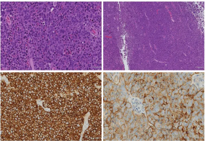
The patient arrived to the emergency department with complaints of postmenopausal abnormal uterine bleeding (AUB) since the previous month, with no other symptoms. Upon physical examination, there were no signs of hemody-namic instability. Upon the speculum examination, a mod-erate amount of necrotic tissue and blood with fetid odor was found in the vagina and through the external cervical orifice; they were sent for a histological test. The blood work was normal, with a hemoglobin level of 11.7 g/dL. The pelvic ultrasound showed a heterogenic endometrial thickening of 21 mm, atrophic ovaries, and no adnexal masses. The histo-logical study revealed a neoplasm with diffuse infiltration of atypical plasma cells, suggestive of myeloma. Immunohis-tochemistry: CD138 and CD56 positive, cytokeratins, S100, estrogen receptors, actin, desmin, CD20 and CD79a negative (►Fig. 1). The kappa and lambda light chain analysis was inconclusive. She had an antigen Ki-67 level of 60%.
Shortly after, the patient returned to the emergency with a history of severe pain and functional disability of the right arm after brushing her teeth. Radiography identified a supracondylar pathological fracture of the right humerus, which reinforced the diagnosis hypothesis of MM. Additionally, she maintained the
Fig. 1 Histological study of fragments of the endometrium. (AandB) Hematoxylin-eosin (HE). Immunohistochemistry with positivity for CD56 (C) and CD138 (D) antibodies.
AUB, but with associated anemia (hemoglobin level of 7.2 g/dL). The patient was then admitted to the Department of Orthope-dics. During hospitalization and a complementary study, a rise in the β-2-microglobulin and kappa chain levels was noted. Computed tomography (CT) showed diffuse osteopenia and two expansive lesions: one with 6.97.9 cm, solid and hetero-geneous, well-delimited, with moderate contrast-enhanced, located on the left hypochondrium, between the diaphragmatic cupula, stomach, pancreas and spleen, with apparent cleavage plan with adjacent structures; the other lesion measured 10119 cm, and was heterogeneous, with macrolobulated contours, and intense contrast-enhanced, located on the pelvis, probably of neoformative origin, and was responsible for an increase in the uterine volume (►Fig. 2).
Conservative treatment for the fracture was decided, and the patient was referred to a Hematology-Oncology appoint-ment. In the follow-up, the diagnosis of MM was assumed, considering that the patient had a histologically-confirmed plasmacytoma, bone lesion of target organ and increase in biomarkers. However, as the patient was totally dependent, bedridden, without conditions for intensive chemotherapy regimens and presented progressive worsening of the gen-eral condition with performance status 4 at the time of the appointment, we decided to immediately start a treatment with melphalan combined with prednisolone, without wait-ing for other tests, namely the bone biopsy.
The patient maintained a gradual worsening of the gen-eral condition, and only had a chemotherapy cycle before dying shortly after, in a palliative care unit.
Discussion
The extramedullary and bone plasmacytomas correspond to localized forms of plasma cells neoplasms,8and result from the proliferation of monoclonal plasma cells.9
The mean age of onset of these lesions is 55 years, with a predominance of females,1and the most common location of extramedullary plasmacytomas is the upper respiratory and digestive tracts (82%), followed by the gastrointestinal tract, the urogenital tract, the skin, the lung and the breast.10The initial form of presentation may correspond to thefinding of one or more localized swellings and/or the onset of nonspe-cific symptoms related to its location.11
The association between plasmacytomas, especially bone plasmacytomas, and MM has been well-described.11 Extra-medullary involvement, however, is less frequent, and it generally presents in more advanced stages of the disease.12 Not only is there a risk of progression of solitary plasmacy-tomas to MM, but plasmacyplasmacy-tomas may occur in naturally as secondary forms of MM.13
In less than 5% of patients with a plasma cell dyscrasia, the onset of the disease is the detection of a plasmacytoma with no manifestations of systemic disease.14 The incidence of extramedullary plasmacytomas at the time of the diagnosis of MM is around 7–18%, and 6–20% of patients will develop this type of tumor during the MM follow-up,15with a better survival prognosis in the latter situation.1
The diagnosis of primary plasmacytoma (bone or extra-medullary) differs from MM because there is a histological confirmation but no evidence of plasma cells involving the bone marrow, with no evidence of lytic lesions in the bone study, and absence of hypercalcemia, anemia or insufficiency associated renal disease.10
Due to its important association with MM and prognosis implications,10 the initial investigation of patients with extramedullary plasmacytoma should include a detailed study to confirm or exclude this diagnosis.8Similarly, the follow-up of patients with the diagnosis of plasmacytoma should include adequate surveillance to allow the early detection of MM, although the duration and frequency of such follow-up have not yet been well-established.8
The occurrence of plasmacytomas in the female genital tract is rare, with few cases described.3–7,14,16,17Due to the scarcity of available information, the optimal follow-up and treatment are also to be clarified.
Regarding the treatment, the distinction between prima-ry plasmacytoma and MM is essential, as the approaches are quite different. While the former has a good response to radiotherapy (the first-line treatment), in the latter, the systemic treatment is the choice.
We describe a rare diagnosis of a uterine extramedullary plasmacytoma detected by postmenopausal AUB. In this case, although the complementary study was not concluded due to the rapid worsening of the general health state, the finding of bone lesions and histologically-confirmed plas-macytoma led to the diagnosis of MM. This report is relevant because it constitutes a differential diagnosis to be presented in the study of pelvic masses with important management and prognosis implications.
Conflicts to Interest
The authors have no conflicts of interest to declare. Fig. 2 Enlarged uterus with a heterogeneous uterine lesion of
References
1 Schols SE, Tick LL. Recurrent extramedullary plasmacytoma in asymptomatic multiple myeloma: a case report. J Med Case Reports 2015;9:37
2 Ruiz Santiago F, Tello Moreno M, Martín Castro A, Guzmán Alvarez L, Navarrete González P. Soft tissue extramedullary plasmacy-toma. Case Rep Med 2010;2010:307902
3 Fischer EG, Bocklage TJ, Rabinowitz I, Smith HO, Viswanatha DS. Primary plasmacytoma arising in an endocervical polyp with detec-tion of neoplastic cells on papanicolaou test. A case report and review of the literature. Arch Pathol Lab Med 2003;127(01):e28–e31
4 Mondal SK, Chatterjee S, Mandal S, Bhattacharjee D. Primary extramedullary plasmacytoma of ovary: Report of a rare neo-plasm. J Cancer Res Ther 2015;11(04):923–924
5 Zhong YP, Zhang JJ, Huang XN. Multiple myeloma with rupture of ovarian plasmacytoma. Chin Med J (Engl) 2012;125(16):2948–2950
6 Emery JD, Kennedy AW, Tubbs RR, Castellani WJ, Hussein MA. Plasmacytoma of the ovary: a case report and literature review. Gynecol Oncol 1999;73(01):151–154
7 Shakuntala P, Praveen S, Shankaranand B, Rajshekar K, Umadevi K, Bafna U. A rare case of plasmacytoma of the ovary: a case report and literature review. Ecancermedicalscience 2013;7:288
8 Pinto JA, Sônego TB, Artico MS, Leal CFA, Bellotto S. Extramedul-lary plasmacytoma of the Extramedul-larynx. Int Arch OtorhinoExtramedul-laryngol 2012; 16(3):410–413
9 Kilciksiz S, Karakoyun-Celik O, Agaoglu FY, Haydaroglu A. A review for solitary plasmacytoma of bone and extramedullary plasmacytoma. Sci World J 2012;2012:895765
10 Ooi GC, Chim JC, Au WY, Khong PL. Radiologic manifestations of primary solitary extramedullary and multiple solitary plasma-cytomas. AJR Am J Roentgenol 2006;186(03):821–827
11 Guo SQ, Zhang L, Wang YF, et al. Prognostic factors associated with solitary plasmacytoma. Onco Targets Ther 2013;6:1659–1666
12 Huang H, Bazerbachi F, Mesa H, Gupta P. Asymptomatic multiple myeloma presenting as a nodular hepatic lesion: a case report and review of the literature. Ochsner J 2015;15(04):457–467
13 Park YM. Imagingfindings of plasmacytoma of both breasts as a preceding manifestation of multiple myeloma. Case Rep Med 2016;2016:6595610
14 Huang CC, Liu MT, Pi CP, Chung CY. Primary plasmacytoma of the uterine cervix treated with three-dimensional conformal radio-therapy. Singapore Med J 2008;49(12):e361–e364
15 Bladé J, de Larrea CF, Rosiñol L. Extramedullary involvement in multiple myeloma. Haematologica 2012;97(11):1618–1619
16 Johansen B, Ahlbom G, Ostergård B. Extramedullary solitary plasmocytoma at the uterine cervix as a cause of postcoital bleeding. Acta Obstet Gynecol Scand 1989;68(03):279–280
17 Sun N, Wang L, Li W. A case of extramedullary solitary plasma-cytoma arising at the uterine cervix. Eur J Gynaecol Oncol 2012; 33(04):423–424