Novel functional methods in the evaluation of breast lesions
*
Novos métodos funcionais na avaliação de lesões mamáriasFilipe Ramos Barra1, Renato Ramos Barra2, Alaor Barra Sobrinho3
Breast cancer is one of the most prevalent cancers in women. Mammography is an excellent method with proven impact on breast cancer mortality reduction. Nevertheless, mammography is not a perfect method, with some weaknesses, especially in screening women with dense breasts, preoperative staging and treatment evaluation. Breast magnetic resonance imaging has shown the necessity and relevance of functional evaluation of breast lesions. The authors describe two methods of functional evaluation of breasts and describe their own experience with contrast-enhanced digital mammography and with breast-specific gamma imaging. Both methods are already available in our country and have shown promising results in the detection of mammographically occult lesions, confirmation of suspicious lesions and in the reduction of unnecessary biopsies and may supplement the weaknesses of mammography, particularly in the investigation of dense breasts.
Keywords: Mammography; Breast scintigraphy; Breast neoplasms; Magnetic resonance imaging.
O câncer de mama é um dos mais prevalentes nas mulheres. A mamografia é um excelente método com impacto comprovado na redução da mortalidade pelo câncer de mama. Porém, não é um método perfeito, apresentando al-guns pontos fracos, principalmente no rastreamento de mulheres com mamas densas, estadiamento e avaliação de tratamento. A ressonância magnética mamária mostrou a necessidade e importância da avaliação funcional mamá-ria. Descrevemos dois métodos de avaliação funcional mamária e demonstramos nossa experiência com a mamogra-fia digital com contraste e com a imagem molecular mamária realizada em gama-câmara específica. Estes dois mé-todos já estão disponíveis em nosso meio e apresentam resultados promissores na detecção de lesões mamográficas ocultas, confirmação de lesões suspeitas e redução de biópsias desnecessárias, podendo assim melhorar o estudo mamário, principalmente nos pontos falhos da mamografia.
Unitermos: Mamografia; Cintilografia mamária; Neoplasias da mama; Ressonância magnética.
Abstract
Resumo
* Study developed at IMEB – Imagens Médicas de Brasília, Brasília, DF, Brazil.
1. Master, MD, Radiologist, IMEB – Imagens Médicas de Brasília, Brasília, DF, Brazil.
2. Fellow Master degree, Nuclear Physician, IMEB – Imagens Médicas de Brasília, Brasília, DF, Brazil.
3. Nuclear Physician, Technical Director, IMEB – Imagens Mé-dicas de Brasília, Brasília, DF, Brazil.
Mailing Address: Dr. Filipe Ramos Barra. SMHN, Quadra 2, Bloco C, Ed. Dr. Crispim, Sobreloja 18, IMEB. Brasília, DF, Brazil, 70710-100. E-mail: [email protected]
Received May 20, 2012. Accepted after revision September 25, 2012.
Barra FR, Barra RR, Barra Sobrinho A. Novel functional methods in the evaluation of breast lesions. Radiol Bras. 2012 Nov/Dez; 45(6):340–344.
The breasts density is directly related to the utilization of hormone replacement therapy, and indirectly related to age, par-ity and body mass index(8). In women with dense breasts, there is an increase in the number of dubious (probably benign) find-ings, and in the rate of false-positive results determining unnecessary biopsies(9). Even with the utilization of computer-aided de-tection (CAD) systems, the mammography performance is not perfect, particularly for detecting architectural distortion and nod-ules(10).
Ultrasonography plays a relevant role as a tool complementary to mammography and clinical examination. Besides detecting mammographically occult lesions, ultra-sonography is useful in the differentiation between cystic and solid nodules and in the characterization of the degree of suspicion of solid nodules. The utilization of mor-phometric parameters provides a better dif-ferentiation between benign and malignant wide breast cancer mortality rates have
decreased but remain high, probably be-cause the diagnosis is made only at late stages of the disease. In developed coun-tries, the average five-year survival rate is about 85%, while in developing countries it is around 60%(1). One of the factors con-tributing to a longer survival is the correct staging and early detection of metastases, which can be achieved with PET/CT, bone scintigraphy or multidetector computed tomography(2,3).
Mammography is the only morphologi-cal imaging method with a proved impact on the reduction of breast cancer mortal-ity(4). However, as a two-dimensional method, mammography is less sensitive to differentiate tissues, particularly in young women and in those with dense breasts(5,6). Additionally, it does not present a good performance in surgical planning, with a rate of postoperative residual lesion rang-ing between 30% and 60%(7).
INTRODUCTION
Breast cancer is the most prevalent type of cancer in women worldwide, and its in-cidence has increased over the last years. Except for non-melanoma skin tumors, breast cancer is the most frequent type of cancer in women among the population of the Southeastern, Center-Western and Northeastern regions Brazil(1).
country-lesions. Amongst such parameters, the depth-to-width ratio should be highlighted for its good performance and facility of application(11).
Magnetic resonance imaging (MRI) has shown excellent results in the detection and characterization of breast lesions. Such method has brought along new concepts related to angiogenesis and vascular sup-ply. In the daily clinical practice, the main indications for MRI include evaluation of inconclusive sonographic or mammo-graphic findings, surgical planning and evaluation of treatment response(12). Diffu-sion-weighted sequences allow the differ-entiation between some benign and malig-nant lesions(13). So one questions whether only the morphological analysis provided by mammography is sufficient or whether an evaluation of the vascular and metabolic behavior of the lesions is required(14,15).
Currently, MRI is the best method to assess dense breasts, women at high (per-sonal or familial) risk, dubious mammo-graphic findings, pre-neoadjuvant chemo-therapy staging, and treatment response(15– 17). Limitations of MRI include high cost,
long interpretation time, contraindications, besides the not always easy correlation of the method with mammography and ultra-sonography because of different posi-tionings.
Novel functional evaluation technolo-gies have been developed to supply the needs in this field. The authors have re-viewed studies published about contrast-enhanced digital mammography and mo-lecular imaging, describing the techniques, commenting their main features, advan-tages and disadvanadvan-tages, besides briefly reporting their initial experience.
CONTRAST-ENHANCED DIGITAL MAMMOGRAPHY
Technological developments of digital detectors and X-ray tubes, as well as stud-ies about the dual-energy technique have allowed the development of contrast-en-hanced digital mammography, also called contrast-enhanced spectral mammography or simply contrast-enhanced mammogra-phy(18).
Two contrast-enhanced mammography techniques are described, namely the
dual-energy technique and the temporal subtrac-tion technique(18,19). Independently from the technique, iodinated contrast agent is utilized at the same dose utilized for com-puted tomography (1–2 ml/kg), preferably injected by an injection pump at a flowing rate of 3 to 5 ml/s, with the same contra-indications and risks described for that method, and requiring similar prepara-tion(20,21).
Contrast-enhanced mammography with temporal subtraction is similar to angiog-raphy. Before contrast injection, a mask image is acquired for subtraction of the subsequent contrast-enhanced images. The contrast agent is administered with the breast compressed. The whole procedure lasts about five minutes and may be suscep-tible to motion artifacts. Similarly to MRI, such technique allows the analysis of the kinetic curve of breast lesions enhance-ment, but only one breast can be evaluated at a single view(22).
The dual-energy technique explores the different X-ray attenuations produced by the different materials. The digital mam-mography system is adapted with insertion of a copper filter, so it is possible to obtain an X-ray spectrum with energy above the K edge of the iodine (33.2 keV) for high-energy images, generally between 45 and 49 kV.
The iodinated contrast agent is injected with the patient seated and breasts without compression. A pair of low- and high-en-ergy images is acquired for each view, with a mean duration of 10 seconds per view. The images are acquired over a period of 2 to 7 minutes, with basis on the previous MRI findings. The high-energy image ra-diation dose corresponds to about 20% of the dose obtained at conventional mam-mography, thus there is a total increment of only 20% over the final dose of the proce-dure(20).
The technique advantage is represented by the possibility of acquiring images on different planes, besides compressions and magnifications from both breasts with a single contrast injection. However, the analysis of the kinetic curve of enhance-ment is not feasible with this technique.
As compared with digital mammogra-phy, contrast-enhanced mammography pre-sents a higher sensitivity (93% versus
78%), with good specificity (83%)(20). As the images were analyzed by multiple ob-servers, an increase of 20% in sensitivity could be observed, particularly in the cases of dense breasts. Some tumors only could be identified on the contrast-enhanced im-ages(23).
In the authors’ experience with the GE SenoBright system (GE Healhtcare; Buc, France) a considerable enhancement of suspicious lesions (ductal carcinoma in situ, invasive carcinoma, and lobular car-cinoma) was observed, with a good corre-lation with ultrasonography and princi-pally with MRI. In women with dense breasts, high-energy images highlight suspicious lesions, as demonstrated on Figures 1 and 2, allowing their retrospec-tive identification at non-contrast-en-hanced mammography.
MOLECULAR BREAST IMAGING
The traditional breast scintigraphy has been utilized since the decade of the 1990’s, when Tc-99 sestamibi tracer uptake by breast lesions was observed. Such a tracer is utilized in myocardial scintigra-phy(24). This is a good method to evaluate both benign and malignant breast lesions, since its performance is not affected by breast density. Because of the large dis-tance between the detector and the breast, this method presents low sensitivity for lesions < 10 mm and for those adjacent to the chest wall.
New gamma camera dedicated to breast evaluation have been developed to improve the traditional scintigraphy resolution(25). Such apparatuses present a common char-acteristic – the positioning of detectors on parallel plates –, allowing images acquisi-tion with posiacquisi-tioning analogous to mam-mography and, as a result with increased sensitivity for detecting small-sized le-sions.
Figure 1. Contrast-enhanced digi-tal mammography of a 65-year-old woman. The upper traditional digi-tal mammographic images demon-strate density of focal asymmetry on the upper lateral quadrant (bold arrow) and presence of a nodule in the lower inner quadrant (thin ar-row), both in the right breast. Lower recombined images (high-energy less low-energy) demonstrate ab-sence of suspicious enhancement, suggesting benignity.
named it high-resolution breast scintigra-phy, in an attempt to connect the traditional breast scintigraphy with the new high-reso-lution technology.
The examination is performed with the woman seated and with the breasts immo-bilized with slight compression, about one third of the compression in mammography. The images may be acquired at any mam-mographic view. In order to facilitate the correlation with mammographic images, cranio-caudal and mediolateral-oblique views are included as a routine. In the litera-ture, the images acquisition time is variable, ranging between 5 and 10 minutes(28,29).
As the radiation dose is considered, there is a substantial difference between scintigraphy and mammography. At mam-mography, the radiation affects only the breasts, whereas at scintigraphy the whole body is involved with particular emphasis on the digestive and urinary systems. The technetium dose reported in the greatest majority of studies was 20–30 mCi, which corresponds to about 7 mSv (whole body
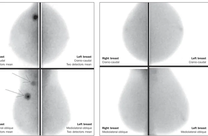
Figure 3. MBI study of a 59-year-old woman with dense breasts whose mam-mogram was inconclusive (BI-RADS 0). Strongly intense tracer uptake is ob-served in the upper lateral quadrant of her right breast with intense uptake by the ipsilateral axillary lymph node. Biopsy demonstrated the presence of an invasive ductal carcinoma with lymph node metastasis.
Right breast Cranio-caudal Two detectors mean
Left breast Cranio-caudal Two detectors mean
Right breast Mediolateral-oblique Two detectors mean
Left breast Mediolateral-oblique Two detectors mean
dose), 2–7 times the radiation dose in a mammographic study (1–3 mSv). Studies have been developed with lower radiation doses (8 mCi and up to 4 mCi) in order to reach a final radiation dose similar to the dose at mammography(26).
In patients with suspicious mammo-graphic and sonomammo-graphic findings the method presented a global sensitivity of 91% (97% for lesions > 10 mm, 91% for lesions measuring between 5 and 10 mm, and 69% for lesions < 5 mm)(30). Addition-ally, further lesions were found in 10% of the cases(31). As the method is compared with MRI, there was 97% agreement, with MRI sensitivity of 98% and 94% for MBI(30). Such a difference is principally due to lesions measuring between 0.2 and 0.4 cm(30).
In the authors experience with the GE Discovery NM 750B system (GE Health-care; Haifa, Israel), with an acquisition pro-tocol of 10 minutes per view, contrast up-take was observed in ductal carcinomas in situ, invasive carcinomas, lobular
carcino-mas, phylloid tumors and fibroadenomas (Figures 3 and 4). At MRI studies of some women, the authors observed a similar sen-sitivity for lesions > 0.5 cm.
DISCUSSION
Novel vascular/functional evaluation techniques for breast lesions have recently emerged. Their best clinical indications are still to be established but, theoretically they should be similar to the MRI clinical indi-cations.
At screening, such techniques might detect occult lesions in dense breasts, clear-ing up dubious findclear-ings (asymmetry or fo-cal distortions, asymmetry seen on a single view); in the diagnosis, they would reduce the number of unnecessary biopsies; in the treatment planning, they would be useful in the evaluation of the local disease extent and in the detection of other foci; in both chemo and radiotherapy treatment follow-up, they would allow an evaluation of the treatment response.
Figure 4. 55-year-old women submitted to conservative surgery in her right breast. At the MBI study, reduction in volume of the right breast is observed, but no suspicious tracer uptake is present, suggesting absence of residual or recurrent lesion.
Right breast Cranio-caudal
Left breast Cranio-caudal
Right breast Mediolateral-oblique
Contrast-enhanced mammography, as well as MRI, evaluates the morphology and vascularization of lesions. The major ad-vantages in relation to MRI include a per-fect correlation with mammographic im-ages, lower cost and shorter acquisition time. Disadvantages include the current unfeasibility of performing biopsies and marking of enhancement areas, besides possible adverse effects of iodinated con-trast agents.
As compared with MRI, molecular breast imaging presents the following ad-vantages: positioning analogous to mam-mography, facilitating less complex com-parisons and interpretations, and lower cost. As compared with mammography, molecular breast imaging applies lower breast compression force and its sensitiv-ity does not depend on the breast denssensitiv-ity. Disadvantages include longer acquisition time, a still high total radiation dose, and unfeasibility of morphological analysis.
The false-positive results described in the literature and observed in the authors experience are similar to those at MRI, namely, fibroadenoma, adenosis, fibro-cystic changes, papilloma, phylloid tumor fibrosis, inflammatory fat necrosis, radial scar, focal inflammation and pseudoangio-matous hyperplasia(20,21,26,29).
It is important to note that initial stud-ies, the present one included, do not exactly represent the actual population. The ob-servers involved in the images interpreta-tion are more careful in their analyses and, in several studies, the findings are more valued, since their populations present a high index of cancer.
As MRI is taken as a basis for a com-parative cost analysis, molecular breast im-aging costs about 70–80%, and contrast-enhanced mammography, 50% of the MRI cost.
CONCLUSION
Novel techniques for functional evalu-ation of breasts are currently available, pre-senting promising results and, in some cases, a performance similar to MRI. Their
indications might be the same as for MRI, with the advantage of lower cost. Further results should be expected in order to de-fine a procedure flowchart and thus mak-ing a good use of the advantages of each technology with minimum injury and risk for the population.
REFERENCES
1. Brasil. Ministério da Saúde. Instituto Nacional de Câncer. Câncer: incidência de câncer no Brasil. Rio de Janeiro, RJ: INCA; 2012.
2. Miranda CMNR, Santos CJJ, Maranhão CPM, et al. A tomografia computadorizada multislice é uma ferramenta importante para o estadiamento e seguimento do câncer de mama? Radiol Bras. 2012;45:105–12.
3. Brennan ME, Houssami N. Evaluation of the evi-dence on staging imaging for detection of asymp-tomatic distant metastases in newly diagnosed breast cancer. Breast. 2012;21:112–23. 4. Chala LF, Barros N. Avaliação das mamas com
métodos de imagem. Radiol Bras. 2007;40(1):iv– vi.
5. Boyd NF, Guo H, Martin LJ, et al. Mammographic density and the risk and detection of breast can-cer. N Engl J Med. 2007;356:227–36. 6. Buist DSM, Porter PL, Lehman C, et al. Factors
contributing to mammography failure in women aged 40-49 years. J Natl Cancer Inst. 2004;96: 1432–40.
7. Gwin JL, Eisenberg BL, Hoffman JP, et al. Inci-dence of gross and microscopic carcinoma in specimens from patients with breast cancer after re-excision lumpectomy. Ann Surg. 1993;218: 729–34.
8. Alvares BR, Freitas CHA, Jales RM, et al. Den-sidade mamográfica em mulheres menopausadas assintomáticas: correlação com dados clínicos e exames ultrassonográficos. Radiol Bras. 2012;45: 149–54.
9. Prado GLM, Guerra MTPM. Valor preditivo positivo das categorias 3, 4 e 5 do Breast Imag-ing ReportImag-ing and Data System (BI-RADS®). Radiol Bras. 2010;43:171–4.
10. Calas MJG, Gutfilen B, Pereira WCA. CAD e mamografia: por que usar esta ferramenta? Radiol Bras. 2012;45:46–52.
11. Calas MJG, Alvarenga AV, Gutfilen B, et al. Ava-liação de parâmetros morfométricos calculados a partir do contorno de lesões de mama em ultras-sonografias na distinção das categorias do sistema BI-RADS. Radiol Bras. 2011;44:289–96. 12. Marques EF, Medeiros MLL, Souza JA, et al.
Indicações de ressonância magnética das mamas em um centro de referência em oncologia. Radiol Bras. 2011;44:363–6.
13. Pereira FPA, Martins G, Figueiredo E, et al. O uso da difusão por ressonância magnética na diferen-ciação das lesões mamárias benignas e malignas. Radiol Bras. 2009;42:283–8.
14. Kuhl C. The current status of breast MR imaging.
Part I. Choice of technique, image interpretation, diagnostic accuracy, and transfer to clinical prac-tice. Radiology. 2007;244:356–78.
15. Kuhl CK. Current status of breast MR imaging. Part 2. Clinical applications. Radiology. 2007; 244:672–91.
16. Yeh ED. Breast magnetic resonance imaging: cur-rent clinical indications. Obstet Gynecol Clin North Am. 2011;38:159–77, ix.
17. Mameri CTS. Impacto da ressonância magnética mamária no tratamento cirúrgico, abordagem axilar e terapêutica sistêmica do câncer de mama. Radiol Bras. 2008;41:422.
18. Dromain C, Balleyguier C, Adler G, et al. Con-trast-enhanced digital mammography. Eur J Radiol. 2009;69:34–42.
19. Helvie MA. Digital mammography imaging: breast tomosynthesis and advanced applications. Radiol Clin North Am. 2010;48:917–29. 20. Dromain C, Thibault F, Muller S, et al.
Dual-en-ergy contrast-enhanced digital mammography: initial clinical results. Eur Radiol. 2011;21:565– 74.
21. Lewin JM, Isaacs PK, Vance V, et al. Dual-energy contrast-enhanced digital subtraction mammog-raphy: feasibility. Radiology. 2003;229:261–8. 22. Jong RA, Yaffe MJ, Skarpathiotakis M, et al.
Con-trast-enhanced digital mammography: initial clini-cal experience. Radiology. 2003;228:842–50. 23. Diekmann F, Freyer M, Diekmann S, et al.
Evalu-ation of contrast-enhanced digital mammography. Eur J Radiol. 2011;78:112–21.
24. Khalkhali I, Vargas HI. The role of nuclear medi-cine in breast cancer detection: functional breast imaging. Radiol Clin North Am. 2001;39:1053– 68.
25. Majewski S, Curran E, Keppel C, et al. Optimi-zation of dedicated scintimammography proce-dure using detector prototypes and compressible phantoms. Nuclear Science Symposium Confer-ence Record, 2000 IEEE; 2000.
26. Rhodes DJ, Hruska CB, Phillips SW, et al. Dedi-cated dual-head gamma imaging for breast can-cer screening in women with mammographically dense breasts. Radiology. 2011;258:106–18. 27. Tafreshi NK, Kumar V, Morse DL, et al.
Molecu-lar and functional imaging of breast cancer. Can-cer Control. 2010;17:143–55.
28. Hruska CB, Rhodes DJ, Collins DA, et al. Evalu-ation of molecular breast imaging in women un-dergoing myocardial perfusion imaging with Tc-99m sestamibi. J Womens Health (Larchmt). 2012;21:730–8.
29. Kim BS. Usefulness of breast-specific gamma imaging as an adjunct modality in breast cancer patients with dense breast: a comparative study with MRI. Ann Nucl Med. 2012;26:131–7. 30. Hruska C, Boughey J, Phillips S, et al.
Molecu-lar breast imaging: a review of the Mayo Clinic experience. Am J Surg. 2008;196:470–6. 31. Rhodes DJ, O’Connor MK, Phillips SW, et al.