Fetal and placental Doppler velocimetry in hypertensive
pregnant women and perinatal outcomes according
to gestational age*
Dopplervelocimetria fetoplacentária em gestantes hipertensas e resultados perinatais segundo a idade gestacional
Pedro Pires1, Aníbal Eusébio Faúndes Latham2, Suellene Keylla de Magalhães Mabessone3, Ana de Fátima de Azevedo Ferreira3, Fabiana Gomes de Souza Rodrigues3, Janaina Souza Leon3, Juliana Limeira de Moura Ramos3
OBJECTIVE: To evaluate the pulsatility index of umbilical artery (UAPI) and middle cerebral artery (MCAPI), as well as the umbilical-cerebral pulsatility (UAPI/MCAPI) ratio in fetuses of hypertensive pregnant women and associated adverse perinatal outcomes. MATERIALS AND METHODS: The authors have analyzed UAPI, MCAPI and UAPI/MCAPI ratio in 289 fetuses of hypertensive women, correlating the results with the presence of adverse perinatal outcomes. Results were compared with and without adjustment for gestational age. RESULTS: Apgar score < 7 at the 5th minute was associated with altered outcomes after adjustment for gestational age. The risk for small-for-gestational-age infant increased three times after such adjustment, with statistical significance for all the Doppler parameters. The increase in risk for neonatal hypoxia after adjustment for gestational age was statistically significant for UAPI and UAPI/MCAPI ratio. No increase was observed in the risk for respiratory distress syndrome in the adjusted analysis. A three-time higher risk for perinatal mortality and altered UAPI with statistical significance was observed after adjustment. CONCLUSION: In fetuses of hypertensive pregnant women, UAPI demonstrated better correlations with perinatal outcomes than MCAPI and UAPI/MCAPI ratio. The risk for adverse gestational outcome should be evaluated taking the gestational age into consideration.
Keywords: Doppler velocimetry; Hypertension; Perinatal outcome.
OBJETIVO: Avaliar índices de pulsatilidade das artérias umbilical (IPAU) e cerebral média (IPACM) e relação do índice de pulsatilidade umbilico-cerebral (IPAU/IPACM) em fetos de gestantes hipertensas e presença de resultados perinatais adversos. MATERIAIS E MÉTODOS: Analisamos IPAU, IPACM e IPAU/IPACM de 289 fetos de gestantes hipertensas quanto à previsão dos resultados perinatais adversos. Os resultados foram comparados sem e com ajuste pela idade gestacional. RESULTADOS: O índice de Apgar < 7 no 5º minuto foi associado com resultados alterados após o ajuste por idade gestacional. O risco para recém-nascidos pequenos para a idade gestacional aumentou em três vezes após o ajuste, com significância estatística em todos os parâmetros do Doppler. Na síndrome da hipóxia neonatal o aumento do risco ajustado pela idade gestacional foi estatisticamente significante no IPAU e IPAU/IPACM. Não houve aumento no risco de síndrome do desconforto respiratório na análise ajustada. A mortalidade perinatal e o IPAU alterado apresentaram um risco três vezes maior e foram estatisticamente significantes após o ajuste. CONCLUSÃO. Em gestantes hipertensas, o IPAU apresentou melhor correlação com os resultados perinatais do que o IPACM ou relação IPAU/IPACM. O risco de resultados adversos deve considerar a idade gestacional.
Unitermos: Dopplervelocimetria; Hipertensão; Resultados perinatais.
Abstract
Resumo
* Study developed at Faculdade de Ciências Médicas da Uni-versidade de Pernambuco (UPE), Recife, PE, Brazil.
1. PhD, Coordinator for Unit of Fetal Medicine, Maternal-Neo-natal Department – Faculdade de Ciências Médicas da Univer-sidade de Pernambuco (UPE), Recife, PE, Brazil.
2. PhD, Professor, Voluntary Collaborator of Obstetrics at Uni-versidade Estadual de Campinas (Unicamp), Campinas, SP, Brazil. 3. Specialists in Ultrasonography and Tocogynecology, MDs, Obstetricians, Maternal-Neonatal Department – Faculdade de Ciências Médicas da Universidade de Pernambuco (UPE), Recife, PE, Brazil.
Mailing address: Dr. Pedro Pires Ferreira Neto. Avenida
Dou-occupation is even greater in the presence of maternal diseases that are known to cause placental insufficiency and intrauter-ine growth restriction (IUGR) as is the case, mainly, of arterial hypertension(1). This is
the clinical entity that is mostly associated with IUGR and placental insufficiency(2–7).
The Doppler technique allows the noninvasive study of the uterine and Pires P, Latham AEF, Mabessone SKM, Ferreira AFA, Rodrigues FGS, Leon JS, Ramos JLM. Fetal and placental Doppler velocimetry in hypertensive pregnant women and perinatal outcomes according to gestational age. Radiol Bras. 2010;43(3): 155–160.
INTRODUCTION
Currently, one of the main preoccupa-tions in obstetrics is to assure good vitality conditions for the fetus at risk. Such
pre-tor Malaquias, 195, ap. 1401, Graças. Recife, PE, Brazil, 52050-060. E-mail: [email protected]
fetoplacental circulation, allowing the early diagnosis of hypoxia states and the predic-tion of adverse perinatal outcomes(6,8-10).
The Doppler scan allows the analysis of the main vessels resistance index, with the most utilized ones being the umbilical ar-tery and the middle cerebral arar-tery(11–14). In
the compensation phase of hypoxia, the placental resistance increases and the um-bilical arteries resistance indices rise. Sub-sequently, a progressive reduction of the cerebral vascular resistance is observed, progressing to “centralization”. Such phe-nomenon precedes severe fetal involve-ment by 10 to 12 days, with fetal acidosis and higher perinatal morbimortality(6).
Alterations in the Doppler pattern of the umbilical artery, particularly absent or re-versed diastolic flow, are indicative of pla-cental dysfunction(15), with high risk of
fe-tal distress and, particularly, vulnerability to prematurity complications(16), which
de-termines a balance between fetal risk ver-sus neonatal risk in the identification of the appropriate moment for intervention(17–19).
For some authors, obstetric manage-ment is still predominantly based on the umbilical artery Doppler analysis(4,18,20–22),
although some studies point towards the inclusion of more than one vessel in the assessment of vitality of fetuses at risk(7,23).
It is important to highlight that severe dopplervelocimetric alterations indicative of intensive fetal monitoring or interruption occur in premature fetuses, particularly in cases of extreme prematurity(24).
With the purpose of clarifying the capa-bility of different Doppler indicators to pre-dict the risk of perinatal complications, the authors have simultaneously evaluated the risk of bad perinatal outcomes in the pres-ence of dopplervelocimetric changes in the umbilical artery, middle cerebral artery and in the umbilical-cerebral indices. At the same time, the authors have evaluated the occurrence of substantial changes in results after control according to gestational age at birth.
MATERIALS AND METHODS
The initial series comprised 497 hyper-tensive pregnant women in the period from January 2002 to August 2006. For the stud-ies of the fetoplacental circulation, a
Shi-madzu ultrasonography unit model SDU-2200 (Shimadzu; Kyoto, Japan) with a Doppler device with color blood flow map-ping and a 3.5 MHz convex transducer, was utilized. The window filter was set between 50 and 100 Hz. With the patient lying in semi-Fowler position and in the absence of body motion and fetal breathing move-ments, utilizing real-time images, the blood flow color mapping was initiated, thus obtaining the color mapping of the vessels to be studied, which were evaluated by means of the Doppler device, with adjust-ments of the sample volume for each ves-sel, in the absence of fetal movements.
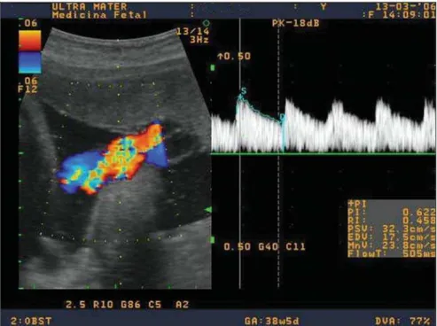
The Doppler scan of the umbilical artery was performed in open loop, close to the placental insertion and insonation angle always below 60°. The spectral analysis was considered appropriate in cases where at least three similar velocity waves were ob-served in the same spectrum (Figure 1). The analysis was considered as abnormal in cases where the diastole was absent or re-versed in the umbilical artery, or the pulsa-tility index was above the 95th percentile(25).
The middle cerebral artery was visual-ized from the circle of Willis and was insonated immediately after its origin in the internal carotid artery. The angle between the sound beam and the flow was captured
the closest possible to 0°, with the pulsa-tility index being measured (Figure 2). A middle cerebral artery pulsatility index (MCAPI) < 5th percentile and umbilical artery/cerebral artery (UAPI/MCAPI) ratio > 95th percentile for the gestational age were considered as abnormal(25).
Exclusion criteria were the following: cases with intervals between the last dopplervelocimetric study and the labor > 7 days, absence of data on perinatal out-comes at birth, presence of congenital de-fects or fetal or neonatal chromosomal ab-normalities, maternal disease leading to congenital infection, and gestational age at birth < 24 weeks. With these criteria, 208 patients were excluded, so the final sample included the remaining 289 pregnant women.
The software EPI-Info, version 1.0 was utilized for data organization. The cases were divided into three gestational age groups (< 33 weeks, 33 to 36 weeks and
≥ 37 weeks) and classified according to the presence or absence of each adverse peri-natal outcome as follows: Apgar score at the 5th minute < 7; newborn small for ges-tational age (SGA); occurrence of neona-tal hypoxic-ischemic syndrome (HIS); oc-currence of respiratory distress syndrome (RDS) and perinatal death.
A prospective analysis of risk of bad perinatal outcomes calculated by the rela-tive estimated risk (odds ratio) of the stud-ied outcomes was performed, in accor-dance with results of Doppler velocimetry performed up to seven days before the la-bor. Later, the same calculation was carried out, this time adjusted according to gesta-tional age.
The present study was developed in compliance with Resolution 196/1996 of Conselho Nacional de Saúde (Brazilian Council of Health), and the recommenda-tions of the Helsinki Declaration VI for research with humans, and was duly sub-mitted and approved by the Committee for Ethics in Research of Centro Integrado de Saúde Amaury de Medeiros of Univer-sidade de Pernambuco.
RESULTS
Sample characteristics
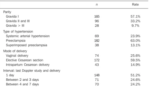
The patients’ ages ranged from 14 to 43 years (mean age, 26 years); with respect to parity, 57.1% of the women were primi-gravidas. Nearly two thirds of cases corre-sponded to preeclampsia. Vaginal deliver-ies comprised only 25.6% of the cases. The last Doppler scan was performed up to one
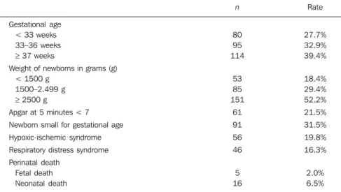
in 2%, and neonatal death in 6.5% of cases (Table 2).
Association between
the dopplervelocimetric results and perinatal outcomes
No association was observed between UAPI, MCAPI and of the UAPI/MCAPI ratio and Apgar score < 7 at the 5th minute before the adjustment for gestational age. After the adjustment, the risk of Apgar < 7 at the 5th minute was more than two times and almost twice higher in fetuses in whom the UAPI and MCAPI, respectively, were altered. However, only the association with UAPI alteration reached statistical signifi-cance (Table 3).
The authors observed that alterations in UAPI, MCAPI and UAPI/MCAPI ratio increased from five to seven times the risk of the occurrence of SGA newborns in the analysis without adjustment and around three times in the adjusted analysis accord-ing to gestational age (Table 4). The in-crease in risk was statistically significant for all studied dopplervelocimetric param-eters.
Alterations in UAPI, MCAPI and in the UAPI/MCAPI ratio increased two to five times the risk of HIS before adjustment, and between one and one half and more than three and a half times after the adjust-ment according to gestational age. The in-crease in the adjusted risk was significant only for UAPI and the UAPI/MCAPI ratio (Table 5).
Figure 2. Doppler velocimetry of the cerebral artery visualized from the circle of Willis and insonated immediately after its origin in the internal carotid artery. The angle between the sound beam and the flow was captured as close as possible to 0°.
Table 1 Characteristics of the 289 pregnant women.
Rate
57.1% 33.2% 9.7%
23.9% 63.0% 13.1%
25.6% 59.5% 14.9%
51.2% 24.6% 24.2% n
165 96 28
69 182 38
74 172 43
148 71 70 Parity
Gravida I Gravida II and III Gravida > III
Type of hypertension
Systemic arterial hypertension Preeclampsia
Superimposed preeclampsia
Mode of delivery Vaginal delivery Elective Cesarean section Intrapartum Cesarean delivery
Interval: last Doppler study and delivery 1 day
Between 2 and 3 days Between 4 and 7 days
day before delivery in 51.2% of the cases (Table 1).
The observed increase in the risk of RDS in cases with altered UAPI, MCAPI and UAPI/MCAPI ratio was between two and six times in the analysis without adjust-ment. However, after adjustment by gesta-tional age, no increase was observed in the risk of this neonatal complication in the cases with these alterations at Doppler (Table 6).
In spite of the fact that the gross risk of perinatal mortality having been between three to ten times greater in cases with al-terations in UAPI, MCAPI and UAPI/ MCAPI ratio in the analysis adjusted ac-cording to gestational age, only the altered UAPI presented a statistically significant risk almost three times higher for perina-tal death (Table 7).
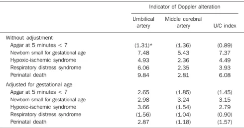
Table 8 shows the summarized results for a better visualization and comparison of the different indicators of Doppler alter-ations. It can be observed that the UAPI is the best indicator o risk of bad perinatal outcomes, particularly when the risk evalu-ation is adjusted by gestevalu-ational age of the neonate.
DISCUSSION
The results of the present study suggest that the evaluation of blood flow in the umbilical artery was the best indicator of risk of adverse perinatal outcome, as com-pared with the evaluation of the middle cerebral artery and with the IAPI/MCAPI ratio. Before the adjustment for gestational age, the three indicators seem to have a similar capability of predicting adverse perinatal outcomes. It is important to note that the increase in risk was always greater when one considers the Doppler results for the umbilical artery compared with the ones for the middle cerebral artery. It should be highlighted that the difference between these two indicators is more no-ticeable when one observes the risks of adverse perinatal outcomes after adjust-ment for gestational age. Various authors have not considered the factor of gesta-tional age in their studies(7,9,11,13,18,26).
In the present study, alterations in UAPI allowed the prediction of a higher risk (about three times higher) in four out of five indicators of the adverse perinatal out-comes studied. For RDS, the risk was 50%
Table 2 Perinatal outcomes.
n
80 95 114
53 85 151
61
91
56
46
5 16
Rate
27.7% 32.9% 39.4%
18.4% 29.4% 52.2%
21.5%
31.5%
19.8%
16.3%
2.0% 6.5% Gestational age
< 33 weeks 33–36 weeks ≥ 37 weeks
Weight of newborns in grams (g) < 1500 g
1500–2.499 g ≥ 2500 g
Apgar at 5 minutes < 7
Newborn small for gestational age
Hypoxic-ischemic syndrome
Respiratory distress syndrome
Perinatal death Fetal death Neonatal death
Table 3 Relative estimated risk (odds ratio) for Apgar score < 7 at 5 minutes, without adjustment and after adjustment for gestational age according to altered indicator at Doppler velocimetry.
Altered indicator
UAPI
MCAPI
UAPI/MCAPI
Gross odds ratio
1.31
1.36
0.89
CI 95%
(0.55–2.89)
(0.71–2.57)
(0.44–1.37)
Adjusted odds ratio
2.65
1.85
1.45
CI 95%
(1.03–6.85)
(0.96–3.58)
(0.66–5.99)
UAPI, umbilical artery pulsatility index; MCAPI, middle cerebral artery pulsatility index; IUAPI/MCAPI, umbilical/ cerebral pulsatility index.
Table 4 Relative estimated risk (odds ratio) for small gestational age, without adjustment and after adjustment for gestational age according to altered indicator at Doppler velocimetry.
Altered indicator
UAPI
MCAPI
UAPI/MCAPI
Gross odds ratio
7.48
5.43
7.37
CI 95%
(3.57–16.09)
(3.05–9.70)
(4.06–13.41)
Adjusted odds ratio
2.98
3.24
3.15
CI 95%
(1.36–6.56)
(1.78–5.89)
(1.66–5.99)
UAPI, umbilical artery pulsatility index; MCAPI, middle cerebral artery pulsatility index; IUAPI/MCAPI, umbilical/ cerebral pulsatility index.
Table 5 Relative estimated risk (odds ratio) for newborn hypoxic-ischemic syndrome, without adjustment and after adjustment for gestational age according to altered indicator at Doppler velocimetry.
Altered indicator
UAPI
MCAPI
UAPI/MCAPI
Gross odds ratio
4.93
2.36
4.49
CI 95%
(2.30–10.43)
(1.23–4.49)
(2.32–8.68)
Adjusted odds ratio
3.66
1.54
2.79
CI 95%
(1.77–7.57)
(0.80–2.99)
(1.34–5.82)
UAPI, umbilical artery pulsatility index; MCAPI, middle cerebral artery pulsatility index; IUAPI/MCAPI, umbilical/ cerebral pulsatility index.
Table 6 Relative estimated risk (odds ratio) for respiratory distress syndrome, without adjustment and after adjustment for gestational age according to altered indicator at Doppler velocimetry.
Altered indicator
UAPI
MCAPI
UAPI/MCAPI
Gross odds ratio
6.06
2.35
3.93
CI 95%
(2.69–13.39)
(1.15–4.74)
(1.92–8.03)
Adjusted odds ratio
1.56
1.04
0.90
CI 95%
(0.68–3.60)
(0.48–2.25)
(0.38–2.09)
higher, but it was not statistically signifi-cant. In contrast, MCAPI alterations al-lowed the prediction of a higher risk only for SGA, and alterations in the UAPI/ MCAPI ratio allowed predictions only for SGA and HIS.
For some authors, the study of more than one vessel contributes to improve the diagnosis of the fetal situation, evaluating the Doppler velocimetry capability to pre-dict perinatal outcomes, including only fetuses with confirmed IURG diagno-sis(3,6,7,12,27,28). In the present study,
hyper-tensive pregnant women were included, as it may lead to placental insufficiency po-tentially presenting a risk of fetal damage, without being necessarily present.
Another important information is re-lated to the evaluation of perinatal out-comes in cases with zero diastole or re-versed flow, which are severe alterations in the dopplervelocimetric pattern(9,10,13,26).
The present study considered not only ab-sent or reversed diastole as altered results of the umbilical artery, but also cases in which the pulsatility indices were above the 95th percentile.
Another factor considered as relevant in the present study was that approximately 75% of the cases had an interval of up to three days between the Doppler study and the labor, and in 50% the interval was one day. In the study developed by Baschat et al.(7) the mean interval between the last
Doppler study and delivery has not been informed, while in other studies the mean interval was > 7 days(9,29). Variations in the
Doppler study-delivery interval may ex-plain differences in results, considering that severe alterations in the Doppler pattern of the umbilical and cerebral arteries precede the worsening in the fetal status(6) by 10–
14 days.
Prematurity remains as the single most important determinant of neonatal compli-cations and mortality(9,27,28,30). In the
litera-ture, some authors have analyzed only pre-mature fetuses, particularly at gestational ages < 32 weeks, differently from the cases in the present study, in which different ges-tational ages were included, allowing a separate analysis of outcomes in extremely premature neonates, premature neonates and term neonates, and evaluate the impact
of prematurity on the perinatal out-comes(3,7,11,13).
The change in results after adjustment for gestational age was significant. It is known that in cases where placental insuf-ficiency and fetal hemodynamic alterations are diagnosed, the tendency is to interrupt the pregnancy to avoid intrauterine fetal death. Therefore, the association between alterations at Doppler and lower gestational ages is evident. Additionally, considering that the lower the gestational age is, the worst the perinatal outcomes are, it is un-derstood that gestational age is a confusing variable in the study of the association be-tween alterations at Doppler and perinatal outcomes.
The most typical example is RDS. Be-fore adjusting by gestational age, the Dop-pler alterations of the umbilical artery seemed to be associated with a six-time higher risk of RDS, but after the adjust-ment, this association was not significant. Among the studied adverse perinatal out-comes indicators, RDS is exactly the one more closely related to gestational age, more so than the situation of chronic intrau-terine hypoxemia. It is therefore clear that the increase in risk of RDS in cases with alterations at Doppler is mostly due to the premature interruption of pregnancy in-duced by the Doppler result than the hy-poxia condition of the fetus.
It would be very attractive if the assess-ment of the fetal status were more impor-tant than the gestational age effects on peri-natal outcomes, so that the moment of de-livery could be based on the tests of fetal assessment.
Therefore, the authors’ opinion is that the gestational age should not be ignored in the analysis of the data evaluated in the present study. This is the most important contribution of the present study, and hope-fully the publication of such results will contribute with future studies to confirm the relevance of the gestational age factor in their analyses to the performance of Doppler velocimetry for assessing fetal vitality.
REFERENCES
1. Sebire NJ. Umbilical artery Doppler revisited: pathophysiology of changes in intrauterine growth restriction revealed. Ultrasound Obstet Gynecol. 2003;21:419–22.
Table 7 Relative estimated risk (odds ratio) for perinatal death, without adjustment and after adjustment for gestational age according to altered indicator at Doppler velocimetry.
Altered indicator
UAPI
MCAPI
UAPI/MCAPI
Gross odds ratio
9.84
2.81
6.08
IC 95%
(3.80–25.67)
(1.15–6.98)
(2.37–16.08)
Adjusted odds ratio
2.87
1.18
1.57
CI 95%
(1.10–7.45)
(0.48–2.94)
(0.56–4.35)
UAPI, umbilical artery pulsatility index; MCAPI, middle cerebral artery pulsatility index; IUAPI/MCAPI, umbilical/ cerebral pulsatility index.
Table 8 Relative estimated risk of adverse perinatal outcomes according to altered indicator at Doppler velocimetry.
Indicator of Doppler alteration
Umbilical artery
(1.31)* 7.48 4.93 6.06 9.84
2.65 2.98 3.66 (1.56)
2.87
Middle cerebral artery
(1.36) 5.43 2.36 2.35 2.81
(1.85) 3.24 (1.54) (1.04) (1.18)
U/C index
(0.89) 7.37 4.49 3.93 6.08
(1.45) 3.15 2.79 (0.90) (1.57) Without adjustment
Apgar at 5 minutes < 7 Newborn small for gestational age Hypoxic-ischemic syndrome Respiratory distress syndrome Perinatal death
Adjusted for gestational age Apgar at 5 minutes < 7 Newborn small for gestational age Hypoxic-ischemic syndrome Respiratory distress syndrome Perinatal death
2. Campbell S, Pearce JM, Hackett G, et al. Quali-tative assessment of uteroplacental blood flow: early screening for high-risk pregnancies. Obstet Gynecol. 1986;68:649–53.
3. Ozcan T, Sbracia M, d’Ancona RL, et al. Arterial and venous Doppler velocimetry in the severely growth-restricted fetus and associations with ad-verse perinatal outcome. Ultrasound Obstet Gynecol. 1998;12:39–44.
4. Nomura RMY, Francisco RPV, Sakamoto K, et al. Centralização da circulação fetal em gestações de alto risco: avaliação da vitalidade fetal e resulta-dos perinatais. Rev Bras Ginecol Obstet. 2001; 23:137–43.
5. Sebire NJ, Goldin RD, Regan L. Histomorpho-logical evidence for chronic vasoconstriction of placental stem vessels in pregnancies with intrau-terine growth restriction and abnormal umbilical artery Doppler velocimetry indices. J Pathol. 2001;195:19A.
6. Baschat AA, Gembruch U, Weiner CP, et al. Qualitative venous Doppler waveform analysis improves prediction of critical perinatal outcomes in premature growth-restricted fetuses. Ultra-sound Obstet Gynecol. 2003;22:240–5. 7. Baschat AA, Galan HL, Bhide A, et al. Doppler
and biophysical assessment in growth restricted fetuses: distribution of test results. Ultrasound Obstet Gynecol. 2006;27:41–7.
8. Soothill PW, Ajayi RA, Campbell S, et al. Rela-tionship between fetal acidemia at cordocentesis and subsequent neurodevelopment. Ultrasound Obstet Gynecol. 1992;2:80–3.
9. Bilardo CM, Wolf H, Stigter RH, et al. Relation-ship between monitoring parameters and perina-tal outcome in severe, early intrauterine growth restriction. Ultrasound Obstet Gynecol. 2004;23: 119–25.
10. Hartung J, Kalache KD, Heyna C, et al. Outcome of 60 neonates who had ARED flow prenatally compared with a matched control group of appro-priate-for-gestational age preterm neonates. Ul-trasound Obstet Gynecol. 2005;25:566–72. 11. Hecher K, Bilardo CM, Stigter RH, et al.
Moni-toring of fetuses with intrauterine growth restric-tion: a longitudinal study. Ultrasound Obstet Gynecol. 2001;18:564–70.
12. Harman CR, Baschat AA, Gembruch U. Venous Doppler in IUGR. Which vessel? Which param-eter? Am J Obstet Gynecol. 2001;185:53.
13. Ferrazzi E, Bozzo M, Rigano S, et al. Temporal sequence of abnormal Doppler changes in the peripheral and central circulatory systems of the severely growth-restricted fetus. Ultrasound Obstet Gynecol. 2002;19:140–6.
14. Schreuder AM, McDonnell M, Gaffney G, et al. Outcome at school age following antenatal detec-tion of absent or reversed end diastolic flow ve-locity in the umbilical artery. Arch Dis Child Fe-tal NeonaFe-tal Ed. 2002;86:F108–14.
15. Baschat AA, Harman CR. Antenatal assessment of the growth restricted fetus. Curr Opin Obstet Gynecol. 2001;13:161–8.
16. Bernstein IM, Horbar JD, Badger GJ, et al. Mor-bidity and mortality among very-low-birth-weight neonates with intrauterine growth restriction. The Vermont Oxford Network. Am J Obstet Gynecol. 2000;182(1 Pt 1):198–206.
17. Zeitlin J, Ancel PY, Saurel-Cubizolles MJ, et al. The relationship between intrauterine growth re-striction and preterm delivery: an empirical ap-proach using data from a European case-control study. BJOG. 2000;107:750–8.
18. Yamamoto RM, Francisco RPV, Miyadahira S, et al. Fatores prognósticos para o óbito perinatal em gestações com diástole zero ou reversa na dop-plervelocimetria das artérias umbilicais. Rev Bras Ginecol Obstet. 2000;22:353–63.
19. GRIT Study Group. A randomised trial of timed delivery for the compromised preterm fetus: short term outcomes and Bayesian interpretation. BJOG. 2003;110:27–32.
20. Francisco RPV, Nomura RMY, Miyadahira S, et al. Diástole zero ou reversa na dopplervelocime-tria das artérias umbilicais. Rev Assoc Med Bras. 2001;47:30–6.
21. Andrade JQ, Miyadahira S, Nomura RMY, et al. Dopplervelocimetria dos compartimentos arterial
e venoso da circulação fetal e umbilical em ges-tação de alto-risco: análise dos resultados perina-tais. Rev Bras Ginecol Obstet. 2002;24:153–60. 22. Romero R, Kalache KD, Kadar N. Timing the delivery of the preterm severely growth- restricted fetus: venous Doppler, cardiotocography or the biophysical profile? Ultrasound Obstet Ginecol. 2002;19:118–21.
23. Pires P. Aplicações da dopplervelocimetria em gestação de alto risco. In: Pires P. Doppler no 2º e 3º trimestres da gestação & hemodinâmica feto-placentária. 1ª ed. Recife: Edupe; 2006. p. 67–82.
24. Pires P, Faundes A. Dopplervelocimetria na ava-liação hemodinâmica materno-fetal. Femina. 2007;35:383–90.
25. Arduini D, Rizzo G. Normal values of pulsatility index from fetal vessels: a cross-sectional study on 1556 healthy fetuses. J Perinat Med. 1990;18: 165–72.
26. Schwarze A, Gembruch U, Krapp M, et al. Quali-tative venous Doppler flow waveform analysis in preterm intrauterine growth-restricted fetuses with ARED flow in the umbilical artery – corre-lation with short-term outcome. Ultrasound Obstet Gynecol. 2005;25:573–9.
27. Baschat AA, Gembruch U, Reiss I, et al. Relation-ship between arterial and venous Doppler and perinatal outcome in fetal growth restriction. Ul-trasound Obstet Gynecol. 2000;16:407–13. 28. Müller T, Nanan R, Rehn M, et al. Arterial and
ductus venosus Doppler in fetuses with absent or reverse end-diastolic flow in the umbilical artery: correlation with short-term perinatal outcome. Acta Obstet Gynecol Scand. 2002;22:786–91.
29. Meyberg GC, Solomayer EF, Grischke EM, et al. Does the measurement of four fetal arteries pro-vide more information than the measurement of just two arteries in prenatal Doppler sonography? Ultrasound Obstet Gynecol. 1999;13:407–14. 30. Zelop CM, Richardson DK, Heffner LJ.