Brachyuran crabs are among the most important groups of crustaceans in coastal ecosystems, and one of the most abun-dant macroinvertebrate groups that inhabit tropical mangroves and tidal flats (CLARK & PAULA 2003). The taxonomic classifica-tion of brachyuran crabs is still mostly based on the morpho-logical characteristics of the adults. However, it has been hypothesized that adults share many convergent adaptations to their specific benthic environment (FLORES et al. 2003, CLARK 2009). A valuable, but frequently overlooked alternative to adult characters is larval morphology (CLARK 2009). Taxonomically useful characters found in larvae are believed to be less prob-lematic, because immature forms inhabit a planktonic envi-ronment with relatively uniform characteristics (RICE 1980). Phylogenetic studies on brachyuran crabs show that larval morphology is more congruent with molecular data than with adult morphology (HULTGREN et al. 2009).
Larval morphology can be used in ecological studies to evaluate species diversity and reproductive period (KORNIENKO & KORN 2009), population estimates, spatial and temporal distri-bution, dispersal, recruitment and other ecological aspects (AN-GER et al. 1995, QUEIROGA et al. 1994, CLARK & PAULA 2003). Such studies depend on the availability of taxonomic identification tools such as identification keys (HART 1971, MARTIN 1984, PAULA 1996, BÁEZ 1997, FRANSOZO et al. 1998, PESSANI et al. 1998, SCHUBART & CUESTA 1998, POHLE et al. 1999, ANOSOV 2000, GONZÁLEZ-GORDILLO et al. 2000, KO & YANG 2003, DOS SANTOS & GONZÁLEZ-GORDILLO 2004, RICE & TSUKIMURA 2007, LEE & KO 2008, GONZALES et al. 2009, KORNIENKO & KORN 2009, KORN & KORNIENKO 2010, VIEIRA & CALAZANS 2010). Alternatively, and to get the most up to date knowledge,
one would have to consult the publications where larvae of in-dividual species are described. However, with many crab larvae still unknown, such resources are presently limited.
Larval morphology may also prove useful in phyloge-netic studies within and among taxonomic groups (RICE 1980, 1983, CLARK & WEBBER 1991, MARQUES & POHLE 1998, 2003, SANTANA et al. 2003, 2004a, b, ANGER 2006, KORNIENKO & KORN 2009) help-ing us to understand species evolution (BÁEZ 1997).
According to CLARK et al. (1998), detailed, standardized descriptions of larvae obtained in the laboratory are necessary in many kinds of studies. As an example, the patterns of setae on the appendages can be used in systematic analyses because they are very conservative (FLORES et al. 2003, VIEIRA & CALAZANS 2010).
Studies based on larval morphology facilitate direct com-parison and help to discern consistent morphological patterns among taxa. However, some old descriptions need to be re-vised, as they sometimes lack the necessary details (SCHUBART & CUESTA 1998). The aim of the present study is to investigate the larval diversity of crustaceans that inhabit an estuary of the Amazon region. We describe the morphology of the first larval stage of the following species: P. americanus Saussure, 1857, Eurytium limosum (Say, 1818), Sesarma curacaoense De Man, 1892, S. rectum Randall, 1840, Armases rubripes (Rathbun, 1897), Aratus pisonii (H. Milne Edwards, 1837), Ocypode quadrata (Fab-ricius, 1787), Uca rapax (Smith, 1870), U. maracoani (Latreille, 1802), U. thayeri Rathbun, 1900, Ucides cordatus (Linnaeus, 1763) and Pachygrapsus gracilis (Saussure, 1858), and provide an iden-tification key for them.
Comparative morphology of the first zoea of twelve brachyuran species
(Crustacea: Decapoda) from the Amazon region
Adelson S. de Souza
1, Rauquírio M. da Costa
2& Fernando A. Abrunhosa
11 Laboratorio de Carcinologia, Universidade Federal do Pará. Alameda Leandro Ribeiro, Aldeia, 68600-000 Bragança, PA,
Brazil. E-mail: adelsonssouza@gmail.com; raucosta@ufpa.br; faraujo@ufpa.br
2 Laboratório de Plâncton e Cultivo de Microalga, Universidade Federal do Pará. Alameda Leandro Ribeiro, Aldeia,
68600-000 Bragança, PA, Brazil.
ABSTRACT. The laboratory-hatched first zoeal stage of twelve brachyuran species collected in the estuarine area of the Caeté River in the Amazonian region are described and illustrated in the present study: P. americanus Saussure, 1857, Eurytium limosum (Say, 1818), Sesarma curacaoense De Man, 1892, S. rectum Randall, 1840, Armases rubripes (Rathbun, 1897), Aratus pisonii (H. Milne Edwards, 1837), Ocypode quadrata (Fabricius, 1787), Uca rapax (Smith, 1870), U. maracoani (Latreille, 1802), U. thayeri Rathbun, 1900, Ucides cordatus (Linnaeus, 1763) and Pachygrapsus gracilis (Saussure, 1858). Through intraspecific comparisons of the respective larval stage, an identification key was generated and provided. Most of the studied species presented morphological differences (e.g. type and presence or absence of setae) when compared to the same species previously described in the literature.
MATERIAL AND METHODS
Field-collecting was conducted in different localities along the mangrove estuary of the Caeté River (Pará, Brazil). Oviger-ous females were captured manually and later taken to the labo-ratory. Next, two females of each species were carefully washed and identified according to RODRIGUEZ (1980) and MELO (1996). Species and their locations, as well as their time of hatching, are shown in Table I.
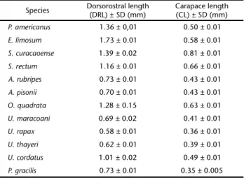
Morphometric data and illustrations are based on about 10 larvae of each species. Carapace length (CL) was obtained by measuring from the base of the rostral spine to the poste-rior margin of the carapace; dorsorostral length (DRL) was ob-tained by measuring from the tip of the rostral spine to the tip of the dorsal spine. Mean values and standard deviation were calculated for each species (Table II).
Table I. Collecting locality and hatching date of species obtained at the Caeté River estuary, Pará, Brazil.
Species Collectinglocality Hatchingdate
Panopeidae
Panopeus americanus Furo Grande (00º50’421”S, 046º38’398”W)
Jan. 2011
Eurytium limosum Ilha de Canela (00°46’571”S, 046°43’524”W)
Aug. 2010
Sesarmidae
Sesarma curacaoense Furo da Ostra (00º53’494”S, 046º39’387”W)
Aug. 2010
S. rectum Furo Grande
(00º50’421”S, 046º38’398”W)
Jan. 2011
Armases rubripes Furo da Ostra (00º53’494”S, 046º39’387”W)
Jan. 2011
Aratus pisonii Furo do Meio (00º52’470”S, 046º39’008”W)
Feb. 2010
Ocypodidae
Ocypode quadrata Praia de Ajuruteua (00º49’751”S, 46º36’248”W)
Jul. 2010
Uca maracoani Ilha de Canela (00°46’571”S, 046°43’524”W)
Apr. 2010
U. rapax Furo do Café
(00º50’752”S, 046º38’892”W)
Mar. 2010
U. thayeri Canal de Chavascal (00º48’963”S, 46º36’974”W)
Apr. 2010
Ucididae
Ucides cordatus Furo da Ostra (00º53’494”S, 046º39’387”W)
Mar. 2009
Grapsidae
Pachygrapsus gracilis Furo do Taici (00º58’138”S, 046º44’264”W)
Dec. 2010
The females were isolated and stored in 5 L-tanks con-taining seawater (salinity = 30) with constant aeration, and subjected to an artificial photoperiod of 12/12 hours until the larvae hatched.
After hatching, individuals of the first larval stage display-ing active swimmdisplay-ing were preserved in glycerine + 70% ethanol (1:1). Larvae were dissected with fine needles, measured, and then illustrated using a Coleman microscope equipped with cam-era lucida and a Zeiss Axioskop 40 compound microscope, both with a micrometer eyepiece. For improved visualization some structures were stained using methylene blue (0.5%).
Table II. Measurements of zoea I of the species described in this study.
Species Dorsorostral length(DRL) ± SD (mm) Carapace length(CL) ± SD (mm)
P. americanus 1.36 ± 0,01 0.50 ± 0.01
E. limosum 1.73 ± 0.01 0.58 ± 0.01
S. curacaoense 1.39 ± 0.02 0.81 ± 0.01
S. rectum 1.16 ± 0.01 0.66 ± 0.01
A. rubripes 0.73 ± 0.01 0.43 ± 0.01
A. pisonii 0.70 ± 0.01 0.43 ± 0.01
O. quadrata 1.28 ± 0.15 0.63 ± 0.01
U. maracoani 0.69 ± 0.02 0.41 ± 0.01
U. rapax 0.58 ± 0.01 0.36 ± 0.01
U. thayeri 0.62 ± 0.01 0.39 ± 0.01
U. cordatus 1.01 ± 0.02 0.49 ± 0.01
P. gracilis 0.73 ± 0.01 0.35 ± 0.005
The illustrations and morphological characteristics were used to construct a dichotomous key. Characters in the key are based mainly on external characters that can be viewed with-out dissection. The terminology used in the descriptions fol-low FACTOR (1978), POHLE & TELFORD (1981), CLARK et al. (1998), RIEGER & SANTOS (2001), GARM (2004), HORN & BUCKUP (2004). Samples of larvae and females were deposited in the Museum Emilio Goeldi (Pará, Brazil) under the following catalog num-bers: P. americanus 1108, E. limosum 1109, S. curacaoense 1110, S. rectum 1111, A. rubripes 1112, A. pisonii 1113, O. quadrata 1114, U. maracoani 1115, U. rapax 1117, U. thayeri 1116, U. cordatus 1118 and P. gracilis 1119.
RESULTS
Panopeidae
Panopeus americanus
Saussure, 1857
developed armed distally with rows of spines. Exopod minute, with 1 terminal simple seta. Mandible. Symmetric, palp ab-sent, incisor process with a prominent tooth associated with about seven small and two teeth arranged separately on the inner margin. Molar process with a large tooth in the base and circular regions provided of small and irregular teeth. Maxillule
(Fig. 4). Coxal endite with 6 plumodenticulate setae (5 terminal and 1 lateral subterminal) and 1 simple subterminal lateral seta. Basial endite with 2 cuspidate and 3 plumodenticulate (1 sub-terminal) setae. Endopod 2-segmented, with 1 sparsely plumose seta in proximal segment and 6 (2 subterminal) sparsely plu-mose setae in distal segment. Maxilla (Fig. 5). Coxal endite bi-lobed with 4 (2 subterminal) + 4 (2 subterminal) plumose setae. Basial endite bilobed with 5 (1 plumodenticulate and 2 subter-minal plumose) setae + 4 (1 subtersubter-minal) plumose setae. Endopod bilobed with 3 (1 subterminal) + 5 sparsely plumose setae. Scaphognathite with 4 marginal densely plumose setae and a long posterior process with microtrichias. First maxil-liped (Fig. 6). Basis with 2, 2, 3, 3 sparsely plumose setae in the inner margin. Endopod 5-segmented with 3, 2, 1, 2 and 5 (1 subterminal simple seta + 4 terminal) sparsely plumose setae from proximal to distal segment. Exopod 2-segmented with 4 terminal long plumose natatory setae. Second maxilliped (Fig. 7). Basis with 1, 1, 1, 1 sparsely plumose setae in the inner mar-gin. Endopod 3-segmented with 1, 1 (denticulate), 5 (1 den-ticulate + 2 simple) sparsely plumose setae from proximal to distal segment. Exopod 2-segmented with 4 long plumose na-tatory terminal setae. Abdomen (Fig. 8). With 5 somites and telson. Somites 2 and 3 with a pair of dorsolateral spines. Somites 2-5 with a pair of small simple posterodorsal setae. Somites 3-5 with a pair of long posterolateral spines. Telson bifurcated, dor-sally curved with 2 small unequal lateral spines and 1 dorsal spine. Inner margin with 6 (3+3) plumodenticulate setae sepa-rated by a medial arch.
Eurytium limosum
(Say, 1818)
Carapace (Fig. 9). Globose, smooth, with a pair of posterodorsal simple seta. Small protuberance in the anterior and posterior regions. Dorsal spine present, long and distally curved. Rostral spine as long as antennal protopod. Lateral spines present, small, about 1/10 antennal length and projected perpendicularly. Eyes sessile. Antennule (Fig. 10). Uniramous, conical-shaped and unsegmented with 2 aesthetascs and 2 simple setae. Antenna (Fig. 11). Protopod well-developed, armed distally with rows of spines. Exopod small, with 1 ter-minal simple seta.
Mandible. Symmetric, palp absent, incisor process with a prominent distal tooth and two teeth arranged separately on the inner margin. Molar process with a large tooth in the base and circular regions provided of small and irregular teeth.
Maxillule (Fig. 12). Coxal endite with 3 plumodenticulate, 1 subterminal plumose and 2 subterminal (1 plumose + 1 simple) lateral setae. Basial endite with 2 cuspidate setae and 3
plumodenticulate (1 subterminal) setae. Endopod 2-segmented, with 1 sparsely plumose seta in the proximal segment and 6 (2 subterminal) sparsely plumose setae in the distal segment.
Maxilla (Fig. 13). Coxal endite bilobed with 4+ 4 (2 subtermi-nal, 1 similar to hamate) plumose setae. Basial endite bilobed with 5 (1 subterminal) + 4 (1 subterminal plumose + 3 plumodenticulate) setae. Endopod bilobed with 3 (1 subtermi-nal) + 5 (1 subtermisubtermi-nal) plumose setae. Scaphognathite with 4 marginal plumose setae and a long distal process with microtrichias. First maxilliped (Fig. 14). Basis with 2+2+3+3 sparsely plumose setae in the inner margin. Endopod 5-seg-mented with 2, 2, 1, 2, 5 (1 subterminal lateral simple setae + 4 terminal) sparsely plumose setae from proximal to distal seg-ment. Exopod 2-segmented with 4 terminallong plumose na-tatory setae. Second maxilliped (Fig. 15). Basis with 1+1+1+1 sparsely plumose setae in the inner margin. Endopod 3-seg-mented with 1, 1 (denticulate), 5 (1 denticulate + 2 simple) sparsely plumose setae from proximal to distal segment. Exopod 2-segmented with 4 terminal long plumose natatory setae.
Abdomen (Fig. 16). With 5 somites and telson. Somites 2 and 3 with a pair of dorsolateral spines. Somites 2-5 with a pair of small simple posterodorsal setae. Somites 3-5 with a pair of posterolateral spines. Telson bifurcated, dorsally curved with 2 (1 fine spine) unequal lateral and 1 dorsal spines. Inner margin with 6 (3+3) plumodenticulate setae separated by a medial arch.
Sersarmidae
Sesarma curacaoense
De Man, 1892
Carapace (Fig. 17). Globose, smooth, with a small pro-tuberance on anterior region, 1 pair of posterodorsal simple setae and 2 pairs of anterodorsal simple setae. Pereiopods present. Dorsal spine present and distally curved. Lateral spines absent. Rostral spine slightly smaller than antenna. Eyes sessile.
Antennule (Fig. 18). Uniramous conical-shaped and unseg-mented with 3 aesthetascs and 2 distal simple seta. Antenna
(Fig. 19). Protopod well-developed longer than rostral spine, armed with 2 rows of unequal spines. Exopod with 2 simple setae of different sizes terminally. Endopod present and extend-ing beyond the half of exopod. Mandible. Symmetric, palp absent, incisor process with a prominent tooth associated with about 7 small teeth. Molar process with prominent terminal dentiform. Maxillule (Fig. 20). Coxal endite with 5 setae (4 plumodenticulate + 1 subterminal plumose) and 2 plumose subterminal lateral setae. Basial endite with 2 cuspidate and 3 plumodenticulate (1 subterminal) setae. Endopod 2-segmented, with 1 simple seta on the proximal segment, distal segment with 1 simple medial and 4 terminal sparsely plumose setae.
setae. Scaphognathite with 4 marginal plumose setae and a long distal process with microtrichias. First maxilliped (Fig. 22). Basis with 2+2+3+3 sparsely plumose setae on the inner mar-gin. Endopod 5-segmented with 2,2,1,2 and 5 (4 sparsely plu-mose terminal setae + 1 subterminal lateral pluplu-mose seta) from proximal to distal segment. Exopod 2-segmented with 4 termi-nal long plumose natatory setae. Second maxilliped (Fig. 23). Basis with 1 (sparsely plumose)+ 1+1+1 simple setae on the inner margin. Exopod 3-segmented with 0,1 (denticulate), 5 (1 denticulate) simple setae from proximal to distal segment. Exopod 2-segmented with 4 long plumose natatory terminal setae. Abdomen (Fig. 24). With 5 somites and telson. First somite with 2 simple medial setae. Somites 2 and 3 with a pair of dorsolateral spines. Somites 2-5 with a pair of small simple posterodorsal setae. Somites 3-5 with a pair of posterolateral spines. Pleopods (under developing) present on somites 2-5. Telson bifurcated, with inner margin with 6 (3+3)
plumodenti-culate setae separated by a medial arch. Each furca with 2 long rows of minute spines (Fig. 25).
Sesarma rectum
Randall, 1840
Carapace (Fig. 26). Globose, smooth with 2 pairs anterodorsal and 1 pair of posterolateral setae. Dorsal spine present and curved. Rostral spine only slightly longer than an-tenna.Mediolateral inferior region with a small protuberance. Lateral spines absent. Eyes sessile. Antennule (Fig. 27). Uniramous, conical-shaped and unsegmented with 3 aesthetascs and 2 unequal simple setae. Antenna (Fig. 28). Protopod well-developed, armed with 2 rows of unequal spines. Endopod present, longer than half of exopod. Exopod reaching half dis-tance of the protopod, bearing 2 unequal simple setae. Man-dible. Symmetric, palp absent, incisor process with a prominent tooth associated with five small and two teeth arranged sepa-Figures 1-16. (1-8) Panopeus americanus: lateral view (1); antennule (2); antenna (3); maxillule (4); maxilla (5); first maxilliped (6); second maxilliped (7); abdomen and telson (8). (9-16) Eurytium limosum: lateral view (9); antennule (10); antenna (11); maxillule (12); maxilla (13); first maxilliped (14); second maxilliped (15); abdomen and telson (16). Scale bars: 1, 9, 16 = 0.13 mm; 2, 6, 7 = 0.08 mm; 10 = 0.06 mm; 3,8, 11,14,15 = 0.1 mm; 4, 5, 12,13 = 0.03 mm.
1
2 3
5
4
7
8
6
13
9
10
11
12
14
rately on the inner margin. Molar process with a large tooth in the base and circular regions provided of small and irregular teeth.
Maxillule (Fig. 29). Coxal endite with 5 setae (3 plumodenticulate + 2 simple, 1 subterminal) and 1 plumose subterminal lateral seta. Basial endite with 2 cuspidate setae and 3 plumodenticulate (1 subterminal). Endopod 2-segmented, with 1 simple seta in the proximal segment, distal segment with 1 simple medial seta and 4 sparsely plumose terminal setae. Maxilla (Fig. 30). Coxal endite bilobed with 5 (3 subterminal) plumose setae + 4 (1 sub-terminal + 1 similar to hamate) plumose setae. Basial endite bi-lobed with 5 (1 plumodenticulate + 4 plumose, 1 subterminal) setae + 4 (2 plumodenticulate + 1 simple + 1 plumose subtermi-nal) setae. Endopod bilobed with 2 (1 subtermisubtermi-nal) + 3 sparsely plumose setae. Scaphognathite with 4 marginal plumose setae and a long distal process with microtrichias. First maxilliped
(Fig. 31). Basis with 2+2+3+3 sparsely plumose setae in the inner margin. Endopod 5-segmented with 2,2,1,2,5 (4 terminal + 1 plumose subterminal lateral) sparsely plumose setae from proxi-mal to distal segment. Exopod 2-segmented with 4 terminal long plumose natatory setae. Second maxilliped (Fig. 32). Basis with 1 (sparsely plumose)+1+1+1 simple setae in the inner margin. Endopod 3-segmented with 0,1(denticulate), 5 (4 simple + 1 den-ticulate) setae from proximal to distal segment. Exopod 2-seg-mented with 4 terminal long plumose natatory setae. Abdomen
(Fig. 33). With 5 somites and telson. The first somite with 1 simple seta. Somites 2 and 3 with a pair of dorsolateral spine. Somites 2-5 with a pair of small simple posterodorsal setae. Telson bifur-cated, with inner margin with 6 (3+3) plumodenticulate setae separated by a medial arch. Each furca with 2 rows of minute spines on the inner margin.
Figures 17-33. (17-25) Sesarma curacaoense: lateral view (17); antennule (18); antenna (19); maxillule (20); maxilla (21); first maxilliped (22); second maxilliped (23); abdomen and telson (24); furca (25); (26-33) Sesarma rectum: lateral view (26); antennule (27); antenna (28); maxillule (29); maxilla (30); first maxilliped (31); second maxilliped (32); abdomen and telson (33). Scale bars: 17, 33 = 0.13 mm; 18, 19, 27, 28 = 0.1 mm; 20, 21, 29, 30 = 0.03 mm; 22, 23, 31, 32 = 0.08 mm; 24, 26 = 0.2 mm; 25 = 0.05 mm.
17
18
21
22 24 25 23
20
26
27
28
30
31
33
32
Armases rubripes
(Rathbun, 1897)
Carapace (Fig. 34). Globose, smooth with 1 pair of anterodorsal simple setae and 1 pair of posterodorsal simple se-tae. Dorsal spine present and curved, with some minute spines randomly arranged on the anterior surface. Mediolateral infe-rior region with a small protuberance. Lateral spines absent. Rostral spine slightly shorter than antenna. Eyes sessile. Anten-nule (Fig. 35). Uniramous, conical-shaped, unsegmented with 3 aesthetascs and 2 unequal simple setae. Antenna (Fig. 36). Protopod well-developed, armed with 2 rows of unequal spines. Exopod about 1/3 protopod size, with 2 simple unequal termi-nal setae. Mandible. Symmetric, palp absent, incisor process with a prominent tooth associated with five small teeth. Molar pro-cess with a prominent tooth and circular region provided of small and irregular teeth. Maxillule (Fig. 37). Coxal endite with 5 (3 plumodenticulate + 2 simple, 1 subterminal) setae and 1 plu-mose subterminal lateral seta. Basial endite with 2 cuspidate and 3 plumodenticulate (1 subterminal) setae. Endopod 2-segmented, with 1 simple seta in the proximal segment, distal segment bear-ing 1 simple medial seta and 4 sparsely plumose terminal setae. Maxilla (Fig. 38). Coxal endite bilobed with 5 (3 subtermi-nal) plumose setae + 4 (2 subterminal + 1 similar to hamate) plumose setae. Basial endite bilobed with 5 (1 subterminal) plu-mose setae + 4 (1 subterminal) pluplu-mose setae. Endopod bilobed with 2 (1 subterminal) + 3 sparsely plumose setae. Scaphognathite with 4 marginal plumose setae and a long distal process with microtrichias. First maxilliped (Fig. 39). Basis with 2+2+3+3 sparsely plumose setae in the inner margin. Endopod 5-seg-mented with 2,2,1,2,5 (4 terminal + 1 lateral subterminal) sparsely plumose setae from proximal to distal segment. Exopod 2-seg-mented with 4 long plumose natatory terminal setae. Second maxilliped (Fig. 40). Basis with 1 (sparsely plumose) + 1+1+1
simple setae in the inner margin. Endopod 3-segmented with 0,1(denticulate), 6 (5 simple + 1 denticulate) setae from proxi-mal to distal segment. Exopod 2-segmented with 4 long plu-mose natatory terminal setae. Abdomen (Fig. 41). With 5 somites and telson. Somites 2 and 3 with a pair of dorsolateral spines. Somites 2-5 with a pair of small simple posterodorsal setae. Somites 3-5 with a pair of small posterolateral spine. Telson bi-furcated with inner margin with 6 (3+3) plumodenticulate setae separated by a medial arch. Each furca with two rows of minute spines on the inner margin.
Aratus pisonii
(H. Milne Edwards, 1837)
Carapace (Fig. 42). Globose, smooth with 1 pair of posterodorsal simple setae and 2 pairs of anterodorsal simple setae. Dorsal spine present and curved, with some minute spines on the anterior surface. Mediolateral inferior region with a dis-tinct protuberance. Lateral spines absent. Rostral spine length equal to antennal protopod. Eyes sessile. Antennule (Fig. 43). Uniramous, conic-shaped and unsegmented with 3 aesthetascs and 2 simple setae of different sizes. Antenna (Fig. 44). Protopod
well-developed armed with 2 rows of unequal spines. Exopod with 2 unequal simple setae and 2-minute terminal spines. Man-dible. Symmetric, palp absent, incisor process with a prominent tooth associated with four small and two teeth arranged sepa-rately on the inner margin. Molar process with a large tooth in base and circular region provided of small and irregular teeth.
Maxillule (Fig. 45). Coxal endite with 5 (3 plumodenticu-late + 2 simple, 1 subterminal) setae and 1 plumose subtermi-nal lateral seta. Basial endite with 2 cuspidate and 3 plumodenticulate (1 subterminal) setae. Endopod 2-segmented with 1 simple seta on proximal segment, distal segment with 1 simple median seta and 4 sparsely plumose terminal setae. Max-illa (Fig. 46). Coxal endite bilobed with 5 (3 subterminal) plu-mose setae + 4 (1 subterminal + 1 similar to hamate) pluplu-mose setae. Basial endite bilobed with 5 (3 plumose, 1 subterminal + 1 plumodenticulate + 1 simple) setae + 4 (1 subterminal) plu-mose setae. Endopod bilobed with 2(1 subterminal) + 3 sparsely plumose setae. Scaphognathite with 4 marginal plumose setae and a long distal process with microtrichias. First maxilliped
(Fig. 47). Basis with 2+2+3+3 sparsely plumose setae in the in-ner margin. Endopod 5-segmented with 2,2,1,2,5 (4 terminal + 1 lateral subterminal) sparsely plumose setae from proximal to distal segment. Exopod 2-segmented with 4 long plumose na-tatory terminal setae. Second maxilliped (Fig. 48). Basis with 1 (sparsely plumose seta) + 1+1+1 simple setae in the inner margin. Endopod 3-segmented with 0,1 (denticulate), 6 (5 simple + 1 denticulate) setae from proximal to distal segment. Exopod 2-segmented with 4 long plumose natatory terminal setae. Abdomen (Fig. 49). With 5 somites and telson. Somites 2 and 3 with a pair of dorsolateral spines. Somites 2-5 with a pair of small simple posterodorsal setae. Somites 3-5 with a pair of short posterolateral spines. Telson bifurcated, inner margin with 6 (3+3) plumodenticulate setae (inner margin of the setae of the central pair lacking plume) separated by a me-dial arch. Each furca with 2-minute lateral spines and 2 rows of minute spines on the inner margin.
Ocypodidae
Ocypode quadrata
(Fabricius, 1787)
Carapace (Fig. 50). Globose, smooth with 1 pair of postero-dorsal simple setae. Anterior region with a small protuberance. Dorsal spine present and curved, bearing minute spines randomly arranged on anterior surface. Rostral spine about length twice of antennal size. Lateral spines present, long and curved downward.
Antennule (Fig. 51). Uniramous, conical-shaped and unseg-mented with 2 aesthetascs and 2 simple setae of different sizes.
Antenna (Fig. 52). Protopod well-developed with 2 rows of small spines. Exopod about 1/5 of protopod length, bearing 2 unequal simple setae and 2 minute terminal spines. Mandible.
circular regions provided of small and irregular teeth. Maxillule
(Fig. 53). Coxal endite with 4 (3 plumodenticulate + 1 simple) setae and 1 lateral subterminal plumodenticulate seta. Basial endite with 2 cuspidate and 3 plumodenticulate (1 subterminal) setae. Endopod 2-segmented, proximal segment lacking setae and distal segment with 4 sparsely plumose setae. Maxilla (Fig. 54). Coxal endite bilobed with 4 (3 subterminal) + 3 (1 similar to hamate) plumose setae. Basial endite bilobed with 5 (3 subter-minal) + 4 (1 subtersubter-minal) plumose setae. Endopod bilobed with 1 sparsely plumose + 2 (1 sparsely plumose + 1 simple) setae. Scaphognathite with 4 marginal plumose setae and a long distal process with microtrichias. First maxilliped (Fig. 55). Basis with 3+2+2+2 sparsely plumose setae in the inner margin. Endopod 5-segmented, with 2,2,1,2,5 (4 terminal + 1 lateral subterminal) sparsely plumose setae from proximal to distal segment. Exopod 2-segmented with 4 long plumose natatory terminal setae.
Sec-ond maxilliped (Fig. 56). Basis with 1+1+1+1 sparsely plumose setae in the inner margin. Endopod 3-segmented with 0,0,5 (1 denticulate, 4 sparsely plumose) setae from proximal to distal segment. Exopod unsegmented with 4 long plumose natatory terminal setae. Abdomen (Fig. 57). With 5 somites and telson. Somites 2 and 3 with a pair of dorsolateral spines. Somites 2-5 with a pair of small simple posterodorsal setae. Posterior margin of the 4th somite laterally enlarged. Telson bifurcated with inner
margin with 6 (3+3) plumodenticulate setae separated by a me-dial arch.
Uca maracoani
(Latreille, 1802)
Carapace (Fig. 58). Globose, smooth with 1 pair of posterodorsal simple setae. Anterior region with a small protu-berance. Dorsal spine present and curved, bearing minute spines randomly arranged on the anterior surface. Lateral spines Figures 34-49. (34-41) Armases rubripes: lateral view (34); antennule (35); antenna (36); maxillule (37); maxilla (38); first maxilliped (39); second maxilliped (40); abdomen and telson (41); (42-49) Aratus pisonii: lateral view (42); antennule (43); antenna (44); maxillule (45); maxilla (46); first maxilliped (47); second maxilliped (48); abdomen and telson (49). Scale bars: 34 = 0.08 mm; 35-38 = 0.03 mm; 39, 40, 47, 48 = 0.06 mm; 43-46 = 0.05 mm; 41, 42, 49 = 0.1 mm.
34
35
38
37
41 39
40 36
46
45 47
48
42 43
present, slightly curved downward. Rostral spines slightly curved. Eyes sessile. Antennule (Fig. 59). Uniramous, conical-shaped and unsegmented with 2 aesthetascs and 2 unequal simple setae. Antenna (Fig. 60). Protopod well-developed, with 2 rows of minutes spines. Exopod with 3 unequal simple setae.
Mandible. Symmetric, palp absent, incisor process with four prominent and two teeth arranged separately on the inner margin. Molar process with a large tooth in base and circular regions provided of small and irregular teeth. Maxillule (Fig. 61). Coxal endite with 4 (3 plumodenticulate + 1 subterminal simple) setae and 1 subterminal lateral plumose seta. Basial endite with 2 cuspidate and 3 plumodenticulate (2 subtermi-nal) setae. Endopod 2-segmented, proximal segment lacking setae, distal segment with 4 sparsely plumose setae. Maxilla
(Fig. 62). Coxal endite bilobed with 4 (3 subterminal) + 3 (1 subterminal + 1 similar to hamate) plumose setae. Basial endite with 5 (2 subterminal) + 4 (1 subterminal) plumose setae.
Endopod bilobed with 1 + 2 sparsely plumose setae. Scaphognathite with 4 marginal plumose setae and a long dis-tal process with microtrichias. First maxilliped (Fig. 63). Basis with 1+1(sparsely plumose)+1+4+2, simple setae in the inner margin. Endopod 5-segmented with 0 (2), 1 (2), 1, 2, 1 (5) se-tae, sparsely plumose, from proximal to distal segment. Exopod 2-segmented with 4 long plumose natatory terminal setae. Sec-ond maxilliped (Fig. 64). Basis with 1+1+1+1 simple setae in the inner margin. Endopod 3-segmented with 0,0,5 (4 simple
+ 1 denticulate) setae from proximal to distal segment. Exopod unsegmented with 4 long plumose natatory setae. Abdomen
(Fig. 65). With 5 somites and telson. Somites 2 and 3 with a pair of dorsolateral spines. Somites 2-5 with a pair of small simple posterodorsal setae. Telson bifurcated with inner mar-gin with 6(3+3) plumodenticulate setae separated by a medial arch. Each furca with 2-minute lateral spines and 2 rows of minute marginal spines.
Figures 50-65. (50-57) Ocypode quadrata: lateral view (50); antennule (51); antenna (52); maxillule (53); maxilla (54); first maxilliped (55); second maxilliped (56); abdomen and telson (57); (58-65) Uca maracoani: lateral view (58); antennule (59); antenna (60); maxillule (61); maxilla (62); first maxilliped (63); second maxilliped (64); abdomen and telson (65). Scale bars: 50, 57 = 0.13 mm; 51, 52, 58, 63, 64 = 0.08 mm; 53, 54, 59-62 = 0.3 mm; 55, 56, 65 = 0.1 mm.
57 52
50
54
53
55
56
65
61 63
64 59
60 62 58
Uca rapax
(Smith, 1870)
Carapace (Fig. 66). Globose, smooth, with 1 pair of posterodorsal simple setae. Dorsal spine present and strongly curved. Lateral spines absent. Rostral spine slightly curved, extending beyond antenna. Eyes sessile. Antennule (Fig. 67). Uniramous, conical-shaped and unsegmented with 2 aesthetascs and 2 simple unequal setae. Antenna (Fig. 68). Protopod well-developed with 2 rows of small equal-sized spines. Exopod about ¼ of protopod length, with 3 unequal simple setae. Mandible. Symmetric, palp absent, incisor pro-cess with a prominent tooth associated with three small teeth of unequal size and two teeth arranged separately on the inner margin. Molar process with a large tooth in base and circular regions provided of small and irregular teeth.
Maxillule (Fig. 69). Coxal endite with 4 (2 plumodenticu-late + 1 simple subterminal + 1 plumose) setae and 1 plumose
subterminal lateral seta. Basial endite with 2 cuspidate setae and 3 (2 subterminal) plumodenticulate setae. Endopod 2-seg-mented with proximal segment without setae and 4 sparsely plumose terminal setae on the distal segment. Maxilla (Fig. 70). Coxal endite bilobed with 4 (3 subterminal) + 3 (1 similar to hamate) plumose setae. Basial endite bilobed with 5 (1 sub-terminal) + 4 (1 subsub-terminal) plumose setae. Endopod bilobed with 1 + 2 sparsely plumose setae. Scaphognathite with 4 mar-ginal plumose setae and a long distal process with microtrichias.
First maxilliped (Fig. 71). Basis with 2+2+3+2 sparsely plu-mose setae in the inner margin. Endopod 5-segmented with 2,2,1,2,5 (1 simple lateral seta) sparsely plumose setae from proximal to distal segment. Exopod 2-segmented with 4 termi-nallong plumose natatory setae. Second maxilliped (Fig. 72). Basis with 1+1+1+1 sparsely plumose setae in the inner mar-gin. Endopod 3-segmented with 0,0,5 (2 simple + 2 sparsely
Figures 66-81. (66-73) Uca rapax: lateral view (66); antennule (67); antenna (68); maxillule (69); maxilla (70); first maxilliped (71); second maxilliped (72); abdomen and telson (73); (74-81) Uca thayeri: lateral view (74); antennule (75); antenna (76); maxillule (77); maxilla (78); first maxilliped (79); second maxilliped (80); abdomen and telson (81). Scale bars: 66, 73, 74, 79, 80 = 0.08 mm; 67, 68, 75-78 = 0.03 mm; 69, 70 = 0.025 mm; 71, 72 = 0.06 mm; 81 = 0.1 mm.
73 68
66
70
67
71 72 69
78
81
80
79 77
76 74
plumose + 1 denticulate) setae from proximal to distal segment. Exopod 2-segmented with 4 terminallong plumose natatory setae. Abdomen (Fig. 73). With 5 somites and telson. Somites 2 and 3 with a pair of dorsolateral spines. Somites 2-5 with a pair of small simple posterodorsal setae. Telson bifurcated with 1 minute lateral spine. Inner margin with 6(3+3) plumodenticu-late setae separated by a medial arch.
Uca thayeri
Rathbun, 1900
Carapace (Fig. 74). Globose, smooth with 1 pair of posterodorsal simple setae. Dorsal spine present and curved, bearing minute spines randomly arranged on the anterior sur-face. Lateral spines absent. Rostral spine slightly curved, ex-tending beyond antenna. Eyes sessile. Antennule (Fig. 75). Uniramous, conical-shaped and unsegmented with 2 aesthetascs and 2 simple setae. Antenna (Fig. 76). Protopod well-developed with 2 rows of small marginal spines. Exopod with 2 simple setae of different sizes and 2 small spines. Man-dible. Symmetric, palp absent, incisor process with a promi-nent tooth associated with two small teeth of unequal size and two teeth arranged separately on the inner margin. Molar cess with a large tooth in the base and circular regions pro-vided of small and irregular teeth. Maxillule (Fig. 77). Coxal endite with 4 apical setae (3 plumodenticulate + 1 simple sub-terminal) and 1 lateral plumose seta. Basial endite with 2 cus-pidate and 3 plumodenticulate (1 subterminal) setae. Endopod 2-segmented, proximal segment lacking setae, 4 sparsely plu-mose terminal setae on the distal segment. Maxilla (Fig. 78). Coxal endite bilobed with 4 setae (2 subterminal) + 3 setae (1 similar to hamate) sparsely plumose. Basial endite bilobed with 5 setae (2 subterminal) + 4 setae (1 subterminal) sparsely plu-mose. Endopod bilobed with 1+2 sparsely plumose setae. Scaphognathite with 4 marginal plumose setae and a long dis-tal process with microtrichias. First maxilliped (Fig. 79). Basis with 2+2+3+2 simple setae in the inner margin. Endopod 5-segmented with 0(2),0(2),0(1),2,4(5) sparsely plumose setae from proximal to distal segment. Exopod 2-segmented with 4 long plumose natatory terminal setae. Second maxilliped (Fig. 80). Basis with 1+1+1+1 simple setae in inner margin. Endopod 3-segmented with 0,0,5 (4 simple + 1 denticulate) simple setae from proximal to distal segment. Exopod 2-segmented with 4 long plumose natatory terminal setae. Abdomen (Fig. 81). With 5 somites and telson. Somites 2 and 3 with a pair of dorsolat-eral spines. Somites 2-5 with a pair of small simple posterodorsal setae. Telson bifurcated, with inner margin with 6 (3+3) plumodenticulate setae separated by a medial arch.
Ucididae
Ucides cordatus
(Linnaeus, 1763)
Carapace (Fig. 82). Globose, smooth with 1 pair of posterodorsal simple setae. Dorsal spine present, sloped poste-riorly. Lateral spines absent. Rostral spine slightly curved,
ex-tending beyond antenna. Eyes sessile. Antennule (Fig. 83). Uniramous, conical-shaped and unsegmented with 2 aesthetascs and 2 simple setae.
Antenna (Fig. 84). Protopod well-developed, bearing 2 rows of small spines equal-sized. Exopod with 2 simple setae of similar size. Mandible. Symmetric, palp absent, incisor pro-cess with a prominent tooth associated with three small teeth of unequal size and two teeth arranged separately on the inner margin. Molar process with a large tooth in base and circular regions provided of small and irregular teeth. Maxillule (Fig. 85). Coxal endite with 4 (3 plumodenticulate + 1 simple sub-terminal) setae and 1 plumodenticulate subterminal lateral seta. Basial endite with 2 cuspidate and 3 plumodenticulate (1 sub-terminal) setae. Endopod 2-segmented with proximal segment lacking setae and 4 sparsely plumose terminal setae in distal segment. Maxilla (Fig. 86). Coxal endite bilobed with 3 (1 sub-terminal) + 4 (2 subterminal, 1 similar to hamate) plumose se-tae. Basial endite bilobed with 5 (2 subterminal) + 4 (1 subterminal) sparsely plumose setae. Endopod bilobed with 1+2 sparsely plumose setae. Scaphognathite with 4 marginal plu-mose setae and a long distal process with microtrichias. First maxilliped (Fig. 87). Basis with 2+2+3+2 sparsely plumose se-tae in the inner margin. Endopod 5-segmented with 2,2,1,2,5 (1 simple lateral) sparsely plumose setae from proximal to dis-tal segment. Exopod 2-segmented with 4 terminal long plu-mose natatory setae. Second maxilliped (Fig. 88). Basis with 1+1+1+1 sparsely plumose setae in the inner margin. Endopod 3-segmented with 0,0,5 (1 denticulate + 2 sparsely plumose + 2 simple) setae from proximal to distal segment. Exopod 2-seg-mented with 4 terminal long plumose natatory setae. Abdo-men (Fig. 89). With 5 somites and telson. Somites 2 and 3 with a pair of dorsolateral spines. Somites 2-5 with short posterolat-eral spines and a pair of small simple posterodorsal setae. Tel-son bifurcated with a minute lateral spine. Inner margin with 6(3+3) plumodenticulate setae separated by a medial arch. Dis-tal inner margin with a serrated furca.
Grapsidae
Pachygrapsus gracilis
(Saussure, 1858)
circular regions provided of small and irregular teeth. Maxillule
(Fig. 93). Coxal endite with 5 (3 plumodenticulate + 1 sparsely plumose + 1 simple) setae and 1 plumose subterminal lateral seta. Basial endite with 2 cuspidate, 2 plumodenticulate and 1 simple subterminal setae. Endopod 2-segmented with 1 simple seta in proximal segment, while the distal segment showing 1 simple medial and 4 sparsely plumose terminal setae. Maxilla
(Fig. 94). Coxal endite bilobed with 5 (2subterminal) + 3 (2 subterminal) plumose setae. Basial endite bilobed, with 5 (sub-terminal) + 4 (1 sub(sub-terminal) plumose setae. Endopod bilobed with 2+2 sparsely plumose setae. Scaphognathite with 4 plu-mose marginal setae with a distal process little developed. First maxilliped (Fig. 95) Basis with 2 (1 sparsely plumose)+ 2+2+2, simple setae in the inner margin. Endopod 5-segmented with
1,2,1,2,5 (4 terminal +1 lateral subterminal) simple setae from proximal to distal segment. Exopod 2-segmented with 4 termi-nal long plumose natatory setae. Second maxilliped (Fig. 96). Basis with 1 (sparsely plumose)+1+1+1 simple setae on the in-ner margin. Endopod 3-segmented with 0,1 (denticulate), 5 (1 denticulate + 2 sparsely plumose) setae from proximal to distal segment. Exopod 2-segmented with 4 terminal long plumose natatory setae. Abdomen (Fig. 97). With 5 somites and telson. Somites 2 and 3 with a pair of dorsolateral spines. Posterior margin of the 4th somite laterally extended with 1 spine. Somites
2-5 with a pair of small posterolateral spines and a pair of small simple posterodorsal setae. Telson bifurcated with the inner margin provided of 6(3+3) plumodenticulate setae separated by a small arch, margins of thefurca serrated.
Figures 82-97. (82-89) Ucides cordatus: lateral view (82); antennule (83); antenna (84); maxillule (85); maxilla (86); first maxilliped (87); second maxilliped (88); abdomen and telson (89); (90-97) Pachygrapsus gracilis: lateral view (90); antennule (91); antenna (92); maxillule (93); maxilla (94); first maxilliped (95); second maxilliped (96); abdomen and telson (97). Scale bars: 82 = 0.13 mm; 83, 95, 96 = 0.06 mm; 84, 91, 92 = 0.05 mm; 85, 86, 94 = 0.03 mm; 95 = 0.025 mm; 87, 88, 90, 97 = 0.08 mm; 89 = 0.1 mm.
82
88 86
85 87
90
97 95
92
96
93
94 91
89 84
Key to identification of Zoea I of the described species
1a. Carapace with lateral spines (Figs 1, 9, 50, 58) ... 2 1b. Carapace lacking lateral spines (Figs 17, 26, 34, 42, 66, 74, 82, 90) ... 5 2a. Antenna with elongate protopod, distally armed with strong
spines in more than 2 rows (Figs 3, 11) ... 3 2b. Antenna with protopod not particularly elongate, and with
2 rows of spines only (Figs 19, 28, 36, 44, 52, 60, 68, 76 and 84) ... 4 3a. Antennule distally with 2 aesthetascs and 2 simple setae
(Figs 9 and 10) ...Eurytium limosum 3b. Antennule distally with 2 aesthetascs and 3 simple setae (Figs 1 and 2) ...Panopeus americanus 4a. Fourth abdominal somite with posterior margin expanded laterally (Figs 50 and 57) ...Ocypode quadrata 4b. Abdominal somites similar, without lateral expansion (Figs 58 and 65) ...Uca maracoani 5a. Abdomen lacking Pleopods (Figs 26, 34, 66, 74, 82, 90) . 6 5b. Abdomen with pleopods (Fig. 17) ...Sesarma curacaoense 6a. Fourth abdominal somite with posterior margin laterally expanded, bearing 1 robust spine (Figs 90 and 97) ...
...Pachygrapsus gracilis 6b. Abdominal somites similar, without lateral expansion and spine (Figs 33, 41, 49, 89) ... 7 7a. Antennal protopod with 2 rows of unequal-sized spines
arranged sparsely and relatively widely apart (Figs 28, 36, 44) ... 8 7b. Antennal protopod with 2 rows of minute equal-size spines,
arranged relatively close to each other (Figs 76, 68, 84) 10 8a. Antennal endopod present (Figs 26 and 28) ...
...Sesarma rectum 8b. Antennal endopod absent (Figs 35, 43, 68, 76, 84) ... 9 9a. Carapace with 2 pairs of simple anterodorsal simple setae between eyes; antennal exopod with 2 minute terminal spines (Figs 42 and 44) ...Aratus pisonii 9b. Carapace with 1 pair of simple anterodorsal setae between eyes; antennal exopod lacking spines (Figs 34 and 36) ....
...Armases rubripes (Fig. 34) 10a. Abdominal somites 3-5 with short posterolateral spines; telson inner margin of distal region with furcal ramous distinctly serrate (Figs 82 and 89) ...Ucides cordatus 10b. Abdominal somites 3-5 with rounded posterolateral knobs, furcal ramous not distinctly serrate using light microscopy (Figs 73 and 81) ... 11 11a. Carapace with dorsal spine posteriorly strongly curved, furcal ramous with 1 minute proximal lateral spine arranged (Figs 66 and 73) ...Uca rapax 11b. Carapace with dorsal spine posteriorly not strongly curved, bearing minute spines randomly arranged on the anterior surface; furcal ramous lacking proximal lateral spines (Fig. 74) ...Uca thayeri
DISCUSSION
The morphological characteristics of brachyuran larvae, especially in the early stages, are essential to systematic and phylogenetic studies (CLARKet al. 1998), because they help to establish character states as primitive or derived (RICE 1983).
According to RICE (1983), the evolutionary trends of Eubrachyuran larvae (zoea) indicate that more derived taxa show a reduction in spines, setae and segmentation, compared with the more primitive taxa. However, such trends, which are presumably associated with a more efficient exploration of the pelagic environment, may evolve independently. Characters of the larval stages, as well as characters of the adults, are liable to convergence (RIEGER 1998).
Nevertheless, the use of larval characteristics in system-atics has been widely accepted and applied by taxonomists (CUESTA & ANGER 2001). Furthermore, as more larvae are being described, morphological comparisons become more feasible (RIEGER 1998).
However, it is important to emphasize that it is difficult to differentiate among the larvae of some species, especially congeners, since their distinctions are based only on minor morphological differences. In such cases, the reliability of spe-cific descriptions may be feasible only when the specimens described are born in the laboratory. Descriptions of larvae collected in the field are often generic due to the difficulty in identifying larvae at the level of species (KORNIENKO & KORN 2009). One example of this problem are Hemigrapsus sanguineus, H. penicillatus and H. longitarsis, described by HWANG et al. (1993), HWANG & KIM (1995) and PARK & KO (2001), respectively. These species have almost identical initial stages, and can only be distinguished when they develop into more advanced stages.
the antennules, the type and quantity of setae on the maxillule, maxilla, as well as maxillipeds, among other characteristics (Table III). While CLARK et al. (1998) argue that taxonomists should be discouraged to establish relationships based on incomplete de-scriptions, the description of KURATA et al. (1981) has much in common with the present one, for instance in the abdomen, antenna, telson, and carapace spines. However, E. limosum is very similar to other species of Panopeidae, and larvae can only be distinguished from the second stage of the zoea on, which has the antenna without any spinous process.
A great similarity among larvae of species in the same genus has also been demonstrated for Sesarmidae (GUERAO et al. 2004, CUESTA et al. 2006b, GUERAO et al. 2007). Although the morphology of species of Aratus, Armases and Sesarma is quite similar in the first zoea, these species can be distinguished, in most cases, by minor differences in the pattern of their setae (SCHUBART & CUESTA 1998). Some species, such as S. curacaoense (ANGER et. al. 1995, SCHUBART & CUESTA 1998), are more clearly differentiated, mainly by the presence of pleopods on the ab-dominal somites and partially differentiated pereiopods (ob-served but not described in this paper) that are not present in other species. These characteristics, together with the number and pattern of setae, maxillule and maxilla (SCHUBART & CUESTA 1998) (Table III) characterize S. curacaoense morphologically as more derived compared to other species of the genus, such as S. reticulatum and S. rectum (ANGER et al. 1995). Both ANGER et al. (1995) and SCHUBART & CUESTA (1998) described a very conser-vative pattern of appendages and setae for S. curacaoense, which is consistent with the results of the present study. However, they differentiated the setae only as simple, plumose and plumodenticulate.
Unlike the description of S. curacaoense, our description of S. rectum differs in several respects from the one previously
given by FRANSOZO & HEBLING (1986). The latter did not report the presence of a pair of posterodorsal setae, and two pairs of simple setae on the anterodorsal portion of the carapace, as well as the following features: two rows of unequal spines on the antennal protopod, cuspidate and plumodenticulate setae on the basial endite, and plumodenticulate and simple setae on the coxal endite of the maxillule. Further differences are present in vari-ous structures, particularly in the maxilla, where the type or the number of setae arranged on the basial and coxal endites vary (Table IV). Discrepancies in the description of those characters, according SCHUBART & CUESTA (1998), are generally due to subject interpretations of various taxonomists, regional, or intraspecific variation. However, caution is needed when making observa-tions on those variaobserva-tions, because descripobserva-tions are often based on features commonly regarded as highly conservative, as the pattern of setae on the appendages (FLORES et al. 2003).
According to CLARK et al. 1998, many recent studies have failed to provide detailed descriptions of some features, mainly involving the pattern of setae of the appendages. This is more common in older contributions, including the description of A. rubripes by DÍAZ & EWALD (1968). The morphological differ-ences between that description and the present study are likely due to DÍAZ & EWALD’s failure to observe the two unequal rows of spines on the antennal protopod, an additional seta on the antennule and coxal endites of the maxillule and maxilla, among other features (Table IV). Possibly the lack of criteria for publishing a description has resulted in such discrepancies. Descriptions of A. pisonii larvae were published by HARTNOLL (1965), WARNER (1968), FRANSOZO et al. (1998) and CUESTA et al. (2006a). When evaluated against modern standards for larval descriptions, the information provided by HARTNOLL (1965) and WARNER (1968) can be considered limited (see CLARK et al. 1998).
Table III. Morphological comparison between the present study and previous descriptions of Panopeusamericanus., Eurytiumlimosum and
Sesarmacuracaoense.
Structure
Panopeusamericanus Eurytiumlimosum Sesarmacuracaoense
NEGREIROS-FRANSOZO
(1986) Present study
KURATAet al.
(1981) Present study
ANGERet al.
(1995)
SCHUBART &CUESTA
(1998) Present study Carapace s nd 1pair s s nd 1pair 2+1pair# s 2+1pair s 2+1pair s Antennule 3aes, 1s 2aes, 3s ? 2aes, 2s 3aes, 2s 3aes, 1s 3aes, 1s Antennal exopod 1s, 0sp 1s, 0sp ? 1s, 0sp 0s, 2sp 2s, 0sp 2s, 0sp Maxillule
CE 5pls 6pls+1s ? 3pds+1pls+1s 6pls 6pds 2pls+4pds
BE 6pls 2cps+3pds ? 2cps+3pds 3pds+2pls+3sps 5pds 2cps+3pds Maxilla
CE 5pls 8pls ? 7pls+1hs 8pls+1sp 9## 8pls+ 1hs
BE 6pls 8pls+1pds ? 6pls+3pds 9-10pds 9## 4pls+3pds+1ds
Telson
Furca 2sp
(1dorsal+1lateral)
3sp (1dorsal+2lateral)
2sp (1dorsal+1lateral)
3sp (1dorsal+2lateral)
2 rows of spines 2 rows of spines 2 rows of spines
Although the descriptions by FRANSOZO et al. (1998) and CUESTA et al. (2006a) are relatively recent, both differ in several aspects from the present description (Table IV), particularly when it comes to setal types. This may be the reason why CUESTA et al. (2006a) have not considered it.
CUESTA et al. (2006b) re-examined the samples described by FRANSOZO et al. (1998), and found several differences between the two descriptions. These data, together with those presented in this study, point to the existence of interspecific variability (CUESTA et al. 2006b). Thus, morphological consistency among popula-tions of A. pisonii may be questioned, and highlights the need for studies that address phylogenetic aspects to assist the resolu-tion of disagreements regarding the morphology of this species.
Although not using current descriptive standards, DIAZ & COSTLOW (1972) described in detail the morphological fea-tures of O. quadrata larvae. Their description did not differ sig-nificantly from the present one, but lacked some information, such as the presence of a pair of posterodorsal simple setae on the carapace, an additional simple seta on the coxal endite of the maxillule and two minute spines on the antennal exopod (Table V). These differences may have to do with methodologi-cal limitations in the preparation of the larvae for microscopy, or result from optical limitations, possibly impairing the de-scription of the appendages. However, other plausible reasons such as population and interspecific variation cannot be ruled out.
Table IV. Morphological comparison between the present study and previous descriptions of Sesarmarectum, Armasesrubripes and Aratus pisonii.
Structure
Sesarmarectum Armasesrubripes Aratuspisonii
FRANSOZO &
HEBLING (1986) Present study
DÍAZ &EWALD
(1968) Present study
FRANSOZOet al.
(1998) CUESTAet al. (2006a) Present study Carapace s nd 2+1pair s s nd 1+1 pair s 2+1pair s 2+1pair s 2+1pair s Antennule 3aes, 2s 3aes, 2s 3aes, seta? 3aes, 2s 3aes, 2s 4aes 3aes, 2s Antennal exopod 2s, 0sp 2s, 0sp 2 ?, 0sp 2s, 0sp 3s, 0sp 2s, 2sp 2s, 2sp Maxillule
CE 5pls 3pds+1ps+2s 5 ser sp 3pds+1pls+2s 5pds 5pds 3pds+1pls+2s BE 5pls 2cps+3pds 5 ser sp 2cps+3pds 5pds 1cps+4pds 2cps+3pds Maxilla
CE 8pls 8cpl+1hs 8 sp 8pls+1hs 8pds 8pds 8pls+11hs
BE 8pls 3pds+5pls+1s 9 sp 8pls+1s 8pds 9pds 1pds+7pls+1s Telson
Furca nd 2 rows of spines nd 2 rows of spines nd 2 lateral sp+2 rows sp 2 lateral sp+2 rows sp (CE) coxal endite, (BE) basial endite, (s) simple seta, (pls) plumose seta, (pds) plumodenticulate seta, (cps) cuspidate seta, (aes) aesthetascs, (sp) spine, (spd) plumodenticulate seta, (ser sp) serrated spine, (sps) sparsely plumose seta, (hs) seta similar to the hamate, (nd) not described, (?) not defined.
Table V. Morphological comparison between the present study and previous descriptions of Ocypodequadrata, Ucamaracoani and Uca rapax.
Structure Ocypodequadrata Ucamaracoani Ucarapax
DIAZ &COSTLOW (1972) Present study NEGREIROS-FRANSOZOet al. (2009) Present study SERBINO (2008) Present study
Carapace s nd 1 pair s s nd 1 pair s s nd 1 pair s
Antennule 2aes, 2s 2aes, 2s 3aes, 1s 2aes, 2s 3aes, 1s 2aes, 2s Antennal exopod 2s, 0sp 2s, 2sp 2s, 0sp 3s, 0sp 2s, 1sp 2s, 0sp Maxillule
CE 5sers+2pro 4pds+1s 3? + 2s 3pds+1pls+1s 5pds 2pds+2pls+1s BE 5sers 2cps+3pds 3cps+4s 2cps+3pds 2pds-cps+3pds 2cps+3pds Maxilla
CE 6pls 6pls+1 hs 6s 6pls+1hs 6pds 6pds+1hs
BE 9pls 9pls 7-10s+2sers 9pls 9pds 9pls
Telson
Table VI. Morphological comparison between the present study and previous descriptions of Ucathayeri, Ucidescordatus and Pachygrapsus gracilis.
Structure
Uca thayeri Ucides cordatus Pachygrapsus gracilis
ANGERet al.
(1990) Present study
RODRIGUES &HEBLING
(1989) Present study
BROSSI-GARCIA &RODRIGUES
(1993) Present study
Carapace s nd 1 pair s s nd 1 pair s s nd 1 pair s
Antennule 2aes, 1s 2aes, 2s 2aes, 1s 2aes, 2s 2aes, 1s 2aes2s
Antennal exopod 3s, 0sp 2s, 2sp 2s, 0sp 2s, 0sp ? 1s
Maxillule
CE 4ser sp 3pds+1pls+1s 4 ? 4pds+1s 5pls 3pds+2pls+1s
BE 4 pl sp+1pls 2cps+3pds 5 ? 2cps+3pds 5-6cps+2-3s 2cps+2pds+1s Maxilla
CE 6 pl sp 6pls+1hs 6pls 6pls+1hs 7pls 8pls
BE 8 pl sp 9pls 9pls 9pls 8pls 9pls
Telson
Furca – – nd 1lateral sp+serrate margin nd serrate margin (CE) coxal endite, (BE) basial endite, (s) simple seta, (pls) plumose seta, (pds) plumodenticulate seta, (cps) cuspidate seta, (aes) aesthetascs, pl sp (plumose spine), (sp) spine, (spd) plumodenticulate seta, sers (serrate seta), (ser sp) serrated spine, (sps) sparsely plumose seta, (hs) seta similar to the hamate, (nd) not described, (?) not defined.
The recent description of U. maracoani by NEGREIROS-FRANSOZO et al. (2009) differs from ours in a number of ways, often related to the omission or classification of setal types, such as coxal and basial endites of the maxillule and maxilla, carapace and abdominal somites (Table V). Such inconsisten-cies have raised questions about the stability of the morpho-logical pattern of this species, establishing the need to investigate possible population variations.
In describing the first larval stage of U. rapax, SERBINO (2008) mentioned more details than NEGREIROS-FRANSOZO et al. (2009) in their description of U. maracoani, and description of U. thayeri by ANGERet al. (1990). However, her description is somewhat different from that in the present study, particularly regarding the number of setae and aesthetascs on the antennule, the types of setae on the basial endite of the maxilla, the coxal and basial endites of the maxillule (Table V), and the type of setae on the basis and endite of the first and second maxilliped.
According ANGER et al. (1990), there is great need for fur-ther laboratory investigations on the larval stages of species of Uca. Such investigations would contribute to the clarification of the taxonomic relationships of the group, and allow the identification of larvae in ecological studies. SERBINO (2008) noted that many species of this genus have unique character-istics, while others species have variable characters, and some do not have any features that can be used to distinguish them. We also found several differences with respect to the de-scription of Uca thayeri by ANGER et al. (1990). However, these were not as pronounced as for U. maracoani. The disagreements involve mainly the terminology used for setae of the maxillule and maxilla, and the number of setae on the antennules, coxal endite of the maxillule and coxal and basial endites of the maxilla (Table VI).
The description of U. cordatus by RODRIGUES & HEBLING (1989) does not differ much from that in the present study. The former did not report the presence of a single seta on the antennule and a small lateral spine on each furca, which also have serrated margins (Table VI). Although few, these differences may inter-fere with the identification of specimens collected in the plank-ton, which may hinder or even jeopardize comparative morphological or ecological studies of zooplankton.
Among all the larval descriptions analysed in the present study, we found that the one of P. gracilis by BROSSI-GARCIA & RODRIGUES (1993) differed the most (Table VI). This relates not only to inadequate descriptions of some appendages, but also to discrepancies in the patterns of setae.
According to CLARK et al. (1998), the format of descriptions of brachyuran larvae may vary considerably among authors. But in some cases this variation is clear, establishing a morphologi-cal pattern, which is not very satisfactory for comparative stud-ies. Therefore, in addition to more detailed studies on the larval morphology relating to variability, we also need the help of mo-lecular analyses to clarify certain taxonomic relationships.
Despite these shortcomings, the present study provides additional information on the morphology of brachyuran lar-vae to assist in the identification of plankton samples from different regions, and to update knowledge on comparative morphology of larvae among the groups investigated here.
ACKNOWLEDGEMENTS
LITERATURE CITED
ANGER, K. 2006 Contributions of larval biology to crustacean research: a review. Invertebrate Reproduction and Development 49 (3): 175-205.
ANGER, K.; M. MONTÚ; C. BAKKER & L. LOUREIRO. 1990. Larval development of Uca thayeri Rathbun, 1900 (Decapoda: Ocypodidae) reared in the laboratory. Meeresforsch32: 276-294.
ANGER, K.; D. SCHREIBER & M. MONTÚ. 1995 [1997]. Abbreviated larval development of Sesarma curacaoense (Rathbun, 1897) (Decapoda: Grapsidae) reared in the laboratory. Nauplius 3: 127-154.
ANOSOV, S.E. 2000. Keys to the identification of brachyuran larvae of the Black Sea. Crustaceana73: 1239-1246. BAÉZ, P. 1997. Key to the families of decapod crustacean larvae
collected off northern Chile during an EI Niño event.
Investigaciones Marinas25: 167-176.
BROSSI-GARCIA, A. L. & M. D. RODRIGUES. 1993. Zoeal morphology of Pachygrapsus gracilis (Saussure, 1858) (Decapoda, Grapsidae) reared in the laboratory. Invertebrate Reproduction and Development24 (3): 197-204.
CLARK, P.F. 2009. The bearing of larval morphology on Brachyuran Phylogeny, p. 221-241. In: J.W. MARTIN; K.A. CRANDALL. & D.L. FELDER. (Eds). Decapoda Crustacea Phylogenetics. Lafayette, Louisiana, USA, 567p.
CLARK, P.F. & J. PAULA. 2003. Descriptions of ten xanthoidean (Crustacea: Decapoda: Brachyura) first stage zoeas from Inha-ca island, Mozambique. The Raffles Bulletin of Zoology 51 (2): 323-378.
CLARK, P.F. & W.R. WEBBER. 1991. A redescription of Macrocheira kaempferi (Temminck, 1836) zoeas with a discussion of the classification of the Majoidea Samouelle, 1819 (Crustacea: Brachyura). Journal of Natural History25: 1259-1279. CLARK, P.F.; D.K. CALAZANS & G.W. POHLE. 1998. Accuracy and
standardization of brachyuran larval descriptions.
Invertebrate Reproduction and Development 33 (2-3): 127-144.
CUESTA, J.A. & K. ANGER. 2001. Larval morphology of the sesarmid crab Armases angustipes Dana, 1852 (Decapoda, Brachyura, Grapsoidea). Journal of Crustacean Biology21 (3): 821-838.
CUESTA, J.A.; M.U. GARCIA-GUERRERO; A. RODRIGUEZ & M.E. HENDRICKX. 2006a. Larval morphology of the Sesarmid crab, Aratus pisonii (H. Milne Edwards, 1837) (Decapoda, Brachyura, Grapsoidea) from laboratory-reared material.
Crustaceana79(2):175-196.
CUESTA, J.A.; G. GUERAO; H.C. LIU & C.D. SCHUBART. 2006b. Morphology of the first zoeal stages of eleven Sesarmidae (Crustacea, Brachyura, Grapsoidea) from the Indo-West Pacific, with a revision of larval characters of the family.
Invertebrate Reproduction and Development49 (3): 151-173.
DÍAZ, H. & J.J. EWALD. 1968. A comparison of the larval development of Metasesarma rubripes (Rathbun) and Sesarma ricordi H. Milne Edwards (Brachyura, Grapsidae) reared under similar laboratory conditions. Crustaceana Supplement 2: 225-248.
DIAZ, H. & J.D. COSTLOW. 1972. Larval development of Ocypode quadrata (Brachyura: Crustacea) under laboratory conditions.
Marine Biology15: 120-131.
DOS SANTOS, A. & J.I. GONZÁLEZ-GORDILLO. 2004. Illustrated key for the identification of the Pleocyemata (Crustacea: Decapoda) zoeal stages, from the coastal region of south-western Europe. Journal of the Marine Biological Association of the United Kingdom84: 205-227.
FACTOR, J.R. 1978. Morphology of the mouthparts of larval lobsters, Homarus americanus (Decapoda: Nephropidae), with special emphasis on their setae. Biological Bulletin154: 383-408.
FLORES, A.A.V.; J. PAULA & T. DRAY. 2003. First zoeal stages of grapsoid crabs (Crustacea: Brachyura) from the East African coast.
Zoological Journal of the Linnean Society137: 355-383. FRANSOZO, A. & N.J. HEBLING. 1986. Desenvolvimento larval de
Sesarma (Holometopus) rectum Randall, 1840 (Decapoda, Grapsidae), em laboratório. RevistaBrasileira de Biologia 46 (2): 353-364.
FRANSOZO, A.; J.A. CUESTA & M.L. NEGREIROS-FRANSOZO. 1998. The first zoeal stage of two species of Grapsidae (Decapoda, Brachyura) and a key to such larvae from the Brazilian coast.
Crustaceana71: 331-343.
GARM, A. 2004. Mechanical functions of setae from the mouth apparatus of seven species of Decapod Crustaceans. Journal Morphology260: 85-100.
GONZALES, V.A.; G.G. CASTAÑEDA & B. TSUKIMURA. 2009. A key to the Brachyuran megalopae of native and invasive species inhabiting the San Francisco bay estuary. Proceedings of the California Academy of Science60 (17): 611-621. GONZÁLEZ-GORDILLO, J.I.; A. DOS SANTOS & A. RODRÍGUEZ. 2000.
Com-plete larval development of Philocheras monacanthus from laboratory culture, with a key to the zoeae of the European species of the genus (Decapoda: Caridea: Crangonidae).
Journal of Crustacean Biology20 (1): 75-88.
GUERAO, G.; K. ANGER; U. NETTELMANN & C.D. SCHUBART. 2004. Complete larval and early juvenile development of the mangrove crab Perisesarma fasciatum (Crustacea: Brachyura: Sesarmidae) from Singapore, with a larval comparison of Parasesarma and Perisesarma. Journal of Plankton Research 26 (12): 1389-1408.
GUERAO, G.; K. ANGER & C.D. SCHUBART. 2007. Larvae and first-stage juveniles of the American genus Armases (Brachyura: Sesarmidae): a morphological description of two complete developments and one first zoeal stage. Journal of Natural History41 (29-32): 1811-1839.
HARTNOLL, R.G. 1965. Notes on the marine grapsid crabs of Jamaica. Proceedings of the Linnean Society of London 176: 113-147.
HORN, A.C.M. & L. BUCKUP. 2004. Morfologia setal de Parastacus brasiliensis (von Martens) (Crustacea, Decapoda, Parastacidae).
Revista Brasileira de Zoologia21: 765-768.
HULTGREN, K.M.; G. GUERAO; F.P.L. MARQUES & F.P. PALERO. 2009. Assessing the contribution of molecular and lar val morphological characters in a combined phylogenetic analysis of the superfamily Majoidea, p. 437-455. In: J.W. MARTIN; K.A. CRANDALL & D.L. FELDER (Eds). Decapod Crustacean Phylogenetics. Boca Raton, Taylor & Francis Group, Crustacean Issues, vol. 18.
HWANG, S.G.; C. LEE & C.H. KIM. 1993. Complete larval development of Hemigrapsus sanguineus (Decapoda, Brachyura, Grapsidae) reared in laboratory. Korean Journal of Systematic Zoology 9: 69-86.
HWANG, S.G. & C.H. KIM. 1995. Zoeal stages and megalopa of Hemigrapsus penicillatus (De Haan, 1835) (Decapoda, Brachyura, Grapsidae) reared in the laboratory. Korean Journal of Systematic Zoology11: 389-409.
KO, H.S. & H.J. YANG. 2003. Zoeal development of Heteropilumnus ciliatus (Decapoda: Pilumnidae), with a key to the known pilumnid zoeas in Korea and adjacent waters. Journal of Crustacean Biology23 (2): 341-351.
KORN, O.M. & E.S. KORNIENKO. 2010. Illustrated key for the identification of brachyuran megalopae (Crustacea: Decapoda) in the plankton of Peter the Great Bay (Sea of Japan).
Invertebrate Reproduction and Development54 (3): 111-119. KORNIENKO, E.S. & O.M. KORN. 2009. Illustrated key for the identification of brachyuran zoeal stages (Crustacea: Decapoda) in the plankton of the Peter Great Bay (Sea of Japan). Journal of the Marine Biological Association of the United Kingdom89 (2): 379-386.
KURATA, H.; R.W. HEARD & J.W. MARTIN. 1981. Larval development under laboratory conditions of the xanthid mud crab Eurytium limosum (Say, 1818) (Brachyura: Xanthidae) from Georgia. Gulf Research Reports7 (1): 19-25.
LEE, S.H. & H.S. KO. 2008. First zoeal stages of six species of Hemigrapsus (Decapoda: Brachyura: Grapsidae) from the northern Pacific including an identification key. Journal of Crustacean Biology28 (4): 675-685.
MARQUES, F. & G. POHLE. 1998. The use of structural reduction in phylogenetic reconstruction of decapods and a phylogenetic hypothesis for 15 genera of Majidae: testing previous larval hypotheses and assumptions. Invertebrate Reproduction and Development22 (2-3): 241-262.
MARQUES, F.P.L. & G. POHLE. 2003. Searching for larval support for majoid families (Crustacea: Brachyura) with particular reference to Inachoididae Dana, 1851. Invertebrate Reproduction and Development43 (1): 71-82.
MARTIN, J.W. 1984. Notes and bibliography on the larvae of Xanthid crabs, with a key to the known Xanthid zoeas of
the western Atlantic and Gulf of Mexico. Bulletin of Marine Science34 (2): 220-239.
MELO, A.G. 1996. Manual de identificação dos Brachyura (ca-ranguejos e siris) do litoral brasileiro. São Paulo, Plêiade, 453p.
NEGREIROS-FRANSOZO, M.L. 1986. Desenvolvimento pós-embrio-nário de Panopeus americanus Saussure, 1857 (Decapoda, Xanthidae) em laboratório. Revista Brasileira de Biologia 46: 173-188.
NEGREIROS-FRANSOZO, M.L.; G.L. HIROSE; A. FRANSOZO & E.A. BOLLA JR. 2009. First zoeal stage and megalopa of Uca (uca) maracoani (Decapoda: Brachyura), with comments on the larval morphology of South-American species of Ocypodidae.
Journal of Crustacean Biology29 (3): 364-372.
PARK, Y.S. & H.S. KO. 2001. Complete larval development of Hemigrapsus longitarsis (Miers, 1879) (Crustacea, Decapoda, Grapsidae), with a key to the known grapsid zoeas of Korea.
Korean Journal of Biological Sciences 6: 107-123. PAULA, J. 1996. A key and bibliography for the identification of
zoeal stages of brachyuran crabs (Crustacea, Decapoda, Brachyura) from the Atlantic coast of Europe. Journal of Plankton Research18 (1): 17-27.
PESSANI, D.; R. BURRI & L. SALTON. 1998. A key for the identification of the known larval stages of the Mediterranean Brachyura.
Invertebrate Reproduction and Development33: 191-199. POHLE, G. & M. TELFORD. 1981. Morphology and classification of decapod crustacean larval setae: a scanning electron microscope study of Dissodactylus crinitichelis Moreira, 1901 (Brachyura: Pinnotheridae). Bulletin of Marine Science31: 736-752. POHLE, G.; F.L.M. MANTELLATTO; M.L. FRANSOZO-NEGREIROS & A.
FRANSOZO. 1999. Decapoda – Brachyura, p. 1281-1351. In: D. BOLTOVSKOY (Ed.). South Atlantic Zooplankton. The Netherlands, Backhuys Publishers.
QUEIROGA, H.; J.D. COSTLOW & M.H. MOREIRA. 1994. Larval abundance patterns of Carcinus maenas (Decapoda, Brachyura) in Canal de Mira (Ria de Aveiro, Portugal).
Marine Ecology Progress Series111: 63-72.
RICE, A.L. 1980. Crab zoeal morphology and its bearing on the classification of the Brachyura. Transactions of the Zoological Society of London35: 271-424.
RICE, A.L. 1983. Zoeal evidence for brachyuran phylogeny, p. 313-339. In: F.R. SCHRAM (Ed.). Crustacean Phylogeny.
Rotterdam, A.A. Balkema, Crustacean Issues 1, 372p. RICE, A. & B. TSUKIMURA. 2007. A key to the identification of
brachyuran zoeae of the San Francisco Bay estuary. Journal of Crustacean Biology27: 74-79.
RIEGER, P.J. 1998. Desenvolvimento larval de Uca (Minuca) burguese Houthuis (Crustacea, Decapoda, Ocypodidae), em laboratório. Revista Brasileira de Zoologia15: 727-756. RIEGER, P.J. & A.L.F. SANTOS. 2001. Desenvolvimento larval de
corporais da fase de megalopa. Revista Brasileira de Zoo-logia18: 1281-1317.
RODRIGUEZ, G. 1980. Los decapodos de Venezuela. Caracas, Instituto Venezolano de Invetigaciones Cientificas, 374p. RODRIGUES, M.D. & N.J. HEBLING. 1989. Ucides cordatus cordatus
(Linnaeus, 1763) (Crustacea, Decapoda). Complete larval development under laboratory conditions and its systematic position. Revista Brasileira de Zoologia6 (1): 147-166. SANTANA, W.; G. POHLE & F. MARQUES. 2003. Zoeal stages and
megalopa of Mithrax hispidus (Herbst, 1790) (Decapoda: Brachyura: Majoidea: Mithracidae): a reappraisal of larval characters from laboratory cultured material and a review of larvae of the Mithrax-Mithraculus species complex.
Invertebrate Reproduction and Development44: 17-32. SANTANA, W.; G. POHLE & F. MARQUES. 2004a. Larval development of Apiomithrax violaceus (A. Milne Edwards, 1868) (Decapoda: Brachyura: Majoidea: Pisidae) reared in laboratory conditions, and a review of larval characters of Pisidae.
Journal of Natural History38: 1773-1797.
Submitted: 21.IV.2012; Accepted: 01.XI.2012. Editorial responsibility: Marcos D.S. Tavares
SANTANA, W.; F. MARQUES & G. POHLE. 2004b. Larval stages of Stenocionops furcatus (Olivier, 1791) (Decapoda: Brachyura: Majoidea). Journal of Plankton Research26: 859-874. SCHUBART, C.D. & J.A. CUESTA. 1998. First zoeal stages of four
Sesarma species from Panama, with identification keys and remarks on the American Sesarminae (Crustacea: Brachyura: Grapsidae). Journal of Plankton Research20: 61-84. SERBINO, N.M.B. 2008. Descrição do primeiro estágio larval de Uca
rapax (Smith, 1870) (Brachyura: Ocypodidae). Revista Intertexo 1: 6-15. Available online at: http://www.revistaintertexto.com.br/ index.asp?pg=ler_artigo&cod=38 [Accessed: 28/VI/2011]. VIEIRA, R.R.R. & D.K. CALAZANS. 2010. Chave ilustrada para
iden-tificação das zoés de Brachyurado estuário da Lagoa dos Patos (RS) e região costeira adjacente. Biota Neotropica10: 431-437.
WARNER, G.F. 1968. The larval development of the mangrove tree crab Aratus pisonii (H. Milne-Edwards, 1837) reared in the laboratory (Brachyura, Grapsidae). Crustaceana 2