online | memorias.ioc.fiocruz.br
Frequency and molecular characterisation of
Entamoeba histolytica
,
Entamoeba dispar
,
Entamoeba moshkovskii
, and
Entamoeba hartmanni
in the context of water scarcity in northeastern Brazil
Deiviane Aparecida Calegar1, Beatriz Coronato Nunes2, Kerla Joeline Lima Monteiro2,
Jéssica Pereira dos Santos3, Helena Keiko Toma4, Tais Ferreira Gomes5,
Marli Maria Lima5, Márcio Neves Bóia1, Filipe Anibal Carvalho-Costa2,3/+
1Fundação Oswaldo Cruz, Instituto Oswaldo Cruz, Laboratório de Biologia e Parasitologia de Mamíferos Silvestres Reservatórios,
Rio de Janeiro, RJ, Brasil 2Fundação Oswaldo Cruz, Instituto Oswaldo Cruz, Laboratório de Epidemiologia e Sistemática Molecular,
Rio de Janeiro, RJ, Brasil 3Fundação Oswaldo Cruz, Escritório Técnico Regional Fiocruz Piauí, Brasil
4Universidade Federal do Rio de Janeiro, Laboratório de Diagnóstico Molecular e Hematologia, Rio de Janeiro, RJ, Brasil 5Fundação Oswaldo Cruz, Instituto Oswaldo Cruz, Laboratório de Ecoepidemiologia da Doença de Chagas, Rio de Janeiro, RJ, Brasil
This study aimed to estimate the frequency, associated factors, and molecular characterisation of Entamoeba histolytica, Entamoeba dispar, Entamoeba moshkovskii, and Entamoeba hartmanni infections. We performed a sur-vey (n = 213 subjects) to obtain parasitological, sanitation, and sociodemographic data. Faecal samples were pro-cessed through flotation and centrifugation methods. E. histolytica, E. dispar, E. moshkovskii, and E. hartmanni were identified by nested-polymerase chain reaction (PCR). The overall prevalence of infection was 22/213 (10.3%). The infection rate among subjects who drink rainwater collected from roofs in tanks was higher than the rate in sub-jects who drink desalinated water pumped from wells; similarly, the infection rate among subsub-jects who practice open defecation was significantly higher than that of subjects with latrines. Out of the 22 samples positive for morphologi-cally indistinguishable Entamoeba species, the differentiation by PCR was successful for 21. The species distribution was as follows: 57.1% to E. dispar, 23.8% to E. histolytica, 14.3% to E. histolytica and E. dispar, and 4.8% E. dispar and E. hartmanni. These data suggest a high prevalence of asymptomatic infection by the group of morphologically indistinguishable Entamoeba histolytica/dispar/moshkovskii complexand E. hartmanni species. In this context of water scarcity, the sanitary and socioenvironmental characteristics of the region appear to favour transmission.
Key words: parasites - protozoa - entamoebiasis - epidemiology - Brazil
doi: 10.1590/0074-02760150383
DAC and BCN contributed equally to this study. + Corresponding author: guaratiba@ioc.fiocruz.br Received 7 October 2015
Accepted 4 January 2016
Intestinal protozoan infections are closely related to a lack of proper sanitation and environmental contamina-tion with faecal matter. Thus, their prevalence is higher in specific environmental scenarios that occur most often in developing countries (Ojha et al. 2014, Turkeltaub et al. 2015). Amoebiasis is a potentially severe and life threat-ening infection caused by enteric protozoa (Ralston & Petri Jr 2011, Skappak et al. 2014), most commonly Enta-moeba histolytica, which is distributed worldwide (WHO 1997, Jackson 1998). The motile (trophozoite) form of E. histolytica inhabits the human colon where it multiplies and differentiates into cysts that are released into the envi-ronment. In turn, these cysts are responsible for transmit-ting the infection to another host via the faecal-oral route. The parasite invades the intestinal mucosa and causes many forms of invasive disease, including dysentery (Lin & Kao 2013). The parasite also exhibits bloodborne spreading and causes extraintestinal lesions, mainly liver
abscesses (Wuerz et al. 2012). The latter form occurs only rarely. Invasive disease occurs when virulent trophozoites disrupt the mucoepithelial barrier by crossing the mucus layer, thereby damaging intestinal cells. This damage leads to inflammation and, consequently, dysentery (Thi-beaux et al. 2013). Nevertheless, the majority of infections seem to be asymptomatic (Chacín-Bonilla 2013).
More recently, dysentery and extraintestinal disease have been proposed to be potentially associated with E. dispar and E. moshkovskii (Parija & Khairnar 2005, Costa et al. 2010). These findings complicated our understanding of the pathogenic behaviour and public health importance of indistinguishable E. histolytica/E. dispar/E. moshkovskii complex and E. hartmanni parasites (Oliveira et al. 2015).
Vast rural areas in northeastern Brazil are charac-terised by deficits in sanitation infrastructure. Moreo-ver, improper disposal of waste occurs frequently. These semiarid regions are also subjected to water stress due to prolonged droughts. Therefore, alternative water man-agement approaches have been applied in this region (Rasella 2013). In this context, specific epidemiological scenarios associated with water scarcity could favour transmission of enteric pathogens. For example, water must be stored for many months during the dry season.
This study aimed to use molecular techniques to es-timate the frequencies of infection with E.histolytica, E. dispar, E. moshkovskii, and E. hartmanni in a population subjected to water scarcity in the Northeast Region of Brazil. This study also aimed to identify factors associ-ated with these infections.
SUBJECTS, MATERIALS AND METHODS Study area and population - This study was per-formed in Russas, a municipality located 165 km from Fortaleza, the capital of the state of Ceará (Fig. 1). This region belongs to the semiarid region of northeastern Brazil, in the Caatinga biome. Russas has 74,243 inhab-itants and a total area of 1,588 km². The study included four rural communities in the municipality: Riacho do Barro (132 inhabitants), Timbaúba do Pitingão (109 in-habitants), Barracão (315 inin-habitants), and Patos de Tito (54 inhabitants). Russas has a hot, dry climate and is subjected to prolonged droughts. The rainy season typi-cally extends from December-June (annual rainfall in 2013 = 418 mm, mean annual rainfall = 792.6 mm).
Nev-ertheless, seasonal rains have been reduced in the last few years and the region has been subjected to severe drought during the field work periods.
Study design and sampling strategy - We performed a cross-sectional survey from August-September 2013. The survey included 213 subjects (70 families): 53 jects (18 families) from Timbaúba do Pitingão, 28 sub-jects (9 families) from Riacho do Barro, 119 subsub-jects (38 families) from Barracão and 13 subjects (5 families) from Patos do Tito. Therefore, our study included 35% of the 610 residents in the four communities. We designed our sampling strategy specifically to include all house-holds with children. During domicile visits, researchers distributed bottles without preservatives for faeces col-lection and obtained sanitation and sociodemographic data. In addition, the field team investigated whether the residents presented symptoms consistent with amoebia-sis, such as diarrhoea, presence of mucus, pus, and/or blood in the stool, and abdominal pain, among others. The baseline characteristics of the study subjects are presented in Table I. Stool samples were collected the next day at each household and were transported to the field laboratory under refrigeration (4ºC). The rates of E. histolytica, E.dispar, E. moshkovskii, and E. hartmanni detection in distinct sociodemographic settings were compared using Fisher’s exact test. Statistical signifi-cance was established at p < 0.05.
Laboratory procedures - Initially, faecal samples were processed through the zinc sulphate flotation (Faust technique) and the formalin-ethyl-acetate cen-trifugation (modified Ritchie technique) methods (Faust et al. 1938, Young et al. 1979). For the Faust technique, 7 mL of gauze-filtered faecal suspension was spun by centrifugation and the resultant pellet was re-suspend-ed in zinc sulphate solution (1,180 g/mL). The suspen-sion was shaken and spun by centrifugation again, after which the resultant supernatant was examined by light
microscopy. For the Ritchie method, gauze-filtered fae-cal suspensions were spun by centrifugation and the re-sultant pellets were re-suspended in 5 mL of water and 3 mL of ethyl-acetate was added to each suspension. The sedimented matter was examined by light micros-copy. It was not possible to perform permanent smear staining for light microscopy or to measure amoebae cysts in the field laboratory; thus, E. hartmanni could not be distinguished from E.histolytica, E.dispar,and E. moshkovskii. Faecal samples were cryopreserved and transported to the city of Rio de Janeiro, Brazil for molecular tests. All indistinguishable E. histolytica/E. dispar/E. moshkovskii complex and E. hartmanni posi-tive faecal samples were subjected to DNA extraction using the ZR Fungal/Bacterial DNA MiniPrep™ kit. Nested-polymerase chain reaction (PCR) was performed according to the protocol described by Paglia and Visca (2004). Initially, 1,076 bp fragment of the small subunit rRNA gene sequence common to the Entamoeba genus was amplified using primers E1 (5-TGCTGTGATTA-AAACGCT-3) and E2 (5-TTAACTATTTCAATCTC-GG-3). Nested-PCR was performed with primers Eh-L
(5-ACATTTTGAAGACTTTATGTAAGTA-3) and Eh-R (5-CAGATCTAGAAACAATGCTTCTCT-3), which are specific for E. histolytica and amplify a 427 bp fragment, Ed-L (5-GTTAGTTATCTAATTTCGATTAGAA-3) and Ed-R (5-ACACCACTTACTATCCCTACC-3), which are specific for E. dispar and amplify a 195 bp product, and Mos 1 (5-GAAACCAAGAGTTTCACAAC-3) and Mos 2 (5-CAATATAAGGCTTGGATGAT-3), which are specific for E. moshkovskii and yield a 553 bp product (Paglia & Visca 2004, Lau et al. 2013). Molecular char-acterisation of E. hartmanni was performed essentially as described by Gomes et al. (2014), but with minor modifications. Briefly, primers EhartR1 mod (5-ATT-GTCTTCACTATTCCATGCC-3) and EhartF mod (5-CCAGCTTTCCAAACATGATG-3) were used to am-plify a 186 bp product. PCR products were resolved on 1.5% agarose gels, stained with ethidium bromide, and visualised via ultraviolet illumination.
Ethics - This study was approved by the Ethi-cal Committee in Research with Humans, Oswaldo Cruz Institute, Oswaldo Cruz Foundation (CAAE: 12125713.5.0000.5248).
RESULTS
The overall prevalence of infection with indistin-guishable E. histolytica/E. dispar/E. moshkovskii com-plex and E. hartmanni organismswas 22/213 (10.3%). Of these 22 positive faecal samples, one was identified only through the flotation (Faust) method, 13 were identified only with the centrifugation (Ritchie 1948) method, and eight were identified with both techniques. The detection rates of nonpathogenic amoebas were as follows: Endoli-max nana,4.2% (n = 9), Entamoeba coli,11.3% (n = 24), and Iodamoeba butschlli,7% (n = 15). Giardiaintestinalis was detected in 30 subjects (14.1%). The age distribution of indistinguishable E. histolytica/E. dispar/E. mosh-kovskii complex and E. hartmanni infections is presented in Fig. 2. Regarding infection positivity according to sex, indistinguishable E. histolytica/E. dispar/E. moshkovskii complex and E. hartmanni infections were found in 12/106 males and 10/107 females (p = 0.704).
As presented in Table II, the detection rate of indis-tinguishable E. histolytica/E. dispar/E. moshkovskii com-plex and E. hartmanni among subjects who drink rain-water collected from roofs in tanks was higher than the rate in people who drink desalinated water pumped from wells. In addition, the detection rate among subjects who practice open defecation was significantly higher than that of inhabitants who have latrines. The positivity rates of subjects in different income strata were similar.
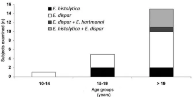
Species-level identification could be performed for 21 of the 22 samples positive for indistinguishable E. histolytica/E. dispar/E. moshkovskii complex and E. hart-manni. The species distribution was as follows: 12 (57.1%) E. dispar, 5 (23.8%) E. histolytica, 3 (14.3%) co-infections with E. histolytica and E. dispar, and one (4.8%) co-infec-tion with E. dispar and E. hartmanni (Fig. 3). No sample was positive for E. moshkovskii. The age distributions of subjects infected with different species are shown in Fig. 4.
TABLE I
Sociodemographic characteristics of the studied population, Russas, state of Ceará, Brazil, 2013
Characteristics n (%)
Gender
Male 106 (49.8)
Female 107 (50.2)
Age group (years)
0-4 18 (8.4)
5-9 27 (12.7)
10-14 37 (17.4)
15-19 14 (6.6)
> 19 117 (54.9)
Community
Barracão 119 (55.9) Patos do Tito 13 (6.1) Riacho do Barro 28 (13.1) Timbaúba do Pitingão 53 (24.9) Income strata
Extreme poverty (< US$a 17) 20 (9.4)
Poverty (US$ 17-34) 27 (12.7) Not poor (> US$ 34) 166 (77.9) Source of drinking water
Desalinated brackish water from wells 138 (64.8) Rain water stored in cisterns 56 (26.3)
Other 19 (8.9)
Sanitation facilities
Latrine 166 (77.9)
Open defecation 47 (22.1)
DISCUSSION
A key issue for understanding the morbidity associ-ated with amoebiasis is to define the proportion of infec-tions associated with the pathogenic species E. histolyti-ca. Interestingly, studies in different regions have shown that many subjects infected with indistinguishable E. histolytica/E. dispar/E. moshkovskii complex and E. hartmanni parasites actually harbour low-pathogenicity species such as E. dispar, E. moshkovskii, or even E. hartmanni (Gomes et al. 2014, Nair & Variyam 2014, Efunshile et al. 2015, Nath et al. 2015). The proportions of these subjects are variable, but can be quite high.
E. dispar and E. moshkovskii are indistinguishable from E. histolytica by light microscopy. Thus, routine parasitological techniques are not suitable for discrimi-nating these organisms. This limitation means that a
significant number of patients being treated with anti-parasitic drugs such as metronidazole may not actually be infected with E. histolytica.
In the present study, approximately two-thirds of all infections were not caused by E. histolytica. We note that all subjects were asymptomatic at the time of the stool test. Even so, we infer that nonpathogenic species are detected more frequently than E. histolytica in the studied area. This observation is particularly relevant because increasing importance has been given to tradi-tionally nonpathogenic species such as E. dispar and E. moshkovskii, since invasive amoebiasis has been dem-onstrated to be associated with these species (Parija & Khairnar 2005). It is likely that the determinants of invasive amoebiasis are complex and also involve host factors (Bosch & Siderovski 2013, Thibeaux et al. 2013).
TABLE II
Rate of detection of Entamoeba histolytica/Entamoeba dispar/Entamoeba moshkovskii complex and Entamoeba hartmanni by source of drinking water, place of defecation, and income, Russas, state of Ceará, Brazil, 2013
Positive/tested subjects
(% of positive) pa
Source of drinking water
Desalinated brackish water from wells 9/138 (6.5) 0.054 Rain water stored in cisterns 9/56 (16.1)
Sanitation facilities
Latrine 13/166 (7.8) 0.032
Open defecation 9/47 (19.1)
Family month income per capita
< US$b 17 4/20 (20) 0.308
US$ 17-34 2/27 (7.4)
> US$ 34 16/166 (9.6)
a: Fisher exact test; b: US$ 1.00 = R$ 4,00 (22 September 2015).
Fig. 4: frequency of identification of Entamoeba histolytica, En- tamoeba moshkovskii, Entamoeba dispar and Entamoeba hartmanni
by nested-polymerase chain reaction by age group in 21 positive sub-jects (Russas, state of Ceará, Brazil, 2013).
Fig. 3: detection and differentiation of Entamoeba histolytica, En- tamoeba moshkovskii, Entamoeba dispar and Entamoeba hartmanni
by nested-polymerase chain reaction. PCR products were visualised in 1.5% agarose gel with EtBr staining. Line 1: 100 bp DNA ladder; 2, 3: one faecal sample with mixed infection by E. dispar and E. histo-lytica, respectively;4, 6, 7: faecal samples positive for E. dispar; 5, 14: empty wells;8, 9: faecal samples positive for E. histolytica; 10: nega-tive control for E. dispar; 11: negative control for E. histolytica; 12: positive control for E. dispar; 13: positive control for E. histolytica; 15: faecal sample positive for E. hartmanni; 16: negative control.
The nonpathogenic species E. hartmanni can be dis-tinguished from E. histolytica, E. dispar,and E. mosh-kovskii by light microscopy. However, this distinction requires detailed observation of nuclear structures, which requires permanent smear staining, an ocular micrometer, and a highly skilled parasitologist. These criteria are hard to meet for many laboratories. We propose that the possi-bility of E. hartmanni infection should also be considered in people who excrete indistinguishable E. histolytica/E. dispar/E. moshkovskii complex and E. hartmanni cysts. In the present study, E. hartmanni was detected in one of the indistinguishable E. histolytica/E. dispar/E. mosh-kovskii complex and E. hartmanni positive samples.
The study population is located in a sociodemograph-ic and environmental setting characterised by defsociodemograph-icits in sanitation infrastructure and water stress. The study area is located in a low-rainfall region in the Caatinga biome that is subjected to prolonged droughts and prone to desertification. Nonpotable water is obtained from a reservoir in the locality and used for livestock water-ing and other suitable applications. In the last decade, a strategy has been implemented in which rainwater is collected during the rainy season from roofs via gutters. This collected rainwater is stored in household tanks for later use during droughts. This strategy has significantly improved access to drinking water in the study area. Ar-tesian wells constructed in the region are another source of drinking water. However, this water is brackish and must be desalinated before consumption. We found that the rate of E. histolytica, E. dispar,and E. hartmanni positivity was almost three times higher in subjects who drink collected rainwater than in subjects who drink de-salinated brackish water drawn from the artesian wells. We hypothesise that the long period (between the dry season and the rainy season) of rainwater storage in tanks favours contamination with amoeba cysts, thereby enabling transmission. Interestingly, consumption of rainwater captured from roofs has been demonstrated to reduce the prevalence of G. intestinalis infection in a semiarid region in northeastern Brazil (Fonseca et al. 2014). Regarding the place of defecation, subjects who practice open defecation exhibited a significantly higher positive rate compared with subjects who defecate in la-trines. Moreover, an even higher positive rate was ob-served in people who deposit faeces directly into the soil compared with subjects with rudimentary tanks.
In some regions of the world, including Latin Amer-ica, inadequate sanitary conditions facilitate the trans-mission of amoebiasis, thereby generating high preva-lence rates (Braga et al. 1998, Ramos et al. 2005). In these scenarios, invasive amebic dysentery and liver ab-scesses are expected to occur. However, these diseases were not observed in the present study. Severe cases of amoebiasis are identified infrequently in Brazil, which may be explained by the relative improvement of living conditions over the past few decades.
Cumulatively, our data suggest a high prevalence of asymptomatic infection with indistinguishable E. histolytica/E. dispar/E. moshkovskii complex and E. hart-manni parasites. These asymptomatic infections appear to be caused by predominantly nonpathogenic species or parasites with low pathogenic potential. In the context of scarce water resources, the sanitary and socioenviron-mental characteristics of the region appear to be associ-ated with transmission.
REFERENCES
Bosch DE, Siderovski DP 2013. G protein signaling in the parasite
Entamoeba histolytica. Exp Mol Med 45: e15.
Brumpt E 1926. Individualité de l’Entamoeba dispar. Présentation de pièces.Bull Soc Pathol Exot 19: 399-404.
Brumpt E 1949. Précis de parasitologie, Masson & Cie, Paris, 2138 pp.
Chacín-Bonilla L 2013. Amebiasis: aspectos clínicos, terapéuticos y de diagnóstico de la infección. Rev Med Chil 5: 609-615.
Costa CA, Brito KN, Gomes MA, Caliari MV 2010. Histopathologi-cal and immunohistochemiHistopathologi-cal study of the hepatic lesions exper-imentally induced by Entamoeba dispar. Eur J Histochem 3: e39.
Efunshile MA, Ngwu BA, Kurtzhals JA, Sahar S, König B, Stensvold CR 2015. Molecular detection of the carriage rate of four intesti-nal protozoa with real-time polymerase chain reaction: possible overdiagnosis of Entamoeba histolytica in Nigeria. Am J Trop Med Hyg 93: 257-262.
Faust EC, D’antoni JS, Odom V, Miller MJ, Peres C, Sawitz W, Thomen LF, Tobie J, Walker JH 1938. A critical study of clinical laboratory techniques of the diagnosis of protozoan cysts and hel-minth eggs in feces. I - Preliminary communication. Am J Trop Med Hyg 18: 169-183.
Fonseca JE, Carneiro M, Pena JL, Colosimo EA, da Silva NB, da Costa AG, Moreira LE, Cairncross S, Heller L 2014. Reducing occurrence of Giardia duodenalis in children living in semiarid regions: impact of a large scale rainwater harvesting initiative.
PLoS Negl Trop Dis 6: 2943.
Goldman M 1969. Entamoeba histolytica-like amoebae occurring in man. Bull World Health Organ 3: 355.
Gomes TDS, Garcia MC, Cunha FDS, de Macedo HW, Peralta JM, Peralta RHS 2014. Differential diagnosis of Entamoeba spp in clinical stool samples using SYBR green real-time polymerase chain reaction. Sci World J 2014: 8 pp.
Jackson TE 1998. Entamoeba histolytica and Entamoeba dispar are distinct species; clinical, epidemiological, and serological evi-dence. Int J Parasitol 28: 181-186.
Lau YL, Anthony C, Fakhrurrazi SA, Ibrahim J, Ithoi I, Mahmud R 2013. Real-time PCR assay in differentiating Entamoeba histol-ytica, Entamoeba dispar, and Entamoeba moshkovskii infections in Orang Asli settlements in Malaysia. Parasit Vectors 6: 250.
Lin CC, Kao KY2013. Ameboma: a colon carcinoma-like lesion in a colonoscopy finding. Case Rep Gastroenterol 7: 438-441.
Nair GV, Variyam EP 2014. Noninvasive intestinal amebiasis: Enta- moeba histolytica colonization without invasion. Curr Opin In-fect Dis 5: 465-469.
Nath J, Hussain G, Singha B, Paul J, Ghosh SK 2015. Burden of major diarrheagenic protozoan parasitic co-infection among amoebic dysentery cases from north east India: a case report. Parasitol-ogy 10: 1318-1325.
Ngui R, Angal L, Fakhrurrazi SA, Lian YL, Ling LY, Ibrahim J, Mahmud R 2012 Differentiating Entamoeba histolytica, Enta-moeba dispar, and Entamoeba moshkovskii using nested poly-merase chain reaction (PCR) in rural communities in Malaysia.
Parasit Vectors 5: 187.
Ojha SC, Jaide C, Jinawath N, Rotjanapan P, Baral P 2014. Geohel-minths: public health significance. J Infect Dev Ctries 8: 5-16.
Oliveira FMS, Neumann E, Gomes MA, Caliari MV 2015. Entamoe-ba dispar: could it be pathogenic. Trop Parasitol 10: e0137327.
Paglia MG, Visca P 2004. An improved PCR-based method for detec-tion and differentiadetec-tion of Entamoeba histolytica and Entamoeba dispar in formalin-fixed stools. Acta Trop 92: 273-277.
Parija SC, Khairnar K 2005. Entamoeba moshkovskii and Entamoe-ba dispar-associated infections in Pondicherry, India. J Health Popul Nutr 23: 292-295.
Pimenta PF, Diamond LS, Mirelman D 2002. Entamoeba histolytica
Schaudinn, 1903 and Entamoeba dispar Brumpt, 1925: differenc-es in their cell surfacdifferenc-es and in the bacteria-containing vacuoldifferenc-es.
J Euk Microbiol 49: 209-219.
Ralston KS, Petri Jr WA 2011. Tissue destruction and invasion by En-tamoeba histolytica.Trends Parasitol 27: 253-263.
Ramos F, Moran P, González E, Garcia G, Ramiro M, Gómez A, León MDCG, Melendro EI, Valadez A, Ximénez C 2005. High preva-lence rate of Entamoeba histolytica asymptomatic infection in a rural Mexican community. Am J Trop Med Hyg 73: 87-91.
Rasella D 2013. Impact of the water for all program (PAT) on child-hood morbidity and mortality from diarrhea in the Bahia state, Brazil. Cad Saude Publica 29: 40-50.
Ritchie L 1948. An ether sedimentation sechnique for outine stool examinations. Bull US Army Med Dep 8: 326.
Skappak C, Akierman S, Belga S, Novak K, Chadee K, Urbanski SJ, Church D, Beck PL 2014. Invasive amoebiasis: a review of Enta-moeba infections highlighted with case reports. Can J Gastroen-terol Hepatol 28: 355-359.
Thibeaux R, Weber C, Hon CC, Dillies MA, Avé P, Coppée JY, Labruyère E, Guillén N 2013. Identification of the virulence landscape essential for Entamoeba histolytica invasion of the hu-man colon. PLos Pathog 9: e1003824.
Tshalaia LE 1941. On a species of Entamoeba detected in sewage ef-fluents. Med Parazitol (Mosk) 10: 244-252.
Turkeltaub JA, McCarty TR, Hotez PJ 2015. The intestinal protozoa: emerging impact on global health and development. Curr Opin Gastroenterol 31: 38-44.
WHO - World Health Organization 1997. WHO/PAHO/UNESCO report. A consultation with experts on amoebiasis. Epidemiol Bull 18: 13-14.
Wuerz T, Kane JB, Boggild AK, Krajden S, Keystone JS, Fuksa M, Kain KC, Warren R, Kempston J, Anderson J 2012. A review of amoebic liver abscess for clinicians in a nonendemic setting. Can J Gastroenterol 26: 729-733.