Value of magnetic resonance imaging for diagnosis of dentigerous cyst
Texto
Imagem


Documentos relacionados
Assim, propõe o BE que sejam introduzidas as seguintes alterações: “A eliminação da condição de se ser casado ou viver em união de facto como critério de
The role of diffusion magnetic resonance imaging in Par- kinson’s disease and in the differential diagnosis with
El trabajo presentado aquí no pretende ser ni exhaustivo ni conclusivo, sino apenas el comienzo de la exploración del marco teórico ofrecido por la Filosofía de la Ciencia de
An immunodiagnostic method based on a direct sandwich enzyme-linked immunosorbent assay (ELISA), using monoclonal antibodies, has been examined in a number of African laboratories
Assim como os alunos conseguem observar dos professores suas qualificações, e o perfil empreendedor dentro de sala de aula, que pode ser um esforço da IES
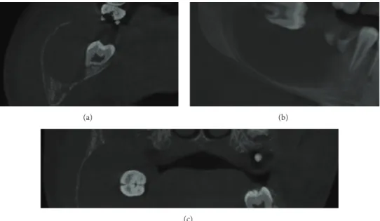
However, the appearance of this cyst on cone beam computed tomography (CBCT) and magnetic resonance imaging (MRI) have received relatively little attention.. CBCT and
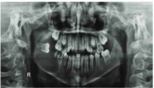
Common non-neoplastic causes of jaw swelling in this age group are apical cyst, dentigerous cyst, calcifying epithelial odontogenic cyst, odontogenic keratocyst, periapical
he magnetic resonance imaging (MRI) showed rims of hypointensity on T2 around the brainstem, cerebellum, and spinal cord, which was consistent with radiological diagnosis
