Leopoldino Capelozza Filho*, Alberto Consolaro**, Mauricio de Almeida Cardoso*, Danilo Furquim Siqueira*
Enamel drilling for canine traction:
Advantages, disadvantages, description
of surgical technique and biomechanics
Introduction: The management of unerupted teeth has always been considered as a chal-lenging procedure in orthodontic practice. Within this perspective, the search for effective-ness in the procedures adopted for the management of unerupted teeth is essential, which explains the purpose of the present paper. When enamel drilling is performed, a natural structure is transixed, which may be restored with composite material and may dispense from risky procedures such as bonding attachment technique and lasso wire technique. Ob-jective: The present paper aims to present protocols for enamel drilling for canine traction (EDCT), speciically for maxillary canines, the most frequent teeth showing tooth position anomalies. In this paper, clinical cases with different degrees of complexities were illus-trated, and, based upon the literature review and the 30 years of expertise with high rate of clinical success, advantages and disadvantages are discussed comparing EDCT and accessory bonding for canine traction (ABCT).
Abstract
Keywords: Canine impaction. Tooth traction. Segmented arch mechanics.
* PhD Professor, Graduate and Postgraduate Program of Specialization and Master’s Degree in Orthodontics at Sagrado Coração University - Bauru/USC.
** Full Professor of Oral Pathology, FOB-USP. Full Professor Postgraduate Program, FORP-USP. How to cite this article: Capelozza Filho L, Consolaro A, Cardoso MA,
Siqueira DF. Enamel drilling for canine traction: Advantages, disadvantages, description of surgical technique and biomechanics. Dental Press J Orthod. 2011 Sept-Oct;16(5):172-205.
» The authors report no commercial, proprietary, or inancial interest in the
IntrOductIOn
The most frequent absence of teeth in the dental arch involves permanent canines, if third molars are not taken into account.21,28,36 In ran-dom samples, the frequency of unerupted canines ranges from 1.5 to 2% in the maxilla, and 0.3% in the mandible10,20,22,23,26,30,35 Conversely, frequency is high (23.5%) in samples previously selected for orthodontic treatment.3,24,37 In female patients the unerupted canines (1.17%) are twice as frequent as in males (0.51%)2 and occur palatally two to three times more often than buccally.1,33
Although hereditary33 factors seem to play a role in the pathogenesis of unerupted teeth, es-pecially in palatal occurrences, the exact causes are still unknown.34 Among the factors most of-ten associated with unerupted teeth are discrep-ancies between tooth size and arch length, ab-normal position of the tooth germ or tooth, pro-longed retention or early loss of deciduous teeth, the occurrence of cystic or neoplastic formation and iatrogenic causes.2,4 Although unfounded, other etiologies are sometimes mentioned, such as systemic causes.
The diagnosis and treatment of unerupted teeth requires competent general practitioners, pediatric dentists, oral and maxillofacial surgeons, periodontists and orthodontists, as well as patient compliance.31,36 Prognosis for the use of tooth traction should be considered with serious reser-vation, or at least rather limited at irst because the chance of failure can never be ruled out as it depends on many variables.38 Parents or legal guardians should be made keenly aware of the odds to avert false expectations.
Conventional radiographic techniques have always presented limitations in locating unerupt-ed maxillary canines, especially panoramic X-rays, which require additional radiographs such as periapical X-rays by the Clark technique10 or occlusal maxillary radiographs. These techniques were limited and could only spot unerupted ca-nines buccally or palatally, but the relationship
between canines and adjacent teeth was not ad-dressed and the potential loss of the root struc-ture of lateral incisors (which are most common-ly affected teeth in these situations) was totalcommon-ly unknown. Orthodontic planning was thereby curtailed since it was only possible to assess these variables and the integrity of the lateral incisor root during the surgical procedure performed in order to access the unerupted canine. Common sense, caution and periodic controls were a nec-essary support to ensure that procedures based on this limited diagnostic came to fruition.
Cone beam computed tomography (CBCT) made the diagnosis of anomalies in the position of maxillary canine, also called dysgenesis, much more effective. CBCT’s various slice planes and the resulting 3D reconstructions, viewable from virtually every angle, allows today’s profession-als to plan orthodontic traction of maxillary canines with greater accuracy and reinement. This allows surgeons to deal with canines, their dental follicle, cervical region and adjacent teeth with the aid of detailed planning, which ultimately reduce the risks of unintended out-comes. In other words, technological advances in imaging have increased the chances of orth-odontic traction being accomplished more safe-ly and accuratesafe-ly. It also eliminates the possi-bility of preexisting processes such as external cervical resorption, aveolodental ankylosis and replacement resorption in the teeth to be sub-mitted to traction.17 When the imaging diagnosis reproduces more faithfully the actual position, the prognosis tends to be more precise and the treatment plan can be tailored to the individual.
present during surgery, it was requested that in-formation was described in an oficial referral to enable safer traction. Maintaining this hypoth-esis, the surgeon must have enough orthodontic knowledge to guide the orthodontist on how best to perform the movement.
Early identification of non-eruption of the canine may reduce the need for complex and expensive orthodontic treatment. It is impor-tant that general practitioners and pediatric dentists be vigilant when monitoring eruption in children in the mixed dentition stage, not just by taking care of oral health, but also by identifying potential disruptions in this pro-cess. Ectopic eruption and impaction of max-illary permanent canines are frequent issues in orthodontic practice. In addition to being regarded as real challenges for the orthodon-tist, these two oral conditions can significantly lengthen total treatment time11 as well as in-crease treatment complexity.8
In planning treatment of an unerupted ca-nine, one is advised to assess the thickness of the dental follicle, bearing it in mind when creating space to accommodate this tooth in the dental arch, aiming at either normal canine eruption or its orthodontic traction. The space required for the physiological eruption of an unerupted canine is, in theory, 1.5 times the mesiodistal size of the canine crown, a neces-sary condition for eruption to occur without orthodontic assistance.12,15
In patients with unerupted canines, the cor-responding deciduous teeth are usually found in the arch and their mesiodistal dimension is much smaller than that of the permanent ca-nine. Creating space in the analogous arch for the mesiodistal dimension of the unerupted canine is a daunting task and often impossible to achieve, especially if the goal is to increase space by 50%.12 It is usually impractical, from a mechanical point of view, to wait for a physi-ological eruption, and this is precisely why
traction is indicated. Monitoring the patient and the risky relations of this tooth with the neighboring teeth will determine the appropri-ate moment for this approach.
When the patient’s face and the transverse dimensions of the upper arch can support it, orthopedic maxillary expansion seems unques-tionable with this protocol – a sine qua non condition for a real increase in bone mass by adding bone to the midpalatal suture – creat-ing space and enablcreat-ing a better eruption path-way. The goal is a real bone gain by placing bone in the region of the midpalatal suture and increasing the perimeter of the arch. This creates favorable conditions for the canine to find eruption space and redirect its pathway, often avoiding surgical approaches and orth-odontic traction. This is only possible within a follow-up perspective with growth monitoring and assisted eruption, where these problems are diagnosed at an early stage, enabling an in-terceptive procedure and subsequent follow-up to assess progress.
According to the literature, several thera-peutic treatment options are available for pa-tients affected by this anomaly, namely: Ab-sence of immediate treatment and long-term monitoring, self-transplantation of the canines, extraction of unerupted canines and closing of spaces with restorative treatment, extraction of unerupted canines and closing of spaces with orthodontic treatment, and finally, surgi-cal exposure of unerupted canines and use of orthodontic forces to bring the tooth into oc-clusion.2,4,31,32,35
There is consensus indicating that the canine should never be tied with wire because of the inherent dificulty posed by this procedure and because it causes cervical resorption as the steel ligature is placed along the cementoenamel junction (CEJ). Historically, the irst protocols used in the traction of unerupted maxillary ca-nines consisted in binding the neck of the tooth with steel wire. The force and displacement of the orthodontic wire on the neck of the tooth would expose the dentin gaps in the CEJ, add-ing to the constant in ammation that resulted from the continuous trauma.17
Accessory Bonding for Canine Traction (ABCT) and Enamel Drilling for Canine Trac-tion (EDCT) are the most common procedures. ABCT is perhaps the technique of choice of most dentists as it prevents erosion of tooth structure. Contrary to the choice of most orth-odontic colleagues, the authors of this article never performed the ABCT procedure for rea-sons that will be presented in this paper, and have always applied the EDCT procedure for this purpose. This technique was successfully applied over thirty years of orthodontic prac-tice and now boasts a caseload with 100% suc-cessful cases, which justifies its disclosure to the scientific community. In addition to this outstanding accomplishment, it should be em-phasized that not a single canine ever required further treatment, which was the main reason for always choosing this option. Over time, a protocol for this procedure was formulated and is presented below. The EDCT technique can be adopted in all cases with no restriction because drilling can be performed in different areas of the crown of the unerupted canine, ac-cording to how one needs to move this tooth.
In light of the above, the aim of this paper is to create protocols for EDCT technique, specii-cally for maxillary canines since these teeth are more often affected by position anomalies, also called dysgeneses. By describing the advantages
and disadvantages of EDCT compared with the ABCT technique, illustrated through case studies of different levels of complexity, the primary in-tention is to create a concise methodology, based on the literature and iltered through clinical ex-perience of over thirty years performing EDCT with a high success rate.
AdVAntAGES And dISAdVAntAGES OF EnAmEl drIllInG FOr cAnInE trAc-tIOn (Edct): cOmpArAtIVE AnAlySIS
Advantages
decreased risk of a new surgical procedure The risk of a new surgical procedure to ac-cess the unerupted canine may occur in the ABCT technique due to immediate bond fail-ure of the accessory after delivery of the trac-tion force. This bond failure may be caused by excessive force and/or contamination during the process of bonding the orthodontic acces-sory. Considering that most patients eligible for traction are children, management may prove more difficult, with increased risk of this oc-currence while exposing them to a new sur-gical procedure, a risk that could certainly be avoided. Therefore, the authors’ preference for EDCT – despite the biological cost involved (wear of enamel, a structure that is not re-placed by the body) – considerably reduces the possibility of reopening for new access to the unerupted canine, since when this tooth is tied the risk is virtually nonexistent.
less tissue manipulation
EGF-activated mediators, induces pericoronal bone resorption, an essential phenomenon in the occurrence of tooth eruption.
The cementoenamel junction lies between enamel and cementum. It is therefore reasonable to assert that the DF in the cervical region over-lies the line formed by the neighboring relation-ship between enamel and cementum. The CEJ has gaps along the cervical circumference of all human teeth in which the tubules are open and exposed to inorganic and organic components, but especially proteins. This cervical region is a sensitive tooth structure due to the fragile junc-tion between enamel and cementum.16,17
During surgical removal of the DF in the cervical region the dentin gaps present in all human teeth, including deciduous teeth, are inevitably exposed to connective tissue after the ap is folded back over the tooth. The ex-posure of these dentin proteins, deined as se-questered antigens, can induce, over weeks or months, an immunological process of elimina-tion that is clinically known as External Cervi-cal Resorption (ECR). This process may occur during orthodontic traction or after the tooth has reached the occlusal plane.16,17 In many such cases a belated detection tends to be the rule. ECR is deined as a slow, painless, insidious pro-cess that does not compromise pulp tissues. In more advanced cases, it can lead to gingival
in-ammation and pulpitis secondary to bacterial contamination. One way to prevent this occur-rence is to leave at least 2 mm of soft tissue from the DF attached to the cervical region.16,17
The ABCT technique requires greater ex-posure of the crown and hence greater need to remove osseous tissue and manipulate the DF, implying a higher risk of trauma to the ECJ. This region should be handled only when ab-solutely necessary.16,17 When this occurs, the chances of external resorption in this region af-ter the traction procedure are increased, which causes loss of structure of the tooth under traction.
This effect can be further compounded by ex-cessive or extensive application of acids and other products used to etch the tooth enamel. Over-application can drain these products into the cervical region, where fixation of the DF to the ECJ occurs, chemically affecting cells and tissues, exposing and even increasing den-tinal gaps and releasing the sequestered anti-gens into the adjacent tissue after the surgical wound has healed.16,17
The surgical procedure must be well planned and carried out with precision, without exagger-ated forces and repetitive handling of the instru-ments used in the procedure.19 Surgical instru-ments should not be anchored or ixed to the cer-vical region of the upper canines because chisels and tips of surgical instruments such as forceps can mechanically damage the follicle and peri-odontal tissues in the cervical region and expose or increase the exposure of the dentin at ECJ, a starting point for ECR.16,17
When drilling is the procedure performed to access the unerupted canine, only a small portion of the tooth crown requires exposure, and only enough to allow the procedure to be performed. This portion of the crown may be the tip of the cusp or any of the proximal surfaces, depending on the anatomical features of the canine, which displays an enamel bridge along the entire crown with suficient strength to withstand anchorage and traction.
Shorter surgery time
The EDCT technique eliminates the need for conventional steps of regular bonding, which involves etching, moisture control, ad-hesive application and bonding of orthodontic accessory. Performing all these steps in an en-vironment with total moisture control requires more time in the trans-surgical phase, consid-ering the difficulty of this procedure, which is carried out through surgical exposure of the canine in an open field. In addition, the surgi-cal procedure must be performed by a compe-tent oral and maxillofacial surgeon, although these professionals, more often than not, have little experience in bonding orthodontic ac-cessories. The EDCT technique eliminates all the steps listed above, which results in shorter surgical time, less bleeding and therefore less postoperative edema.
Application of force in the long axis of the tooth with a better established magnitude
The EDCT technique allows the application of force directly to the long axis of the tooth un-der traction, resulting in increased control over traction direction. When an accessory is bonded to the buccal or lingual surface of an unerupted canine and traction force is applied, the direction of the resultant force should be observed in order to avoid undesired movement.
Moreover, the presence of a bulky body such as a bracket or button on the surface of the ca-nine in an area subjected to a repair process after access surgery probably restricts canine movement making it difficult to determine the amount of force to be applied. Admittedly, the ideal force must be small in magnitude, ranging from a minimum amount of around 35 to 60 grams, when traction copies an eruption move-ment, to greater forces, required when the ca-nine needs to undergo translatory forces in or-der to avert obstacles in its eruption pathway. In either case, determining an adequate force
is rendered more difficult if restrictive factors, such as the ones mentioned above, establish undefined magnitude decreases in the force available to perform the movement of traction.
disadvantages
risk of enamel fracture
The EDCT technique requires care to pre-vent the enamel from fracturing when twisting the ligature wire. Stronger ligatures are often used for this purpose, i.e., so that the risk of fracture and consequent need for reopening are minimized. Twisting the ligature without con-sidering basic precautions, as inserting the ex-plorer probe tip between the ligature and the canine, can cause enamel fracture and require new drilling, further increasing the biological cost of the procedure.
potential pulp damage
Canine drilling should be performed per-pendicular to the long axis of the tooth with a small diameter (¼”) high speed spherical car-bide bur and copious irrigation. This is impor-tant to prevent the bur from reaching the pulp chamber, thereby causing irreversible pulpitis or even requiring endodontic treatment. The competence and experience of a professional surgeon is of paramount importance to avoid such damage.
Esthetics
When the EDCT technique is performed, the hole drilled during the procedure should be filled by means of esthetic restoration follow-ing the emergence of the unerupted canine in the oral cavity. Given the ongoing advances in dental restorative materials and assuming that this procedure is performed by a competent professional – by drilling a big enough hole as to allow the passage of a folded ligature wire – it is unreasonable to suspect that drilling might impair esthetics. As stated earlier, a spherical ¼” diameter carbide bur should suffice.
Greater professional experience
The EDCT procedure requires an expe-rienced, insightful surgeon to determine the actual position of the unerupted canine since this drilling, as previously stated, should be performed perpendicular to the long axis of the tooth, despite the reduced need for re-moval of osseous tissue and manipulation of the DF. The cases that require greater atten-tion are those with severe impacatten-tion because the procedure – which involves a flap in an open field, as well as the presence of bleed-ing – is usually performed in children or ado-lescents, with little if any collaboration, under local anesthesia, protocol usually adopted by the authors’ surgical team.
AdVAntAGES And dISAdVAntAGES OF AccESSOry BOndInG FOr cAnInE trActIOn (ABct): cOmpArAtIVE AnAlySIS
Advantages
lower biological cost
Since the ABCT procedure does not require drilling of the unerupted canine crown, it en-tails a lower biological cost compared to the EDCT technique, i.e., canine structure is ful-ly preserved. It is worth mentioning that this
biological cost is decreased as long as care is ex-ercised in washing the canine crown after etch-ing so as not to allow the acid to remain in the DF when the ABCT technique is performed.
lower risk of pulp damage
When the ABCT procedure is performed, drilling of the unerupted canine crown is not necessary and therefore the risks related to pulp damage are minimized or virtually elimi-nated. The risk of pulp damage is related to a poorly executed EDCT technique, i.e., when drilling is not performed perpendicular to the long axis of the unerupted canine.
disadvantages
Increased manipulation of the dental follicle (dF)
It should be noted that the ABCT technique requires exposure of the unerupted canine crown so as to create a surface large enough to bond the attachment used for traction. Therefore, the need to remove osseous tissue is greater as is the manipulation of the DF during the surgical procedure. Whenever these tissues are over-manipulated the biological costs are higher, as well as the risks of ECR occurring after traction of the unerupted canine.
longer surgery time
A
C D E
B
Force application
Forces induced to perform traction of un-erupted canines should be directed, whenever possible, using the long axis of the tooth as reference. Typically, the bonding of a bracket or lingual button to the mesial region of the clinical crown of an unerupted canine does not allow the traction force to make the tooth copy the eruption movement. Since the bond-ing of this accessory, which will receive the wire and the traction forces are routinely per-formed in less than ideal positions, resulting from unfavorable technical conditions and the need to restrict tissue manipulation, canine
displacement can follow undesired pathways. This may present risks for the adjacent teeth and require more extensive movements for the proper positioning of the canine after its emer-gence in the oral cavity (Fig 1).
Even in this context of inadequate movement of the canine, an additional dificulty lies in dein-ing the level of force, which should be at the same time light and suitable for the traction movement. In other words, copying the eruption movement or predicting the type of displacement that the canine will perform during eruption caused by orthodon-tic traction, seems to be very important and made dificult when the ABCT technique is adopted.
FIGURE 2 - Female patient, nine years and six months old, Caucasian, Pattern I5 brachyfacial was referred by a pediatric dentist for orthodon-tic evaluation due to routine radiographs indicating poor positioning of the maxillary permanent canines. In the second transitional period of mixed dentition, the intraoral examination (A-E) revealed a Class I occlusal relationship bilaterally with adequate overbite and overjet. In occlusion, the upper midline was found slightly deviated to the left relative to the lower midline. In the upper arch, teeth #12 and #22 had distal angulation and labial inclination of the crown, and lower arch exhibited mild crowding in the lower incisor region. Both deciduous canines were present, with no mobility and the permanent successors had no palpable area, nor root apex closure. With regular monitoring and good dental hygiene, the patient had not undergone previous orthodontic treatment. Patient history revealed no oral habits, medical problems or eruption disorders in the family.
clInIcAl cASES tHAt IlluStrAtE tHE EnAmEl drIllInG FOr cAnInE trActIOn (Edct) tEcHnIquE
In this topic, the EDCT surgical technique will be demonstrated through case studies that disclose different levels of complexity, which will be discussed in the captions of each igure.
EDCT ABCT
Decreased risk of new surgical procedure
Risk of new surgical procedure due to accessory bond failure
Less tissue manipulation Greater manipulation of tissues to expose tooth surface
Shorter surgery time Longer surgery time
Direction of force in the long axis of the tooth
Direction of force dependent on accessory positioning
Risk of enamel fracture No risk of enamel fracture
May cause pulp damage Minimal pulp damage
Future need for esthetic restoration
Less potential need for esthetic restoration
Demands a more experienced surgeon
No need for experience in drilling
No acid action on tooth Acid action on tissues at CEJ and DF
TABLE 1 - Comparison between advantages and disadvantages in using Enamel Drilling for Canine Traction (EDCT) versus Accessory Bonding for Canine Traction (ABCT).
A B C
FIGURE 3 - The initial panoramic radio-graph confirmed the presence of all per-manent teeth and evidenced the reason for the consultation: Malposition of teeth #13 and #23 associated with divergence of the crowns of teeth #12 and #22, characteris-tic of the inter-transitional period of mixed dentition, despite the fact that the patient was in the beginning of the second tran-sitional period. Radiographically, the upper permanent canines were mesially angulat-ed, with tooth #23 in a critical position, i.e., near the roots of the maxillary permanent lateral incisors, with the apices of the per-manent canines not yet closed.
FIGURE 4 - A technique of horizontal displacement of the tube (Clark technique)10 using periapical radiographs confirmed that the upper left canine was impacted in a palatal position. This was the reason for the consultation and the need for treatment. The time was entirely appropriate for treatment as it enabled a strategy aimed at improving the prognosis, which is always limited. This possibility was provided by a competent pediatric dentist who per-ceived the problem and made the referral. The treatment for this patient was designed to intercept the pathway of the ectopic tooth #23, which was in a more critical position than tooth #13.
A B C
A B C
A B C
FIGURE 6 - The expansion appliance was cemented with internal and external connecting bars bonded with resin to the buccal and palatal canines and first primary molars in order to enhance anchorage, in addition to extension bars for teeth #16 and #26 (A). Before starting the active phase of expansion the ligature wire tying tooth #23 was attached to the structure of the Haas expander (B and C).
FIGURE 7 - The maxillary orthopedic expansion seems unquestionable in this protocol, in the sense that it is undoubtedly a condition for a real increase in bone mass by adding bone to the midpalatal suture, thereby creating space and enabling a better eruption pathway for tooth #13 as well. The extremely fa-vorable timing with which this procedure was performed improved the prognosis and facilitated treatment. After maxillary expansion and expander screw fixation (A), a segment of TMA wire (0.019 x 0.025-in) was adapted to the Haas appliance through a hole in the acrylic made with a steel round bur and fixed with acrylic resin. After curing the resin, tooth #23 began to be pulled in the palatal and occlusal direction, delivering an amount of force equivalent to 60 grams (B and C). The initial palatal movement was meant to prevent contact between the crown of tooth #23 and the root of tooth #22, thus reducing the risk of root resorption. This is the usual practice to protect the roots of adjacent teeth during induced canine eruption movement.
FIGURE 8 - After five months of traction, a periapical radiograph was taken of the region in which it appeared that the traction movement had allowed for the correction of the eruption pathway of tooth #23. In this phase, the partial removal of the Haas expander was indicated.
A B C
D E F
A B C
D E
F G H
FIGURE 10 - Intraoral photographs (A-E) seven months after initiating mechanical traction of tooth #23 (one month after the force was directed buccally), showing the tip of the canine cusp already in the oral cavity, palatally (D). In this phase, the remaining expander segment was removed and teeth #63 and #64 extracted. Interception of the ectopic eruption pathway of tooth #23 was resolved and, as of this phase, time was allowed to elapse until all teeth had been replaced before starting corrective orthodontic treatment. Prognosis is good for corrective treatment, unlike what was determined at the beginning of treatment, thanks in part to the ectopic eruption pathway of tooth #23. A panoramic radiograph (F) was requested for evaluation at the end of the second transitional period of mixed dentition, pending only the exfoliation of tooth #55, whose extraction was requested. In this phase, at the age of ten years and six months, the patient was instructed to resume corrective orthodontic treatment after complete eruption of teeth #15 and #25.
A B C
D E
A B C
D E
FIGURE 12 - Four months after the beginning of the corrective orthodontic treatment with 0.018” leveling archwires in the upper and lower dental arches (A-E) a lingual button was bonded to the buccal surface of tooth #23, and a 0.019 x 0.025”-in TMA wire segment attached to enable the buc-cal movement of this tooth, both assisted by an open coil Nitinol spring placed between teeth #22 and #24, and biodap in the occlusal surface of tooth #36, to raise the bite.
FIGURE 13 - In the phase of arch leveling using 0.020-in steel archwires in the upper and lower dental arches (A-E), ten months after starting the corrective orthodontic treatment, tooth #23 was no longer in crossbite and had assumed a normal position.
A B C
D E
A B C
D E
FIGURE 15 - Intraoral control photographs two years after removal of the appliance ( A-E) exhibited stability of occlusal relationships achieved by implementing an expansion and protrusion mechanics, and confirmed by long-term follow-up.
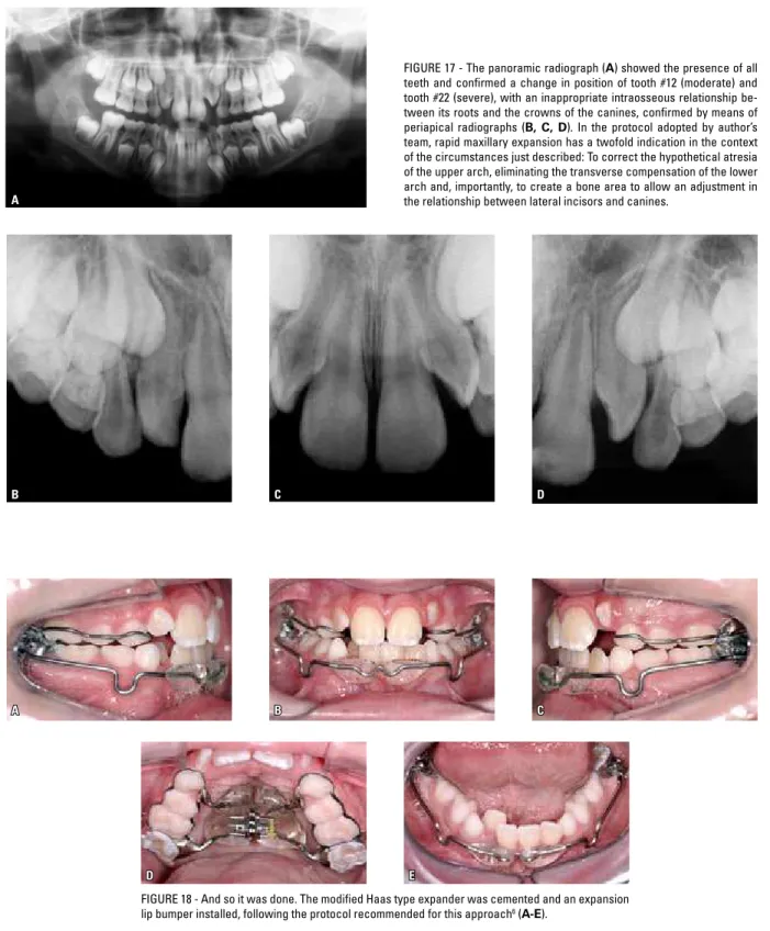
FIGURE 16 - A description of the treatment for traction of a retained canine through the buccal side will be illustrated with the case of a Caucasian girl, prob-ably Pattern I,5 and mesofacial, who participated in a growth and eruption monitoring program and was subjected to an initial assessment when she was nine years and nine months old. At this early time, a lack of motivation to present for the consultations affected the eruption of the upper lateral incisors. At the end of the first period the mixed dentition exhibited a Class I occlusal relationship. Although there were no crossbites, the upper dental arch morphology suggested atresia, a hypothesis which, if true, would lead to a diagnosis of compensatory adjustment atresia in the lower arch as well.
B C D
A B C
D E
A
FIGURE 18 - And so it was done. The modified Haas type expander was cemented and an expansion lip bumper installed, following the protocol recommended for this approach6 (A-E).
A B C
A B C D
D E
FIGURE 19 - The results achieved in the shape of the dental arches – with the obvious advantage of creating space to accommodate the crowns of lateral incisors – can be observed (A-E), hopefully improving the relationship between the roots of these teeth and the canine crowns.
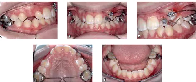
FIGURE 20 - The expander was removed after four months and the lip bumper maintained for night use. Eight months after this intervention of an essentially interceptive and preventive nature, Clark10 technique periapical radiographs showed that tooth #13 had improved and tooth #23 was positioned outside the normal eruption pathway, buccally, interfering with the positioning of tooth #22. A clinical decision was made to request the extraction of teeth #53 and #63 and wait for the eruption of tooth #24.
A B C
A B C
D E
FIGURE 23 - The surgery was performed in strict compliance with the protocol presented in this article. A flap was made on the buccal side (A), with subtle and necessary exposure of the incisal third of the crown of tooth #23 (B), allowing access by drilling at the exact desired spot on the incisal surface, thereby enabling a strategy to achieve maximum traction efficiency. Note how the drilling is minimal (C). The ligature (0.30 mm / 0.12-in thickness) is inserted into the perforation (D) and is carefully twisted (E) so as not to fracture the enamel on the incisal portion. Note the convenient access to tooth #23 and the excellent view of the inadequate relationship between this tooth and the lateral incisor, confirming the etiology of this tooth’s malposition (B).
A B C
A
A
B
B
FIGURE 24 - Immediately after the suture, last phase of the access surgery, a TMA rectangular 0.019 x 0.025-in wire segment was adapted to start moving the canine with the foremost intent of removing it from its position over the root of the lateral incisor (outward and slightly downward, oblique direction labially and incisally).
FIGURE 25 - Action on tooth #22 was post-poned until the position of the canine al-lowed it. When this eventually occurred the canine was already in the mouth, in a higher position than would have been the ideal, but in this case mandatory to enable handling the lateral incisor. Another wire segment with the same specifications adopted for ca-nine traction was adapted to move tooth #22. The initial intention was to upright the root and then more adequately position the crown of the lateral incisor. Note the radiographic image at this stage with tooth #23 still being moved, supported by the ligature inside the perforation, three months after initiating me-chanical traction.
A B C
D E
A B C
D E
FIGURE 28 - The outcome was satisfactory and, within the context, considered likely. After ten months deploying the mechanics to correct the malposition of teeth #22 and #23, a case of moderate complexity emerges to be treated with a fixed orthodontic appliance. Effects resulting from the poor positioning of tooth #22 and resolution of problems regarding the eruption of tooth #13, which were minor, and tooth #23, which were serious, and if not eliminated by an intercep-tive and prevenintercep-tive approach, would result in a severe malocclusion, which would require measures likely to cause tooth loss.
FIGURE 29 - After an eighteen-month treatment with a fixed appliance, the final panoramic radiograph shows adequate and symmetrical root positioning, attesting to the biological efficiency of the suggested proto-col for traction of impacted teeth, in this case somewhat undermined by an inadequate relationship between teeth #22 and #23.
A B C
A B C
D
D
E
FIGURE 30 - The final outcome is a high quality occlusion attained through a simple, conventional orthodontic treatment. With that image in mind, considering the set of advantages offered by this therapy, how can anyone be concerned about drilling a canine?
FIGURE 31 - The treatment of unerupted teeth always has a poor progno-sis, but some cases further compound this limitation. The next case that will be presented comprises what one might call a seriously retained un-erupted tooth, requiring an appropriate protocol, performed with excel-lence, but which nevertheless still carries a poor prognosis. The patient was a Caucasian, Pattern I5, mesofacial boy. The first evaluation was performed at age twelve years and nine months. The reason for seeking treatment was the delay in eruption of the upper right canine and the finding, through a panoramic radiograph, that this tooth was retained in an almost parallel position relative to the palatal plane. Thanks to recent advances in diagnostic technology, computed tomography (CT) was re-quested for this patient.
A
B
C
A B
FIGURE 33 - Given the fact that it provides bone in the anterior maxilla, rapid maxillary expansion (RME) is the standard for treating impacted canines in young patients. As already highlighted before, enough space beyond what is absolutely necessary to accommodate the canine seems to be a mandatory condition to ensure success in this endeavor. Rapid maxillary expansion was performed using a Haas expander, with a triple tube on the band of tooth #16 (A, B), allowing the mesial migration of the anterior teeth toward the midline, an area where new bone is formed after maxillary expansion.
FIGURE 32 - The quality of the images produced by CT are nothing short of impressive. Position of tooth #13 and the relationships it has with the neighboring teeth, specifically with the lateral incisor (A). This allows one to define the strategy and proper approach to perform traction. Thanks to the CT image reconstruction in 3D (B, C), it becomes obvious that the canine cannot be pulled towards the palate given its relationship with the apical region of tooth #22. With this prospect, the treatment can now be fully customized for the patient, including surgical approach and direction of traction.
A B C
D E F
FIGURE 34 - Thus, the buccal traction of the canine with the cantilever – a strategy adopted to move the crown of the canine from a position in which it com-presses the lateral incisor root – is enhanced. Note the images of the active cantilever prior to being inserted for canine traction (A, B) and the cantilever after insertion in the canine hook (C-F) activated for buccal movement, supported by the triple tube on tooth #16 and anchored to the Haas expander structure.
A B C
A B C
D E F
D E
F
G H I
FIGURE 36 - The progress of this movement, which aimed to prevent the canine from impacting the lateral, was completed after nine months of treatment. Note the improvement in the position of the lateral incisor crown (B). At this point, the direction of traction changes while retaining the buccal vector, but now occlusally (A, B, C). The expander continues to provide anchorage and it should therefore be stiff enough to play this part with reliability. Such stiffness – which might otherwise be useful within a general context – is not desirable for tooth #14 since it is adjacent to the movement area and cannot withstand minor movements caused by the canine on being pulled mesially into the bone. For this reason tooth #14 – which was not banded in keeping with the protocol used by the authors’ team when fabricating the Haas appliance – was released from the expansion appliance at this stage.
A B C
D E F
A B C
D E
FIGURE 38 - In this phase, considering the occlusion as a whole and, at the same time, the need to create space in the area with a view to completing the canine traction, the expansion appliance was removed and a fixed orthodontic appliance placed. The treatment was started in the upper dental arch in com-pliance with the protocol used in a patient with bimaxillary retrusion – causing overbite and crowding – with the purpose of deliberately causing protrusion.5,6 Moreover, this can ultimately benefit traction. This is but an overview. However, some details regarding the latter can prove critical, hence the primary goal of this treatment. Note also that the teeth present in the neighborhood of the space created for tooth #13, which is under traction, are managed in very specific ways. Tooth #14 was treated with segmented mechanics using a rectangular wire to position its root in such a way as to not affect the crown of tooth #13, and received the bracket meant for tooth #13. By the same token, tooth #12 received an inverted bracket (the bracket for tooth #22 was bonded to it), thereby reversing the mesial nine-degree angulation and keeping the mesial root angulation.
A B C
D E
A B C
FIGURE 41 - Final intraoral photographs of the patient’s occlusion in the finishing phase just before removal of the appliances. Clinically, this is a very consistent outcome made possible by a protocol that defines the primary actions described earlier in this article, and specific actions for each case. Customization is a set of actions designed specifically for a given patient.6
FIGURE 40 - Panoramic and periapical radiographs taken for the final evaluation attest to the quality of the results. The view of the dental arch in the panoramic radiograph shows a remarkable symmetry of tooth position, considering the original positions (A). A comparison between lateral incisors and canines shows the sequelae resulting from the extensive movement experienced by teeth #12 and #13 (B, C). The apical resorption of tooth #12 was greater than 1 mm and less than 2 mm, while that of tooth #13 was greater than 2 mm and less than one apical third. It can be assumed that such loss will not prevent anyone from considering this treatment not only justifiable but successful.
EnAmEl drIllInG FOr cAnInE trActIOn (Edct) tEcHnIquE: StEp By StEp
The EDCT technique has always been the irst therapeutic option embraced by the au-thors’ team. This procedure has been performed as a protocol for canine traction for over 30 years. This surgical technique involving drilling of the crown of the unerupted canine (EDCT) was irst carried out in the mid-80s by Prof. Dr. Reinaldo Mazzottini in patients with cleft lip
and palate at the HRAC-USP/Bauru hospital, Brazil, and later performed in patients in the authors’ private practice.
A B C
D E
G
F
H
FIGURE 42 - Intraoral photographs (A-E) supplemented by images of panoramic radiograph (F) and periapical radiographs of the upper incisors (G, H) in a young patient (aged fourteen years and five months), Short Face-borderline Pattern I5. In permanent dentition and Class I occlusal relationship (bilateral), the panoramic radiograph (F) revealed that teeth #13 and #23 were impacted, which had motivated the initial consultation, and teeth #53 and #63 were retained. The drill should bore a hole into the crown of
the unerupted canine always perpendicularly to the long axis in order to prevent the bur from ap-proaching the dental pulp. This orientation is not always easy in view of the position of the canine, and requires an experienced surgeon.
Another crucial point is that after passing the folded ligature through the hole in the crown, attention should be paid when twisting the liga-ture wire. An instrument – most commonly an
explorer probe – must always be interposed be-tween the ligature wire and the tooth enamel in order to avoid enamel fracture, which might prompt the need to repeat the drilling.
In this topic, the EDCT surgical technique will be shown step by step using a patient with an indication for traction of teeth #13 and #23, where access and preparation for traction on both unerupted canines was performed in the same surgical procedure.
A B
A
A
B
B
C
FIGURE 43 - After banding the first permanent molars and taking a working impression, a palatal bar was fabricated from 1.2 mm wire to increase anchorage and, consequently, avoid side effects in the maxillary first molars. The transpalatal arch and the upper teeth brack-ets were bonded. On the same day the patient was referred to the surgeon to perform the ex-traction of teeth #53 and #63 and be prepared for access to and traction of teeth #13 and #23 through the alveolar region.
FIGURE 44 - The surgical procedure was started with infiltration anesthesia in the buccal region of tooth #13 and blocking of the nasopalatine nerve lingually. A mucoperiosteal flap was folded down from the mesial side of tooth #11 and mesial side of tooth #14, enough to expose a small portion of the crown of tooth #13 (A). Tooth #53 was extracted and tooth #13 exposed through the removal of bone tissue with a spherical steel bur under copious saline irrigation, always taking care to avoid handling the dental follicle (B) as much as possible.
FIGURE 45 - Drilling of tooth #13 was performed perpendicular to the long axis of the tooth with a small diameter (¼”) high speed bur and copious irrigation. This is important to prevent the bur from reaching the pulp chamber, thereby causing irreversible pulpitis or even requiring endodontic treatment. The competence and experience of a professional surgeon is of paramount importance to avert such damage. A stronger ligature wire (0.30 mm / 0.12-in) was used to minimize the risk of breakage, which might entail the need to reopen the wound. The ligature was inserted in the perforation and twisted with a Mathieu plier (B). Care-lessly twisting the ligature without seeing to it that the explorer probe tip is safely inserted between the ligature and the canine can cause enamel fracture and require new drilling, further increasing the biological cost of the procedure (C).
A B C
A B C
A B C
FIGURE 49 - TMA 0.019 x 0.025-in wire segments were placed in the auxiliary tubes of teeth #16 and #26 in order to pull teeth #13 and #23 in the occlusal and distal direction with the purpose of preventing contact between canines and adjacent lateral incisors. In addition to the direction of traction, it is crucial to measure the intensity of the traction force in moving the canines, always ensuring a magnitude between 35 and 60 grams.25 Note in the intraoral photographs (A, B, C) the cantilevers already positioned in the auxiliary tubes with the activation required for traction, but not yet attached to the hooks fabricated with the ligature wires inserted in the perforations of teeth #13 and #23.
FIGURE 50 - Intraoral photographs (A, B, C) showing active TMA wire segments tied to the hooks made with the ligature wires inserted in the perforations drilled into teeth #13 and #23.
FIGURE 48 - The surgical procedure was completed through bilateral suturing and final adjustment of the ligature wire, which is bent back in the form of a hook while any jutting edges are cut off to avoid hurting the patient. Still under the effects of local anesthesia, the patient re-turned and a leveling 0.014-in Nitinol archwire was inserted between tooth #16 and tooth #26.
C D E
A B
F G H
FIGURE 51 - Drilling of the canine should be performed by means of high speed with copious irrigation, perpendicular to the long axis of the tooth and using a spherical carbide bur with a small diameter (¼”). Note the incorrect insertion of the bur (A) and the proper insertion (B), perpendicular to the long axis of the unerupted canine. This is important to prevent the drilling from reaching the pulp chamber, causing irreversible pulpitis, or the need for endodontic treatment. After drilling the canine (C) with a diameter sufficient as to allow passage of the folded ligature wire (0.30 mm / 0.12-in), the latter is inserted into the perfora-tion (D) aided by the tip of an explorer probe (E). Passing the folded ligature through the perforation enables greater protection against potential fractures (F), when the twisting motion is initiated (G), always at the end of this movement by interposing the tip of an explorer probe between the ligature and dental enamel (H) in order to avoid fracture of the enamel and the consequent need for new drilling.
Edct BIOmEcHAnIcS
Orthodontic traction is intended to redirect the eruption pathway and assist or even replace the eruption force of the unerupted tooth.12 It consists of an extrusive tooth movement and, as such, is determined by the periodontal ligament and its cells.18 Although it is a safe and effective procedure in clinical practice it should only be performed based on biological and up-to-date scientiic knowledge. When performed with con-trolled forces and movement, the pulp is not af-fected and the odontoblasts remain unscathed and do not cause internal resorption.7,13
the resorbed area by new cementoblasts, with deposition of a new layer of cementoblasts and reattachment of periodontal fibers.14,15
Thus, it is advisable to increase the space between the teeth in the upper arch so that the unerupted tooth can lodge itself in the area enclosing the DF and its crown. For the purpose of having a measurable parameter, it is recommended that the mesiodistal distance from the canine crown be calculated and the result multiplied by 1.5. This is not always pos-sible from a clinical point of view, but the use of this criterion and measurement represents a starting point for decision making relevant to each case.14,15 Often, this space cannot be ob-tained. An alternative that might prevent this inconvenience is to divert the eruption path-way of the canine undergoing traction to a site outside the region of conflict with the roots of adjacent teeth. In canines retained on the palate side, this procedure can be performed, whereas canines retained on the buccal side can hardly benefit from this maneuver. The deciding factor in rendering this procedure practicable is the extensive area of attached gingiva provided by the palate, in contrast to a scarcity of attached gingiva on the buccal side.
Since the DF is comprised of soft tissues, it may be physically compressed between the ca-nine crown and the roots of the lateral incisor and first premolar, but performing this maneu-ver during traction may result in the lateral re-sorption of these roots. The opening of space or, as seen above, a temporary change in the erup-tion pathway eliminates compression of the periodontal ligament of adjacent teeth while cementoblasts and cementum re-cover the roots of these teeth. The DF of the unerupted tooth is farthest from the root surface and its mediators no longer act as enhancers of dental resorption, but rather only stimulate pericoro-nal bone resorption to enable the eruption to take place in the desired pathway. By moving
an unerupted canine through orthodontic trac-tion, whenever possible, the dental follicle is also moved away, which is usually sufficient to stop root resorption and repair the surface.14,15
The mechanics of choice for unerupted ca-nine traction should be fully tailored to suit each individual. Thus, straight wire mechanics, admittedly ineffective in this regard, should be avoided for this purpose. Whenever segmented mechanics is employed to enable the mechani-cal traction of an unerupted canine, movement control becomes much more efficient, with greater control over side effects and reduced need for appliance activation.29
These factors together greatly reduce the risk of resorption of the teeth adjacent to the unerupted canine by completely individualizing the direction of traction. This resorption is cer-tainly one of the orthodontists’ greatest fears in carrying out this procedure, which often leads them not to generate these forces for fear of re-sorption, especially in the lateral incisors. Clini-cians with no experience in these movements often discontinue the process for fear of not be-ing able to observe the intraosseous canine, with negative impact on the movement of traction.
The traction force should be continuous and measurable. The amount of force indicated for anterior teeth, according to Graber and Vanars-dal,25 should be between 35 and 60 grams, simi-lar to the movement of the erupting canine. It should be slow and continuous to allow adjacent tissues to accompany the movement, avoiding in-terruptions during this process. This amount of force must be measured using a quality tension gauge, with enough sensitivity to measure small amounts of force, such as reported above.
traction of the unerupted maxillary canines – an extrusive movement – must have forces that are delivered and dissipated slowly, consistent with normal biological tissue. Connective and epithelial tissues are constantly remodeling, which gives them remarkable ability to adapt to new functional demands.16,17
Sometimes, depending on the original posi-tion of the canine and the pathway set for its traction, the force required could be greater than that used only to trigger the movement of the unerupted tooth, which copies the ment of eruption. Translation (bodily) move-ments are often needed, and considering the range of movement, forces of greater magni-tude may be necessary. They must be defined in line with those that would be necessary for it to move with the erupted tooth in its socket.
To obtain low-intensity, continuous forces, technological advances now allow orthodontists to work with good quality resilient wires with moderate formability, which enables the place-ment of irst, second and third order bends. Beta-titanium or titanium-molybdenum (TMA) wires feature half the stiffness and hence double the resilience when compared to steel wires of the same cross section. Moreover, these wires preserve activation for a longer period of time. Frequent activations are no longer necessary and continuous forces are maintained. In addition, one should work with the greatest possible dis-tance between molar tube and canine in order to increase the cantilever and decrease forces.27
In orthodontic traction the bundles of peri-odontal ibers, which are usually inclined toward the apex – from the fascicular bone toward the ce-mentum – are stretched in the occlusal direction and reverse this inclination. Compression of ves-sels and cells will be small but suficient to gener-ate mediators that promote bone resorption in the periodontal surface and reattachment of Sharpey’s ibers in new positions. In the apical region dur-ing orthodontic traction, iber stretchdur-ing occurs
nearly parallel to the long axis of the tooth and the amount of mediators released by the cells amid ibers and extracellular matrix ibers tends to be slightly higher than normal: Apposition due to new bone formation will be almost immediate.16
When the TMA wire is inserted into the aux-iliary tube of the irst permanent molar and the other end is inserted more occlusally in relation to the unerupted canine, this force has an ex-trusive component that causes a reaction in the anchorage molars, i.e., a mesial angulation move-ment of the crown and distal movemove-ment of the root, with a tendency toward mesial intrusion. The canine moves occlusally and tends toward lingual inclination of the crown and buccal incli-nation of the root as a result of the buccal force relative to the center of resistance of the canine.29
In an attempt to minimize side effects dur-ing the movement of traction of the canine, the use of an eficient anchorage system is indicated. In such cases, one should opt to use a welded transpalatal bar with large diameter wires (1.0 to 1.2 mm). Adapted bars should be avoided in these situations because there is some slack be-tween the lingual tube and the palatal bar, which minimizes the control of side effects by allowing greater movement of the anchorage molars.
Even with the use of palatal bars fabricated with large diameter wires, the side effects are never fully controlled, but minimized. Whenever possible, one should band irst molars with triple tubes to perform traction supported on the aux-iliary irst molar tubes, so that the anchorage is enhanced by including these teeth in the upper leveling, with the large caliber leveling arch pass-ing through the main tube.
A B
C
A B C
B
FIGURE 52 - Lateral images illustrating traction of an unerupted canine (tooth #13) by means of straight wire mechanics, with total leveling of the upper dental arch, except for tooth #13, indicated for traction through segmented arch mechanics. The access procedure and preparation for traction had already been performed by the EDCT technique. Anchorage is accomplished with the aid of a transpalatal bar welded with a large diameter wire (1.0 to 1.2 mm) with the intent of minimizing side effects in anchorage molars when pulling an unerupted canine. Tooth #16 is banded with a triple tube, which has two rectangular tubes, one being used for leveling, while the other tube, positioned more cervically, is used for insertion of a TMA 0.019 x 0.025-in wire. At this stage, leveling involves the use of a rectangular 0.019 x 0.025-in steel archwire with an open coil spring placed between teeth #12 and #14, with the aim of maintaining or creating space for canine traction. To illustrate the sequence: Cantilever in position, not yet activated (A); the amount of force is measured from the end of the TMA wire to the surgically accessed canine ligature (B); the use of a tension gauge to measure the force with enough sensitivity to detect low intensity forces, which according to Graber and Vanarsdall25 must range between 35 and 60 grams (C); the movement to pull tooth #13 was initiated by a mechanics that allows individualized control of the targeted teeth while mitigating side effects in adjacent teeth. These are essential conditions for the traction of unerupted canines, something a straight wire mechanics would never have allowed. When the cantilever is activated, it generates an extrusive force on the unerupted canine and an intrusive force on the upper first molar, resulting in distal angulation of the root and mesial angulation of the upper first molar crown (D).
1. Berglund L, Kurol J, Kvint S. Orthodontic pretreatment prior to auto-transplantation of palatally impacted canines: case reports on a new approach. Eur J Orthod. 1996;18:449-56. 2. Bishara SE. Impacted maxillary canines: a review. Am J
Orthod Dentofacial Orthop. 1992;101(2):159-71. 3. Bishara SE. Clinical management of impacted maxillary
canines. Semin Orthod. 1998;4(2):87-98.
4. Bishara SE, Kommer DD, McNeil MH. Management of impacted canines. Am J Orthod. 1976;80:173-90. 5. Capelozza Filho L. Diagnóstico em Ortodontia. Maringá:
Dental Press; 2004.
6. Capelozza Filho L. Metas terapêuticas individualizadas. Maringá: Dental Press; 2011.
7. Capelozza Filho L, Reis SAB, Cardoso Neto J. Uma variação no desenho do aparelho expansor rápido da maxila no tratamento da dentadura decídua ou mista precoce. Rev Dental Press Ortod Ortop Facial. 1999;4(1):69-74. 8. Cardoso MA, Silva SLA, Capelozza Filho L, Consolaro
A, Siqueira DF. Tracionamento de canino permanente superior: relato de caso clínico. Rev Clín Ortod Dental Press. 2011;10(4):108-21.
9. Chambas C. Canine maxillaire incluse et thérapeutique orthodontique. Rev Orthop Dento Faciale. 1993;27:9-28. 10. Clark CA. A method of ascertaining the relative position of the
unerupted teeth by means of ilm radiographs. Proc R Soc Med. 1910;3(Odontol Sect):87-90.
11. Conley RS, Boyd SB, Legan HL, Jernigan CC, Starling C, Potts C. Treatment of a patient with multiple impacted teeth. Angle Orthod. 2007;77(4):735-41.
12. Consolaro A. Tracionamento dentário: mitos, coincidências e fatos - Parte I. Reabsorção interna e reabsorção cervical externa. Rev Clín Ortod Dental Press. 2003;2(5):100.
13. Consolaro A. Tracionamento dentário: mitos, coincidências e fatos - Parte II. Este procedimento provoca anquilose alveolodentária? Rev Clín Ortod Dental Press. 2003 dez-2004 jan;2(6):100. 14. Consolaro A. O folículo pericoronário e suas implicações clínicas
nos tracionamentos dos caninos. Rev Clín Ortod Dental Press. 2010;9(3):105-10.
15. Consolaro A. Tracionamento ortodôntico: possíveis consequências nos caninos superiores e dentes adjacentes – Parte I: reabsorção radicular nos incisivos laterais e pré-molares. Dental Press J Orthod. 2010;15(4):15-23. 16. Consolaro A. O tracionamento ortodôntico representa
um movimento dentário induzido! Os 4 pontos cardeais da prevenção de problemas durante o tracionamento ortodôntico. Rev Clín Ortod Dental Press. 2010;9(4):105-10. 17. Consolaro A. Tracionamento ortodôntico: possíveis
consequências nos caninos superiores e dentes adjacentes – Parte II: reabsorção cervical externa nos caninos tracionados. Dental Press J Orthod. 2010;15(5):23-30. 18. Consolaro A. Consequências e cuidados na luxação
cirúrgica de caninos seguida de tracionamento ortodôntico. O ortodontista deve necessariamente ser comunicado! Rev Clín Ortod Dental Press. 2010 dez-2011 jan;9(6):106-9. 19. Consolaro A, Consolaro RB, Francischone LA.
Tracionamento ortodôntico: possíveis consequências nos caninos superiores e dentes adjacentes - Parte III: anquilose alveolodentária, reabsorção dentária por substituição, metamorfose cálcica da polpa e necrose pulpar asséptica. Dental Press J Orthod. 2010;15(6):18-24.
20. Dachi SF, Howell FV. A survey of 3,874 routine full-mouth radiographs. II. A study of impacted teeth. Oral Surg Oral Med Oral Pathol. 1961;14:1165-9.
21. Erdinc AME. Orthodontic and surgical approach to the treatment of bilaterally impacted maxillary canines: a case report. Quintessence Int. 2008;39(7):587-92.
22. Ericson S, Kurol J. Radiographic assessment of maxillary canine eruption in children with clinical signs of eruption disturbance. Eur J Orthod. 1986;8(3):133-40.
rEFErEncES FInAl cOnSIdErAtIOnS
Clinical experience, cost-effectiveness anal-ysis and the level of risk involved in the trac-tion of unerupted permanent canines led the authors to conclude that the EDCT protocol is more suitable than the ABCT protocol.
AcKnOWlEdGEmEntS
contact address
Leopoldino Capelozza Filho Rua Padre João, nº 14-71 CEP: 17.012-020 – Bauru/SP, Brazil E-mail: lcapelozza@yahoo.com.br
32. McDonald F, Yap WL. The surgical exposure and application of direct traction of unerupted teeth. Am J Orthod. 1986;89(4):331-40.
33. Peck S, Peck L, Kataja M. The palatally displaced canine as a dental anomaly of genetic origin. Angle Orthod. 1994;64(4):249-56.
34. Rebellato J, Schabel B. Treatment of a patient with an impacted transmigrant mandibular canine and a palatally impacted maxillary canine. Angle Orthod. 2003;73(3):328-36. 35. Schubert M, Baumert U. Alignment of impacted maxillary
canines: critical analysis of eruption path and treatment time. J Orofac Orthop. 2009;70(3):200-12.
36. Silva Filho OG, Fugio N, Capelozza Filho L, Cavassan AO. Irrupção ectópica dos caninos permanentes superiores: soluções terapêuticas. Ortodontia. 1994;27(3):50-66. 37. Warford JH Jr, Grandhi RK, Tira DE. Prediction of maxillary
canine impaction using sectors and angular measurement. Am J Orthod Dentofacial Orthop. 2003;124(6):651-5. 38. Zuccati G, Ghobadlu J, Nieri M, Clauser C. Factors
associated with the duration of forced eruption of impacted maxillary canines. A retrospective study. Am J Orthod Dentofacial Orthop. 2006;130(3):349-56.
Submitted: August 16, 2011 Revised and accepted: August 30, 2011
23. Ericson S, Kurol J. Longitudinal study and analysis of clinical supervision of maxillary canine eruption. Community Dent Oral Epidemiol. 1986;14(3):172-6.
24. Ferguson JW. Management of the unerupted maxillary canine. Br Dent J. 1990;169(1):11-7.
25. Graber TM, Vanarsdal RL. Ortodontia: princípios e técnicas atuais. Rio de Janeiro: Guanabara Koogan; 2002.
26. Grover PS, Lorton L. The incidence of unerupted permanent teeth and related clinical cases. Oral Surg Oral Med Oral Pathol. 1985;59:420-5.
27. Gurgel JA, Ramos AL, Kerr SD. Fios ortodônticos. Rev Dental Press Ortod Ortop Facial. 2001;6(4):103-14. 28. Kramer RM, William SAC. The incidence of impacted teeth.
Oral Surg Oral Med Oral Pathol. 1970;29(2):237-41. 29. Lindauer SJ, Isaacson RJ. One-couple orthodontic appliance
systems. Semin Orthod. 1995;1(1):12-24.
30. Lindauer SJ, Rubenstein LK, Hang WM, Andersen WC, Isaacson RJ. Canine impaction identiied early with panoramic radiographs. J Am Dent Assoc. 1992;123(3):91-2, 95-7. 31. Martins DR, Kawakami RY, Henriques JFC, Janson GRP.