Daphne cneorum
L.
Nedeljko T. Manojlović1, Pavle Z. Mašković2,Perica J. Vasiljević3, Ratomir M. Jelić1,Marina Ž. Jusković3, Miroslav Sovrlić1, Leka Mandić2, Marija Radojković4
1University of Kragujevac, Medical Faculty, Kragujevac, Serbia 2University of Kragujevac, Faculty of Agronomy, Čačak, Serbia 3University of Niš, Faculty of Sciences and Mathematics, Niš, Serbia 4University of Novi Sad, Faculty of Technology, Novi Sad, Serbia
Abstract
The present study describes in vitro antimicrobial and antioxidant activities of methanol extracts obtained from the leaves and twigs of the plant Daphne cneorum L. The antimic-robial activity of these extracts was tested against human pathogenic microorganisms using a minimum inhibitory concentration (MIC) values. Total phenolics and flavonoids, as well as the antioxidant activity of the extracts were determined. The two tested extracts showed good antimicrobial and antioxidant activities. The results of a high performance liquid chromatographic (HPLC) method showed that 7,8-dihydroxycoumarin is one of the most abundant secondary metabolite in the tested extracts. The results of this study clearly indicated that the extracts of D. cneorum could beused as apotential source of natural antioxidant and antimicrobial agents.
Keywords: Daphne cneorum; HPLC; daphnetin; antimicrobial activity; antioxidant activity.
SCIENTIFIC PAPER
UDC 58:615.322:543.544.5
Hem. Ind.66 (5) 709–716 (2012)
doi: 10.2298/HEMIND120114029M
Available online at the Journal website: http://www.ache.org.rs/HI/
The genus Daphne (Thymelaeaceae), including about 70 species, is distributed in Europe, Asia, North Africa and Australia. Species Daphne cneorum distri-buted in West, Central and East Europe, Mediterranean region, South–West Asia. In Serbia, D. cneorum is lo-cated on limestone mountains Rtanj and Suva Planina and serpentinite Raška region [1,2].
D. cneorum is an evergreen shrub with long, slen-der, smooth branches. The leaves of D. cneorum are rounded sitting, leathery, stiff, naked, young bright green and then dark green [2].
Previous phytochemicals studies have primarily in-cluded species of the genus Daphne populated Asia. Only some sort of settlement of Europe and the Middle East, such as Daphne mezereum, Daphne oleides, Da-phne gnidioides and Daphne laureola, were the subject of phytochemical research. These studies indicate that genus Daphne contains a large number of classes of se-condary metabolites, dominated by coumarins, flavo-noids, lignans, diterpenes and steroids [3–7].
Species of the genus Daphne are used in natural medicine as a diuretic, laxative, an anticoagulant, in the treatment against skin diseases, toothache, and mala-ria. Previous studies of individual species of the genus
Correspondence: N.T. Manojlović, Department of Pharmacy, Medical Faculty, University of Kragujevac, 34000 Kragujevac, Serbia.
E-mail: [email protected]
Paper received: 14 January, 2012 Paper accepted: 21 March, 2012
Daphne indicate their potential broad application in medicine [7–11].
The aim of this study was to provide information on antimicrobial and antioxidant activities of methanol extracts of the leaves and twigs of D. cneorum L.
MATERIAL AND METHODS
Chemicals used
All chemicals and reagents were of analytical grade and were purchased from Sigma Chemical Co. (St Louis, MQ, USA), Aldrich Chemical Co. (Steinheim, Germany) and Alfa Aesar (Karlsruhe, Germany). 7,8-Dihydroxy-coumarin (Sigma-Aldrich) was used as a standard for HPLC analysis.
Spectrophotometric measurements
Spectrophotometric measurements were performed using a UV–Vis spectrophotometer MA9523-Spekol 211 (Iskra, Horjul, Slovenia).
Plant material
The plant material was collected from Mt. Suva Pla-nina in Serbia during May 2008. A voucher specimen 5496 has been deposited at the Herbarium Moesiacum Niš in the Department of Biology and Ecology, Faculty of Sciences and Mathematics, University of Niš, Serbia.
Preparation of the plant extracts
and extracted with methanol (180 ml) using a Soxhlet apparatus. The mixture was filtered through filter paper (Whatman, No. 1) and evaporated. The residue (5.8 g) was stored in a dark glass bottle for further processing. The extracts were used for chemical and antimicrobial analysis.
Determination of total phenolic content
Total phenols were estimated according to the Fo-lin–Ciocalteu method [12]. The extract was diluted to the concentration of 1 mg/ml, and aliquots of 0.5 ml were mixed with 2.5 ml of Folin–Ciocalteu reagent (pre-viously diluted 10-fold with distilled water) and 2 ml of NaHCO3 (7.5%). After 15 min at 45 °C, the absorbance
was measured at 765 nm using a spectrophotometer against a blank sample. Total phenols were determined as gallic acid equivalents (mg GA/g extract), and the values are presented as means of triplicate analyses.
Determination of flavonoid content
Total flavonoids were determined according to known procedure [13]. A total of 0.5 ml of 2% alumi-num chloride (AlCl3) in methanol was mixed with the
same volume of methanol solution of plant extract. After 1 h of staying at room temperature, the absor-bance was measured at 415 nm in a spectrophoto-meter against the blank sample. Total flavonoids were determined as rutin equivalents (mg RU/g dry extract), and the values are presented as means of triplicate analyses.
HPLC Analysis
High-performance liquid chromatography (HPLC) analysis was carried out on an Agilent 1200 Series HPLC instrument with: C18 column (C18; 25 cm×4.6 mm, 10 m) and UV spectophotometric detector as described previously in the literature [14]. Acetonitrile–water– –phosphoric acid (90:10:0.1, v/v/v) was used as solvent system. Acetonitrile was of HPLC grade, and was pur-chased from Merck (Darmstadt, Germany). Phosphoric acid was analytical grade reagent. Deionized water used throughout the experiments was generated by a Milli-Q academic water purification system (Milford, MA, USA). The sample injection volume was 10 µl. The flow rate was 1.0 ml/min. 7,8-Dihydroxycoumarin was identified on the base of its retention time and absor-ption spectrum (200–400 nm) and by comparison of these results with those of standard.
Test microorganisms
The antimicrobial activity of the plant extracts were tested in vitro against the following bacteria: Staphylo-coccus aureus ATCC 25923, Klebsiella pneumoniae ATCC 13883, Escherichia coli ATCC 25922, Proteus vul-garis ATCC 13315, Proteus mirabilis ATCC 14153, Bacil-lus subtilis ATCC 6633, and fungi: Candida albicans
ATCC 10231 and Aspergillus niger ATCC 16404. The fungi were cultured on potato-glucose agar for 7 days at room temperature of 20 °C under alternating light and dark conditions. They were recultured on a new potato-glucose substrate for another 7 days. The cul-turing procedure was performed four times until pure culture was obtained. The identification of the test mic-roorganisms was confirmed by the Laboratory of Myco-logy, Department of MicrobioMyco-logy, Torlak Institute, Bel-grade, Serbia.
Minimum inhibitory concentration (MIC)
Minimum inhibitory concentrations (MIC) of the ex-tract and cirsimarin against the test bacteria were de-termined by microdilution method in 96 multi-well mic-rotiter plates [15]. All tests were performed in Muller– Hinton broth (MHB) with the exception of the yeast, in which case Sabouraud dextrose broth was used. A vo-lume of 100 µl stock solutions of extract (in methanol, 200 µl/ml) and cirsimarin (in 10% DMSO, 2 mg/ml) were pipetted into the first row of the plate. 50 µl of Mueller–Hinton or Sabouraud dextrose broth (supple-mented with Tween 80 at a final concentration of 0.5% (v/v) for analysis of extract) was added to the other wells. A volume of 50 µl from the first test wells was pipetted into the second well of each microtiter line, and then 50 µl of scalar dilution was transferred from the second to the twelfthwell. 10 μl of resazurin in-dicator solution (prepared by dissolution of a 270 mg tablet in 40 ml of sterile distilled water) and 30 µl of nutrient broth were added to each well. Finally, 10 µl of bacterial suspension (106 CFU/ml) and yeast spore sus-pension (3×10⁴ CFU /ml) was added to each well. For each strain, the growth conditions and the sterility of the medium were checked. Standard antibiotic amracin was used to control the sensitivity of the tested bac-teria, where as ketoconazole was used as control against the tested yeast. Plates were wrapped loosely with cling film to ensure that bacteria did not become dehydrated and prepared in triplicate, and then they were placed in an incubator at 37 °C for 24 h for the bacteria and at 28 °C for 48 h for the yeast. Subse-quently, color change was assessed visually. Any color change from purple to pink or colorless was recorded as positive. The lowest concentration at which color change occurred was taken as the MIC value. The ave-rage of 3 values was calculated, and the obtained value was taken as the MIC for the tested compounds and standard drug.
Antioxidant activity
for-mation of a green phosphate/Mo (V) complex at pH below 7. A total of 0.3 ml of sample extract was com-bined with 3 ml of reagent solution (0.6 M sulfuric acid, 28 mM sodium phosphate and 4 mM ammonium mo-lybdate). The tubes containing the reaction solution wereincubated at 95 °C for 90 min. Then, the absor-bance of the solution was measured at 695 nm using a spectrophotometer against the blank after cooling to room temperature. Methanol (0.3 ml) in place of ex-tract was used as the blank. Ascorbic acid (AA) was used as the standard and total antioxidant capacity was expressed as milligrams of ascorbic acid per gram of dry extract.
Determination of DPPH free radical scavenging acti-vity. The method used by Takao et al. [17] was adapted with suitable modifications from Kumarasamy et al. [18]. DPPH (2,2-diphenyl-1-picrylhydrazyl) (8 mg) was dissolved in methanol (100 ml) to obtain a concentra-tion of 80 µg/ml. Serial diluconcentra-tions were carried out with the stock solution (1 mg/ml) of the extract. Solutions (2 ml each) were then mixed with DPPH (2 ml) and al-lowed to stand for 30 min for any reaction to occur, and the absorbance was measured at 517 nm. Ascorbic acid (AA), gallic acid (GA) and butylated hydroxytoluene (BHT) were used as reference standards and dissolved in methanol to make the stock solution with the same concentration (1 mg/ml). Control sample was prepared containing the same volume without test compounds or reference antioxidants. 95% Methanol was used as a blank. The DPPH free radical scavenging activity (%) was calculated using Eq. (1), where Ac is the absor-bance of control solution and As is the absorbance of the sample solution:
Inhibition = 100(Ac – As)/Ac (1)
The IC50 value, defined as the concentration of the
test material that leads to 50% reduction in the free radical concentration, was calculated as µg/ml through a sigmoidal dose-response curve.
Determination of inhibitory activity against lipid pe-roxidation. Antioxidant activity was determined by the thiocyanate method [19]. Serial dilutions were carried out with the stock solution (1 mg/ml) of the extracts, and 0.5 ml of each solution was added to linoleic acid emulsion (2.5 ml, 40 mM, pH 7.0). The linoleic acid emulsion was prepared by mixing 0.2804 g linoleic acid, 0.2804 g Tween-20 as emulsifier in 50 ml 40 mM phos-phate buffer and the mixture was then homogenized. The final volume was adjusted to 5 ml with 40 mM phosphate buffer, pH 7.0. After incubation at 37 °C in the dark for 72 h, a 0.1 ml aliquot of the reaction solution was mixed with 4.7 ml of ethanol (75%), 0.1 ml FeCl2 (20 mM) and 0.1 ml ammonium thiocyanate
(30%). The absorbance of the mixture was measured at 500 nm and the mixture was stirred for 3 min. Ascorbic
acid, gallic acid, α-tocopherol and BHT were used as reference compounds. To eliminate the solvent effect, the control sample, which contained the same amount of solvent added to the linoleic acid emulsion in the test sample and reference compound, was used. Inhi-bition percent of linoleic acid peroxidation was cal-culated using Eq. (1), where Ac is the absorbance of control solution and As is the absorbance of the sample solution:
Determination of hydroxyl radical scavenging acti-vity. The ability of Daphne cneorum to inhibit non site-specific hydroxyl radical-mediated peroxidation was carried out according to the method described by Hin-neburg et al. [20]. The reaction mixture contained 100 µl of extract dissolved in water, 500 µl of 5.6 mM 2-deoxy-D-ribose in KH2PO4–NaOH buffer (50 mM, pH
7.4), 200 µl of premixed 100 µM FeCl3 and 104 mM
EDTA (1:1, v/v) solution, 100 µl of 1.0 mM H2O2 and 100
µL of 1.0 mM aqueous ascorbic acid. Tubes were vor-texed and incubated at 50 °C for 30 min. Thereafter, 1 ml of 2.8% TCA and 1 ml of 1.0% TBA were added to each tube. The samples were vortexed and heated in a water bath at 50 °C for 30 min. The extent of oxidation of 2-deoxyribose was estimated from the absorbance of the solution at 532 nm. The percentage inhibition values were calculated from the absorbance of the con-trol (Ac) and of the sample (As), where the controls contained all the reaction reagents except the extract or positive control substance. The values are presented as the means of triplicate analyses.
Statistical analysis
The results are presented as mean ± standard de-viations of three determinations. Statistical analyses were performed using Student’s t-test and one way analysis of variance. Multiple comparisons of means were performed by LSD (least significant difference) test. A probability value of 0.05 was considered signi-ficant. All computations were made by employing the statistical software (SPSS, version 11.0). IC50 values
were calculated by nonlinear regression analysis from the sigmoidal dose-response inhibition curve.
RESULTS AND DISCUSSION
HPLC Analysis
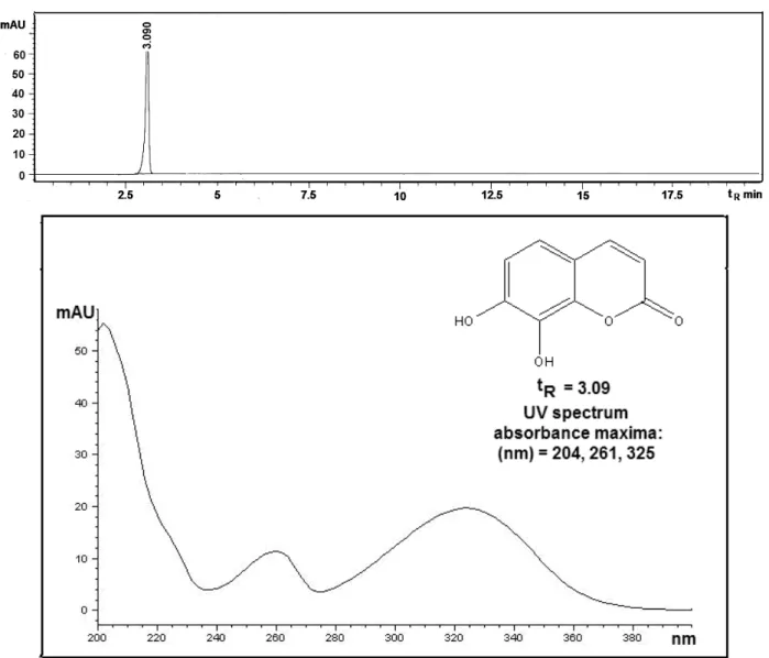
7,8-dihydroxy-coumarin with retention time tR = 3.09 min. Its
pre-sence was confirmed by comparing of the retention time and UV spectrum with the standard. This com-pound has previously been found in some Daphne spe-cies and is known under the name daphnetin [21].
Figure 2 shows the HPLC chromatograms of the methanol extract of twigs of D. cneorum from which it could be seen that daphnetin (tR = 3.09 min) is one of
the major secondary metabolite, as well as in the case of the leaves extract. The HPLC chromatogram and UV
spectrum of standard (with absorbance maxima: 204, 267 and 325 nm) are shown in Figure 3.
Antimicrobial activity
The results presented in Table 1 reveal antimicro-bial activity of the methanol extracts of D. cneorum within the concentration range from 15.62 to 62.50
μg/ml. The highest susceptibility to the methanol ex-tract of D. cneorum branches among the bacteria tested was exhibited by P. vulgaris ATCC 13315 (MIC = 15.62 Figure 1. HPLC Chromatograms of the methanol extract of the leaves of Daphne cneorum at wavelength detection at
254 and 320 nm, respectively.
μg/ml), followed by strains of B. subtilis ATCC 6633 and K. pneumoniae ATCC 13883 (MIC = 31.25 μg/ml) and P. mirabilis ATCC 14153, S. aureus ATCC 25923 and E. coli ATCC 25922 (MIC = 62.5 μg/ml). Among the fungi, C. albicans ATCC 10231 (MIC =31.25 μg/ml) showed the highest susceptibility. On the other hand, A. niger ATCC 16404 (MIC = 62.5 μg/ml) demonstrated the lowest susceptibility.
The highest susceptibility to the methanol extract of D. cneorum newspaper among the bacteria tested was exhibited by B. subtilis ATCC 6633 (MIC = 15.62 μg/ml), followed by strains of S. aureus ATCC 25923, E. coli ATCC 25922 and K. pneumoniae ATCC 13883 (MIC = = 31.25 μg/ml) and P. mirabilis ATCC 14153, P. vulgaris ATCC 13315 (MIC = 62.5 μg/ml). Among the fungi, C. albicans ATCC 10231 (MIC = 15.62 μg/ml) showed the Figure 3. HPLC Chromatogram and UV spectrum of daphnetin (tR = 3.09 min).
Table 1. Minimum inhibitory concentrations, μg/ml, of the methanol extract of Daphne cneorum
Microorganism Methanol extract of leaves Methanol extract of twigs Amracin Ketokonazol
Staphylococcus aureus ATCC 25923 62.5 31.25 0.98 Not tested
Klebsiella pneumoniae ATCC 13883 31.25 31.25 0.49 Not tested
Escherichia coli ATCC 25922 62.5 31.25 0.98 Not tested
Proteus vulgaris ATCC 13315 15.62 6.,5 1.95 Not tested
Proteus mirabilis ATCC 14153 62.5 6.5 1.95 Not tested
Bacillus subtilis ATCC 6633 31.25 15.62 0.24 Not tested
Candida albicans ATCC 10231 31.25 15.62 0.98 Not tested
highest, while A. niger ATCC 16404 (MIC = 62.5 μg/ml) exhibited the lowest susceptibility.
Antioxidant activity
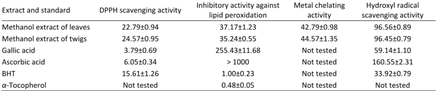
Phenolic compounds and flavonoids have been re-ported to be associated with antioxidant action in bio-logical systems, mainly due to their redox properties, which can play an important role in absorbing and neutralizing free radicals, quenching singlet and triplet oxygen, or decomposing peroxides [22]. One of the more prominent properties of flavonoids is their ex-cellent radical scavenging ability. It is also a valuable aspect for therapeutic and prophylactic applications of flavonoids [23]. The results on total phenolic, flavonoid and total antioxidant capacity of the methanol extracts of leaves and twigs of D. cneorum are given in Table 2. Total phenolic contents were determined and amounted to 76.45±0.79 and 69.67±0.85 mg GA/g, for leaves and twigs, respectively. The contents of flavonoids were determined and amounted to 24.67±0.35 and 34.23± ±0.89mg RU/g, for leaves and twigs, respectively.
Antioxidant activity of the methanol extracts was evaluated using the DPPH and hydroxy radical scaveng-ing, lipid peroxidation and metal chelating assays. The results (Table 3) of the antioxidant activity were com-pared with control antioxidants, ascorbic acid, gallic acid, α-tocopherol and BHT. The results showed that D. cneorum extracts possess antioxidant activity, with to-tal antioxidant capacity of 70.55±0.32 and 70.98±0.35
μg AA/g for leaves and twigs, respectively, and IC50
values of 22.79±0.94 and 24.57±0.95 μg/ml for DPPH free radical scavenging activity, 37.17±1.2 and 35.24± ±0.55 μg/ml for inhibitory activity against lipid peroxi-dation, 96.56±0.89 and 96.45±0.79 μg/ml for hydroxyl radical scavenging activity and 42.79±0.98 and 44.57± ±1.35 μg/ml for metal chelating activity, for leaves and twigs, respectively.
CONCLUSION
Insensitive research for new, unexplored, plant anti-oxidant sources is very significant and can bring new natural products in pharmaceutical and food industry for their every day battle with reactive oxygen species. Finding new antioxidant sources could be important for health benefits considering many diseases that reactive oxygen species induce in biological systems. Discover-ing a natural source of antioxidants could also be sig-nificant for artificial toxic antioxidants replacement in food industry. The results of this study clearly indicated that methanol plant extracts are good scavengers of synthetic DPPH radicals, indicating that they could be used as antioxidant products. Also, they all possess re-ductive capabilities. The rere-ductive capacities of the in-vestigated extracts are lower than those of the inves-tigated standard antioxidant compounds, so they could be considered primarily as antioxidants and then as products with reductive capabilities. All these charac-teristics uphold the use of supplements made this day in everyday free radical damage protection and pre-vention.
The methanol extracts of the leaves and twigs of D. cneorum exhibited good antimicrobial and antioxidant activities. HPLC–UV analysis showed that these two extracts contained 7,8-dihydroxycoumarin as one of the most abundant secondary metabolites. The activity of these extracts is probably derived from a coumarin derivative, a compound that has previously been found to possess various types of biological activity [24]. The results provided evidence that the studied plant might indeed be a potential source of natural antioxidant and antimicrobial agents.
Acknowledgement
The authors acknowledge the financial support of the Ministry of Education, Science and Technological Development of the Republic of Serbia (Grants No.
Table 2. Total phenolics, flavonoids and total antioxidant capacity of the methanol extracts of Daphne cneorum
Extract Total phenolics, mg GA/g Flavonoids, mg RU/g Total antioxidant capacity, μg AA/g
Methanol extract of leaves 76.45±0.79 24.67±0.35 70.55±0.32
Methanol extract of twigs 69.67±0.85 34.23±0.89 70.98±0.35
Table 3. Antioxidant activity (IC50 / µg ml –1
)of the Daphne cneorum extracts tested and standards
Extract and standard DPPH scavenging activity Inhibitory activity against lipid peroxidation
Metal chelating activity
Hydroxyl radical scavenging activity
Methanol extract of leaves 22.79±0.94 37.17±1.23 42.79±0.98 96.56±0.89
Methanol extract of twigs 24.57±0.95 35.24±0.55 44.57±1.35 96.45±0.79
Gallic acid 3.79±0.69 255.43±11.68 Not tested 59.14±1.10
Ascorbic acid 6.05±0.34 > 1000 Not tested 160.55±2.31
BHT 15.61±1.26 1.00±0.23 Not tested 33.92±0.79
172015 and 172047) and Junior Project (No 2011/05) of Medical Faculty University of Kragujevac, Serbia.
REFERENCES
[1] R. Lakušić, Planinske biljke, III izdanje, Zavod za udžbe-nike i nastavna sredstva, Beograd, 1990 (in Serbian). [2] V. Blečić, in: Flora S.R. Srbije 3, M. Josifović (Ed.), Srpska
akademija nauka i umetnosti, Beograd, 1972, p.p. 570– –578 (in Serbian).
[3] L. Pan, X.F. Zhang, Y. Deng, Y. Zhou, H. Wang, L.S. Ding Chemical constituents investigation of Daphne tangu-tica, Fitoterapia 81 (2010) 38–41.
[4] J. Ragot, P. Tubery, M. Carreras-Jansou, A.Lattes, P. Sy-monds, Isolament de la 5 primeverosyl genkwanine des racines de Daphne gnidium, Fitoterapia 59 (1988) 336– –337.
[5] C. Alonso, R. Perez, P.M. Nieto, J. Delgado, Gender di-morphism and altitudinal variation of secondary com-pounds in leaves of the gynodioecious shrub Daphne laureola, J. Chem. Ecol. 31 (2005) 139–150.
[6] A. Ulubelen, B. Terem, E. Tuzlaci, Coumarins and fl
avo-noids from Daphne gnidioides, J. Nat. Prod. 49 (1986) 692–694.
[7] N. Ullah, S. Ahmed, P. Mohammad, H. Rabnawaz, A. Malik, Chemical constituents of Daphne oleoides, Fito-terapia 70 (1999) 214–215.
[8] F. Cottiglia, G. Loy, D. Garau, C. Flori, M. Casu, R. Pom-pei, L. Bonsignore, Antimicrobial evaluation of couma-rins and flavonoids from the stems of Daphne gnidium
L., Phytomedicine 8 (2001) 302–305.
[9] S.M. Kupchan, R.L. Baxter, Mezerein: antileukemic prin-ciple isolated from Daphne mezereum L., Science 187
(1975) 652–653.
[10] L. Iauk, G. Aleo, F. Caccamo, A. Rapisarda, S. Ragusa, A.M. Speciale, Antibacterial and antimycotic activities of
Daphne gnidium L. extracts, Phytoter. Res. 10 (1996) 166–168.
[11] L. Iauk, G. Aleo, F. Caccamo, A., Rapisarda, S. Ragusa, A.M. Speciale, comparative evaluation of antibacterial and antimycotic activities of Daphne gnidium L. leaf and bark extracts, Farmaci Terapia 14 (1997) 37–43. [12] V. Singleton, R. Orthofer, R.M. Lamuela-Raventos,
Anal-ysis of total phenols and other oxidation substrates and antioxidants by means of Folin-Ciocalteu reagent, Me-thods Enzymol. 299 (1999) 152–175.
[13] I.M.C. Brighente, M. Dias, L.G. Verdi, M.G. Pizzolatti, Antioxidant activity and total phenolic content of some Brazilian species, Pharm. Biol.45 (2007) 156–161. [14] N.T. Manojlovic, P.J. Vasiljevic, W. Gritsanapan, R.
Su-pabphol, I. Manojlovic, Phytochemical and antioxidant studies of Laurera benguelensis growing in Thailand, Biol. Res. 43 (2010) 169–176.
[15] D. Satyajit, L.N. Sarker, Y. Kumarasamy, Microtitre plate based antibacterial assay incorporating resazurin as in-dicator of cell growth, and its application in the in vitro
antibacterial screening of phytochemicals, Methods 42
(2007) 321–324.
[16] P. Prieto, M. Pineda, M. Aguilar, Spectrophotometric quantitation of antioxidant capacity through the forma-tion of a phosphomolybdenum complex: specific appli-cation to the determination of vitamin E, Anal. Biochem.
269 (1999) 337–341.
[17] T. Takao, N. Watanabe, I. Yagi, K. Sakata, A simple screening method for antioxidants and isolation of se-veral antioxidants produced by marine bacteria from fish and shellfish, Biosci. Biotechnol. Biochem. 58
(1994) 1780–1783.
[18] Y. Kumarasamy, M. Byres, P. J. Cox, M.Jaspars, L. Nahar, S.D. Sarker, Screening seeds of some Scottish plants for free-radical scavenging activity, Phytother. Res. 21
(2007)615–621.
[19] C.K. Hsu, B.H. Chiang, Y.S. Chen, J.H. Yang, C.L. Liu, Im-proving the antioxidant activity of buckwheat ( Fago-pyrum tataricum Gaertn) sprout with trace element water, Food. Chem. 108 (2008) 633–641.
[20] I. Hinneburg, H.J.D. Dorman, R. Hiltunen, Antioxidant ac-tivities of extracts from selected culinary herbs and spices, Food. Chem. 97 (2006) 122–129.
[21] W. Xu, H. Jin, W. Zhang, X. Hu, W. Zhang, J. Fu, J. Su, S. Yan, Y. Shen, Chemical constituents from Daphne pe-dunculata, Chem. Nat. Comp. 45 (2009) 417–419. [22] M.R. Saha, S.M.R. Hasan, R. Akter, M.M. Hossain, M.S.
Alam, M.A. Alam, M.E.H. Mazumder, In vitro free radical scavenging activity of methanol extract of the leaves of
Mimusops elengi Linn., Bangl. J. Vet. Med. 6 (2008) 197– –202.
[23] B.H. Havsteen, The biochemistry and medical signifi-cance of the flavonoids, Pharmacol. Therapeut. 96
(2002) 67–202.
IZVOD
HPLC ANALIZA, ANTIMIKROBNA I ANTIOKSIDANTNA AKTIVNOST Daphne cneorum L.
Nedeljko T. Manojlović1, Pavle Z. Mašković2,Perica J. Vasiljević3, Ratomir M. Jelić1,Marina Ž. Jusković3, Miroslav
Sovrlić1, Leka Mandić2, Marija Radojković4 1
Univerzitet u Kragujevcu, Medicinski fakultet, Kragujevac, Srbija
2
Univerzitet u Kragujevcu, Agronomski fakultet, Čačak, Srbija
3
Univerzitet u Nišu, Prirodno–matematički fakultet, Niš, Srbija
4
Univerzitet u Novom Sadu, Tehnološki fakultet, Novi Sad, Srbija
(Naučni rad)
Ovaj rad prikazuje in vitro istraživanje antimikrobne i antioksidantne aktivnosti metanolskih ekstrakata lišća i grančica biljne vrste Daphne cneorum L. Antimik-robna aktivnost ovih ekstrakata testirana je na patogenim mikroorganizmima i određene su vrednosti minimalne inhibitorne koncentracije (MIC). Takođe je od-ređen ukupan sadržaj fenola i flavonoida, kao i antioksidantna svojstva ekstrakata. Testirani ekstrakti ispoljili su dobru antimikrobnu i antioksidantnu aktivnost. Re-zultati analize visokoefikasnom tečnom hromatografijom (HPLC) pokazali su da je 7,8-dihidroksikumarin jedan od najzastupljenijih sekundarnih metabolita u oba testirana ekstrakta ove vrste. Rezulatati ove studije jasno ukazuju da ekstrakati biljke D. cneorum mogu biti korišćeni kao potencijali izvoriprirodnih antioksida-nasa i antimikrobnih agensa.
Ključne reči: Daphne cneorum • HPLC •
Dafnetin • Antioksidantna aktivnost •