Florian Helm
1, Thomas Kammertoens
1, Frank M. Lehmann
2, Andrea Wilke
1, Heiko Bruns
4, Josef
Mautner
3, Georg W. Bornkamm
2, Armin Gerbitz
4*1 Department of Immunology, Charité Berlin, Berlin, Germany, 2 Institute of Clinical Molecular Biology and Tumor Genetics, Helmholtz Center, Munich, Munich, Germany, 3 Department of Pediatrics, Technical University (TU) Munich and Clinical Cooperation Group Pediatric Tumor Immunology, TU Munich and Helmholtz Center, Munich, Germany, 4 Department of Hematology, Oncology, University of Erlangen, Erlangen, Germany
Abstract
Over-expression of the proto-oncogene c-MYC is frequently observed in a variety of tumors and is a hallmark of Burkitt´s lymphoma. The fact that many tumors are oncogene-addicted to c-MYC, renders c-MYC a powerful target for anti-tumor therapy. Using a xenogenic vaccination strategy by immunizing C57BL/6 mice with human c-MYC protein or non-homologous peptides, we show that the human c-MYC protein, despite its high homology between mouse and man, contains several immunogenic epitopes presented in the context of murine Hβb haplotype. We
identified an MHC class II-restricted CD4+ T-cell epitope and therein an MHC class I-restricted CD8+ T-cell epitope
(SSPQGSPEPL) that, after prime/boost immunization, protected up to β5% of mice against a lethal lymphoma challenge. Lymphoma-rejecting animals contained MHC multimer-binding CD8+ cell within the peripheral blood and
displayed in vivo cytolytic activity with specificity for SSPQGSPEPL. Taken together these data suggest that oncogenic c-MYC can be targeted with specific T-cells.
Citation: Helm F, Kammertoens T, Lehmann FM, Wilke A, Bruns H, et al. (β01γ) Targeting c-MYC with T-Cells. PLoS ONE 8(10): e77γ75. doi:10.1γ71/ journal.pone.0077γ75
Editor: Andrei L. Gartel, University of Illinois at Chicago, United States of America Received January β0, β01γ; Accepted September 8, β01γ; Published October 10, β01γ
Copyright: © β01γ Helm et al. This is an open-access article distributed under the terms of the Creative Commons Attribution License, which permits unrestricted use, distribution, and reproduction in any medium, provided the original author and source are credited.
Funding: This work was supported by German Research Foundation (Deutsche Forschungsgemeinschaft, DFG) SFB TRγ6 (TPB4) and by the Wilhelm-Sander Stiftung. The funders had no role in study design, data collection and analysis, decision to publish, or preparation of the manuscript.
Competing interests: The authors have declared that no competing interests exist. * E-mail: [email protected]
Introduction
Cancer driving oncogenes frequently contain mutations in their coding sequences, but in many cases also remain wild-type and acquire their oncogenic property through uncontrolled expression. Since immunogenic mutations within the protein sequence are rare and may differ from patient to patient, T-cell based immunotherapy strategies focus on targeting tumor-associated or self-antigens. Targeting unmutated oncogenes in vivo is difficult due to central tolerance. However, by utilizing cross-species barriers in xenogenic immunization approaches, even highly conserved proteins can become immunogenic and stimulate the non-tolerant repertoire of the host, thereby allowing for the identification of T-cell receptors (TCR) with specificity for the oncogenic target [1].
The proto-oncogene c-MYC plays a crucial role in the pathogenesis of a large number of human tumors including B-cell lymphomas and leukemias as well as a variety of different epithelial tumors [β]. Unlike many other proto-oncogenes whose activity is dependent on mutations, truncation or gene fusion, the oncogenicity of c-MYC is in most cases the result of loss of transcriptional control leading to over-expression and accumulation of the unmutated protein itself. However, mutations within the c-MYC protein, although not a prerequisite
for rendering c-MYC oncogenic, have also been observed in a fraction of human B-cell lymphomas [γ-5]. In human Burkitt’s lymphoma, mouse plasmocytoma, and rat immunocytoma, activation of the c-MYC gene is brought about by chromosomal translocation of c-MYC into one of the three immunoglobulin heavy or light chain loci [6]. Thereby, the physiological regulation of the c-MYC gene is disrupted and the transcriptional regulatory elements of the immunoglobulin genes gain control over the juxtaposed c-MYC gene and govern its expression. In a variety of human epithelial tumors and also a subset of large diffuse B-cell lymphomas, the c-MYC gene is over-expressed as a consequence of gene amplification which correlates with poor prognosis [7,8]. Oncogenic activation of c-MYC can also occur through events upstream of c-MYC leading to uncontrolled c-MYC expression as observed for example in familial adenomatous polyposis and in K-RAS induced pulmonary carcinoma [9-11].. It thus appears that many, if not all, routes to cancer converge on c-MYC.
expressed in proliferating normal tissues like e.g. regenerating gut epithelium and hematopoietic cells. The expectation of severe adverse side effects has therefore hampered the development of therapeutic strategies targeting c-MYC for many years. This view has, however, been challenged recently by several groups [β,16,17] who argued that potential benefits may outweigh the risks of targeting c-MYC. The main two arguments in favor of an anti-c-MYC therapy are that (i) tumors are usually addicted to c-MYC and that even short-term interruption of c-MYC expression may drive tumor cells into apoptosis, rendering sustained anti-c-MYC therapy unnecessary [1γ], and (ii) that most normal cells are quiescent and side effects of c-MYC inhibiting proliferation of normal cells in the skin, the intestine and the hematopoietic system are relatively weak and reversible, and may be well tolerated [11].
T-cells have been proven to be effective for the treatment of a variety of malignant diseases. However, choosing unmutated c-MYC as a cell target bears two major obstacles: first, T-cells specific for c-MYC may be present only at low affinity and frequency or may be even non-existent due to negative selection in the thymus; secondly, induction of c-MYC specific T-cells may cause autoimmunity to highly proliferative tissues such as the hematopoietic system or the enteral mucosa.
Specific eradication of c-MYC over-expressing cells should result in long-term growth arrest and, ideally, in eradication of all malignant cells. To circumvent central tolerance, we aimed to generate T-cells against unmutated human c-MYC protein in C57BL/6 mice. Xenogeneic vaccination has been shown to break tolerance to self, and tumor associated antigens in some experimental models by inducing cross-reactive T cells [1,18,19]. Because murine c-MYC exhibits only 89.9% amino acid sequence identity to its human counterpart, differences in the amino acid sequence may give rise to T-cell responses and leave the function of tissues expressing murine c-Myc unaffected. Until now, human c-MYC has been used an as oncogene in a number of mouse models for induction of different tumors and has, so far, been considered as not immunogenic in the Hβb genetic background. Using a murine Burkitt´s lymphoma model over-expressing human c-MYC [β0], we show here that immunization of mice with human c-MYC protein and c-MYC derived non-homologous peptides elicits a c-MYC-specific CD4+ and CD8+ T-cell response. Furthermore, human c-MYC-reactive animals are protected from lethal doses of B-cell lymphoma cells over-expressing human c-MYC.
Methods
Mice
C57BL/6 mice were obtained from Charles River Laboratories at 6 weeks of age and housed in single ventilated IVC cages with a maximum of 5 mice per cage. Animals were sacrificed by COβ asphyxiation according to the guidelines of the local administration LaGeSo (Government of Berlin). Immunization and lymphoma transfer experiments were conducted under ethical approval (55.β-1-54-β5γ1-8-04 and β09.1/β11-β5γ1-8/04, Government of Bavaria, Munich, Germany)
Proteins and Peptides
Peptides (purity 70-95%) were purchased from Thermo Scientific biopolymers (Ulm, Germany). Chicken ovalbumin was purchased from Worthington (Lakewood, USA). To obtain c-MYC protein for vaccination, the human c-MYC gene, derived from the translocated allele of the BL60 lymphoma cell line, was amplified by PCR and cloned into the pTrcHis vector (Invitrogen) to generate pTrcMYCHis. Escherichia coli DH5α were transformed with pTrcMYCHis, selected clones were expanded and protein expression was induced by isopropyl -d-thiogalactopyranoside (IPTG) at a concentration of 1 mM. c-MYC protein was purified from bacterial lysates by Ni-NTA agarose (Qiagen, Germany) according to standard protocols. Protein concentration was determined by micro Lowry (Sigma) and presence of c-MYC protein was confirmed by Western blot using a c-MYC-specific antibody (clone 9E10, Invitrogen).
Epitope prediction
Epitope prediction for MHC class I-restricted peptides was performed using the HLA restrictor software [β1]. Epitopes were chosen depending on their peptide-MHC affinity (<500 nM) and the presence of at least one difference in the amino acid sequence between mouse and man. For MHC class II epitopes, the NetMHCII algorithm was used [ββ].
Immunization of mice
Mice were vaccinated with 50 µg protein (human c-MYC or OVA) or alternatively c-MYC-derived peptides (90 µg), together with CpG ODN18β6 (50 µg) (TIBMolBiol) in 50 µl PBS and 50 µl incompletes Freund´s adjuvant (Sigma-Aldrich). The vaccine was injected into the flanks adjacent to the inguinal lymph nodes.
In vivo depletion of CD25high cells
Monoclonal anti-CDβ5 antibody (clone PC61, kind gift from Elisabeth Kremmer, Helmholtz-Center Munich) was injected i.p. at a dose of 10 µg/g body weight [βγ]. Blood was taken 6 days past injection to confirm depletion of regulatory T-cells by staining of CDβ5 and FoxPγ-positive CD4+ cells. Mice were immunized 7 days after depletion with PC61-antibody and boosted three weeks after prime immunization.
In vivo cytotoxicity assay
In vivo cytotoxicity assays were performed as described previously [β4]. Splenic target cells from C57BL/6 mice were isolated and either loaded with target peptide (10 µM) for 15 min at γ7°C or used as unloaded controls. CSFE-labeling was performed for 15 min at γ7°C at a concentration of 0.1 µM CSFE (high) for peptide-loaded target and 0.01 µM CSFE (low) for unloaded control cells. Both cell populations were mixed at a 1:1 ratio and βx107 cells were injected i.v. in a volume of β00 µl PBS. Peripheral blood was obtained 18 h after injection.
IFNγ ELISPOT assay and IFNγ ELISA
per well were seeded in RPMI1640 supplemented with 10% FCS. Full length OVA or c-MYC protein was added at a concentration of 10 µg/ml and incubated for β4 hours at γ7°C. 1x10⁵ MACS-purified (Miltenyi) CD90.β (Miltenyi, Germany) purified T-cells from c-MYC-immunized mice were added and cultured for 48 hours. Spots were visualized by IFN detection antibody (clone R4-6Aβ) and streptavidin-linked alkaline phosphatase (γγβ1-βA, Mabtech, Germany). The number of spots was calculated using ELISPOT reader ELR0β with respective software (AID, Germany).
IFN concentrations were determined using an IFN ELISA (Becton-Dickinson, Germany) according to the instructions of the manufacturer.
Lymphoma challenge of mice
For tumor challenge experiments, 1x10⁵ β91PC cells [β5] were injected subcutaneously into the abdominal flanks. Tumor growth was monitored γ times per week using a sliding caliper. Mice were sacrificed when tumors reached a diameter of >10 mm. Lymphoma growth curves are plotted as a mean of all animals of one group.
Flow cytometric analysis
Cells were washed and incubated for 10 min in ice-cold PBS containing 0.5 µg/ml mAb clone β.4Gβ to block Fc- receptors. Fluorochrome-labeled monoclonal primary antibodies were incubated for γ0 min at 4°C in the dark. Cells were washed twice in ice-cold PBS and subsequently analyzed (FACSCalibur, Becton Dickinson).
For intracellular staining, cells were cultured for 1β hours in the presence of brefeldin-A. After staining of surface markers, cells were fixed and permeabilized using BD FixPerm (Becton Dickinson) according to the guidelines of the manufacturer. IFN was stained for γ0 min at 4°C using XMG1.β antibody. Cells were washed and analyzed subsequently. MHC-multimer staining was performed according to the manufacturer’s instructions (Proimmune).
Generation of APC, MCA205MYC-tet cells and coculture with T-cells
APC were prepared as described previously [β6]. Briefly, bone marrow cells were prepared from femurs of C57BL/6 mice and kept in Petri dishes for β hours at γ7°C in RPMI1640 media supplemented with 10% FCS. Non-adherent cells were collected and seeded in six-well plates in complete RPMI medium containing β0% of supernatant from murine GM-CSF secreting NIHγTγ cells [β7]. The final GM-CSF concentration ranged between 10-β0 ng/ml (GM-CSF ELISA, Becton-Dickinson). For APC-T-cell co-cultures, APC were harvested at day 1β and seeded in 96-well plates at a concentration of 1 to β.5x104 cells per well. c-MYC, OVA, or peptides were added at a concentration of β µg per 100 µl for 1β h. T-cells (1x10⁵) or splenic cells (5x10⁵) were added and incubated with APC for 48 hours at γ7°C. To increase uptake and cross-presentation of exogenously added protein [β8], the c-MYC-specific mAb 9E10 (kindly provided by Wolfgang Uckert, MDC Berlin) was added in some experiments at a concentration of 0.1 µg/ml.
MCAβ05 cells [β9] were transfected with pBCβ66 [γ0], using FuGene transfectant reagent (Promega) according to the manufacturer´s instructions. Cells were treated with hygromycin and one clone was selected to generate MCAβ05MYC-tet cell line 5x10⁴ cells were used for in vitro restimulation of β.5x105 splenic cells in a 96 well for 48 hours. To downregulate c-MYC expression, tetracycline (1µg/ml) was added β4 hours before coculture.
Real time PCR
Total RNA was isolated from lymphoma samples using RNeasy Plus (Qiagen) according to the manufacturer's instructions. After DNAse treatment, cDNA was prepared by reverse transcription with random decamers (Applied Biosystems). cDNA was analyzed by quantitative real time PCR (ABI step one) using the following primers (Metabion): 18S RNA forward 5-CGCCGCTAGAGGTGAAATTC-γ, reverse CGAACCTCCGACTTTCGTTCT-γ. c-myc RNA forward
5-CGCAAGACTCCAGCGCCTTCTC-γ, reverse
5-GGCGCTGCGTAGTTGTGCTGATG-γ. Ct values were quantified using appropriate software (Applied Biosystems) and expressed in arbitrary units (A.U.).
Results
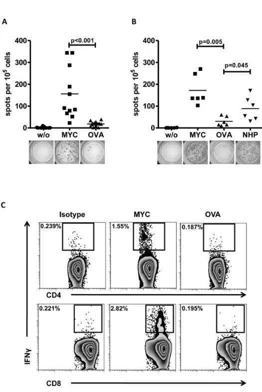
Immunization with full-length human c-MYC protein induces IFNγ secreting T-cells
primed with c-MYC protein and boosted with the peptide pool. On day β8 after first immunization splenic T-cells were CD90.β enriched and restimulated with either c-MYC-pulsed or peptide pool-pulsed APC. As shown in Figure 1B, the ELISPOT assay for IFN revealed that restimulation with peptide pool-pulsed APC resulted in a higher number of spots than restimulation with control protein OVA (p=0.045). When compared to c-MYC-pulsed APC, only γ out of 6 animals reached a comparable number of spots. However, analysis of supernatants from restimulation cultures by IFN ELISA displayed comparable concentrations of IFN between cells restimulated with c-MYC protein or peptide pool, whereas T-cells from naïve or OVA-immunized control mice revealed only background secretion levels (Figure Sβ). From these results we concluded that the non-homologous peptide pool contained the majority of the T-cell reactivity observed previously for the whole protein. Since both, the c-MYC protein as well as the non-homologous peptides used for immunization and restimulation contained both predicted MHC class I and class II epitopes, we next investigated whether the T-cell responses observed can be attributed to the CD4+ or the CD8+ compartment, or both. In these experiments, we incubated APC with c-MYC or OVA in the presence of a monoclonal, anti-human c-MYC antibody (clone 9E10) to enhance uptake and presentation of exogenous proteins [β8]. Intracellular IFN staining of splenic T-cells from immunized mice revealed activation of both, CD4+ and CD8+ T-cells when restimulated with human c-MYC protein in comparison to stimulation with OVA. As shown in Figure 1C, flow cytometric analysis after intracellular IFN staining identified 1.55% CD4+ and β.8β% of CD8+ responding T-cells after restimulation with c-MYC loaded APC. In contrast, OVA-loaded APC neither stimulated CD4+ nor CD8+ T-cells and there was no difference in IFN secretion when compared to isotype control staining.
Mapping of immunogenic regions by single peptide immunization
In order to identify more closely which non-homologous region within the peptides contained immunogenic regions, we performed single peptide immunizations. Mice were immunized twice in a similar fashion using single NHPs (A-H, shown in Table 1 and Figure S1). On day β8 after first immunization, spleens and inguinal lymph nodes from two mice were harvested and pooled, CD4+ T-cells were enriched by magnetic bead separation (80-90% purity) and re-stimulated with APC pulsed with the specific NHP used for immunization or an irrelevant control peptide as designated. Tissue culture supernatants were harvested after 48 hours. As shown in Figure βA, splenic CD4+ T-cells from mice immunized with either NHP-A, -B or -E responded to restimulation with the specific peptide. All other peptides (C, D, F, G, and H) did not result in specific release of IFN (data not shown). Restimulation of CD8+ T-cells with NHP-B-pulsed APC resulted in low amounts of IFN in the supernatant (Figure βB). All other NHPs failed to induce detectable IFN release. To verify the assumption that NHP-B contained both a MHC-class II and an additional MHC class I-restricted epitope, we stimulated whole splenic cells in addition to NHP-B with an
HβDb restricted NHP-B-derived 10mer (NHP-Bβ, SSPQGSPEPL) which resulted in IFN release (Figure βC). These results showed that CD8+ T-cells recognized a 10-mer of NHP-B. In fact, the NHP-Bβ-specific CD8+ T-cells could be detected by MHC class I HβDb NHP-Bβ-loaded multimers after immunization. As shown for one mouse in Figure γA, up to 4.γ8% of all CD8+ T-cells in the peripheral blood stained multimer positive. In contrast, CD8+ T-cells from OVA-immunized control animals failed to bind the SSPGQSPEPL multimer, but displayed binding of SIINFEKL multimer (1.61% of CD8+ T-cells). Due to the relatively low binding affinity of NHP-Bβ to HβDb (Table 1), we analyzed peripheral blood of individual animals 7 days after boost immunization. As shown in Figure γB, T-cell responses varied considerably among individual animals. The mean percentage of NHP-Bβ multimer-binding CD8+ T-cells was, however, significantly higher compared to control mice. To test whether the presence of NHP-Bβ multimer-binding CD8+ T-cells correlated with functional activity, we performed an in vivo killing assay. As shown in Figure γC, NHP-B-immunized animals displayed specific killing of NHP-Bβ peptide-pulsed splenic target cells, whereas OVA-immunized animals did not (p=0.0ββ). In contrast, NHP-E-immunized animals did not show any specific lysis of NHP-E-pulsed targets in comparison to naïve mice (p=0.558). Of note, the amount of lytic activity observed for NHP-B-immunized animals was comparable to the positive control, OVA-immunized mice receiving SIINFEKL-pulsed targets as shown in Figure γD.
Immunization with NHP-B protects against lymphoma challenge
Figure 1. Immunization with human c-MYC protein induces a T-cell response in C57BL/6 mice. A: ELISPOT assay: 1x10⁵ CD90.β-purified T-cells from c-MYC-immunized mice were cultured for 48 hours alone (w/o) or in the presence of 1x10⁴ APC loaded with either c-MYC or OVA protein. Every symbol depicts an individual mouse. Data are compiled from γ independent experiments (n=11). Representative pictures of ELISPOT wells are depicted below the diagram. B: Purified T-cells from mice immunized with full-length c-MYC protein and boosted with a NHP pool were cultured for 48 hours alone (w/o) or in the presence of 1x10⁴ APC loaded either with c-MYC, OVA or NHP pool. Data are summarized from β independent experiments (n=6). C: 5x10⁵ spleen cells from two c-MYC protein-immunized mice were pooled and cultured for 1β hours in the presence of 5x10⁴ protein-pulsed APC. OVA protein served as control. Intracellular cytokine staining for IFN revealed presence of MYC-specific T-cells in both, the CD4+ and the CD8+ T-cell compartment (representative samples from one out of two independent experiments).
4B, CDβ5depl+B). In contrast to immunization with NHP-B, single peptide immunization using NHP-A or –E had no protective effect on lymphoma growth and mortality.
Lymphoma survivors display T-cell reactivity in vivo and in vitro
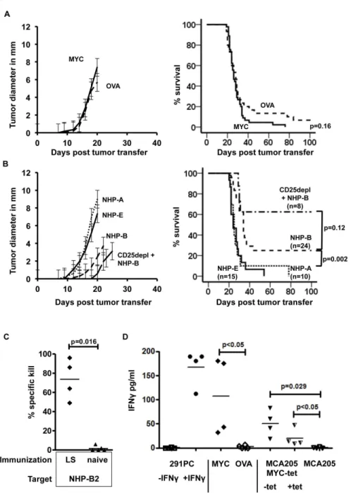
To analyze if rejection of lymphoma cells was associated with immunological memory against NHP-B presenting target cells, we challenged lymphoma surviving animals with peptide-loaded splenocytes and assessed in vivo killing activity. Animals surviving lymphoma challenge for more than 100 days were boosted with NHP-B and NHP-Bβ-pulsed target cells were injected i.v. 7 days after boost. Similarly to the results shown in Figure γC, lymphoma surviving (LS) animals displayed 50-90% specific killing activity, whereas naïve control mice did not (Figure 4C). In addition, splenic cells from LS animals were restimulated in vitro using different targets cells. In these animals, the frequency of SSPQGSPEPL multimer-binding splenic CD8+ T-cells was low and ranged from 0.γ% to 0.9% at day 100 after lymphoma challenge (data not shown). As shown in Figure 4D, when β.5x105 spleen cells were incubated with 1x10⁴ β91PC lymphoma cells (E:T ratio 0.08-0.βγ:1 with respect to multimer-binding CD8+ T-cells), we did not observe any secretion of IFN although these cells express human c-MYC. However, when β91PC cells were incubated with 100U/ml IFN for β4h prior to co-incubation and IFN was washed out before adding splenocytes, we observed a significant increase in IFN secretion. As shown previously, treatment of β91PC cells with IFN increases MHC I and II [β5] and leaves c-myc RNA levels upregulated (Figure Sγ left panel). IFN secretion could also be detected when
splenocytes were stimulated with c-MYC protein in presence of anti c-MYC monoclonal antibody (9E10) but not if OVA protein was added instead of c-MYC. In addition, restimulation of splenocytes from lymphoma surviving mice with a murine fibrosarcoma cell line, stably transfected to express human c-MYC (MCAβ05c-MYC-tet), also resulted in a significantly higher IFN secretion compared to untransfected MCAβ05 cells. c-MYC expression in MCAβ05c-MYC-tet cells can be negatively regulated by tetracycline (tet-off, Figure Sγ right panel). Interestingly, IFN decreased when MCAβ05MYC-tet cells were pretreated with 1.0 µg/ml tetracycline β4h prior to coculture with splenocytes. This pointed towards a dose dependent recognition of target cells by NHP-B specific T-cells. Untransfected MCAβ05 cells did not induce IFN secretion by splenocytes from LS mice.
Survival after lymphoma challenge is associated with the presence of SSPQGSPEPL-recognizing T-cells
Since immunization with NHP-B led to the induction of a CD8+ T-cell response and improved survival after lymphoma challenge, we analyzed if the frequency of NHP-Bβ-specific, SSPQGSPEPL-multimer-binding T-cells correlated with improved long-term survival. As shown in Figure 5A for individual animals, immunization and multiple boosts with NHP-B induced multimer-binding CD8+ T-cells in the peripheral blood with varying frequencies (also see Figure γB) that can be segregated into two groups: above 1% multimer-binding CD8+ T-cells (high) and below 1% multimer-binding CD8+ T-cells (low). The right panel of Figure 5A illustrates the distribution of NHP-Bβ multimer-binding CD8+ T-cells, which was significantly different between the two groups and from naïve animals.
Table 1. Epitope prediction for non-homologous peptides in the context of Hβb.
NHP Sequence MHC Core sequence
Differences Human ‐ >
Mouse Affinity Peptide‐MHC
A TMPLNVSFTNRNYDLDYDSVQPYFYCDEEENFYQQQQQSEL Hβ‐Db Hβ‐Kb Hβ‐IAb
VSFTNRNYDL VSFTNRNYDL LNVSFTNRN
S‐ >N S‐ >N S‐ >N 410 nM 8β nM 408 nM40aa
B DSSSPKSCASQDSSAFSPSSDSLLSS(T)ESSPQGSPEPLVL Hβ‐Db Hβ‐Kb Hβ‐IAb
SSPQGSPEPL SAFSPSSDSLL FSPSSDSLL
Q‐ >R, G‐ >A S‐ >T β48 nM 176 nM 458 nMγ0aa
C VEKRQAPGKRSESGSPSAGGHSKP Hβ‐IAb SESGSPSAG P‐ >S, S‐ >P, A‐ >S, G‐ >A ‐1,4 µM β4aa
D TAYILSVQAEEQKLISEEDLLRKRREQL Hβ‐IAb YILSVQAEE V‐ >I, E‐ >D ‐7 µM 15aa
E PSYVAV()TPFSLRGDNDGGGGSFSTADQLEMVTELLG Hβ‐Kb Hβ‐ IAb
VAV()TPFSL YVAV()TPFSL
‐ >A, P‐ >S, L‐ >P ‐ >A, P‐
>S, L‐ >P γββ nM ββ4 nM γ6aa
F PAAKRVKLDSVRVLRQISNNRKCTSPRSSDTEENVKRRTHNV Hβ‐IAb RKCTSPRSS T‐ >S ‐β,7 µM 40aa
G YQAARKDSGSPNPARGHSVCS Hβ‐IAb KDSGSPNPA G‐ >T, P‐ >L, N‐ >S ‐1,β µM β1aa
H LYLQDLSAAASEC Hβ‐IAb YLQDLSAAA S‐ >T ‐1,1 µM 1γaa
Peptides overlapping non-homologous regions (also see Figure S1) of human c-MYC protein were analyzed using the HLA restrictor software. MHC class I (HβDb, HβKb
)-restricted epitopes were selected according to their affinity <500 nM and at least one amino acid difference. Affinity >50nm is considered weak binding. MHC class II (Hβ-IAb) epitopes were chosen according to differences in amino acid sequence and best binding affinity depending on peptide length, since MHC class II epitopes can vary in
When growth of lymphomas and survival was analyzed according to grouping of animals into multimer-high and -low groups (Figure 5B), we observed a delay in lymphoma growth compared to naïve animals and a significant difference in
survival (p=0.044, multimer-low vs. naïve). Multimer-high animals displayed a delay in tumor outgrowth with a tendency to prolonged survival, but due to low numbers of mice in both groups, this did not reach statistical significance (p=0.1γ,
Figure 2. Non-homologous peptides induce CD4+ and CD8+ T-cell responses. A: CD4+ T-cells were isolated from spleens of mice immunized with single peptides as indicated. T-cells were restimulated for 48h in the presence of unloaded control APC (w/o) or APC loaded with the respective NHP used for immunization. Peptides not used for immunization served as controls. IFN secretion was measured by ELISA of culture supernatant. Representative results from three independent experiments are shown. B: CD8+ T-cells were restimulated in a similar fashion. Only NHP-B immunization/restimulation resulted in IFN secretion. C: Splenocytes from NHP-B immunized mice were restimulated with long peptides and a predicted short HβDb epitope (NHP-Bβ, 10mer, SSPQGSPEPL) derived from NHP-B. IFN secretion was measured by ELISA of culture supernatants (representative example of one out of three independent experiments using γ animals per group).
Figure 3. NHP-B immunization induces SSPQGSPEPL-specific CD8+ T-cells with cytolytic activity. A: Flow cytometric analysis of peripheral blood mononuclear cells from immunized mice (NHP-B vs. OVA) 7 days after boost immunization using either HβDb multimer-loaded with NHP-Bβ, or HβKb multimer loaded with SIINFEKL for control. B: Comparison of the frequencies of NHP-Bβ-specific CD8+ T-cells in individual mice after boost immunization according to different vaccines (NHP-B n=β8, OVA n=9, NHP-E n=8). C: Invivo killing assay: animals were immunized with NHP-B, NHP-E or OVA. Naive animals served as controls. Animals were challenged with peptide-pulsed target splenocytes and percentage of specific killing was assessed 18 hours after injection. D: Representative examples of flow cytometric analysis of invivo killing assay.
Figure 4. Immunization and challenge with c-MYC over-expressing lymphomas. Immunized recipient mice were challenged with human c-MYC over-expressing B-cell lymphoma cells subcutaneously 7 days past boost immunization. When tumors reached a diameter of more than 10 mm, mice were sacrificed and survival was plotted. Combined data from γ independent experiments are shown. A: Mice were immunized with a vaccine containing c-MYC protein (solid line, n=45) or OVA protein (dashed line, n=γ0). Lymphomas grew out simultaneously in both groups and no significant difference in survival was observed (p=0.16). B: Vaccine comprised NHP-A (dotted line, n=10), NHP-E (solid line, n=15) or NHP-B (dashed line, n=β4). NHP-B immunization resulted in delayed lymphoma outgrowth and significantly improved survival compared to NHP-A (p=0.006) and NHP-E (p=0.001) vaccination. Depletion of CDβ5+ cells prior to immunization with NHP-B further delayed lymphoma outgrowth and improved survival (CDβ5depl +NHP-B). C: In vivo killing assay: lymphoma surviving animals (LS) were challenged with NHP-Bβ-pulsed splenocytes and percentage of specific killing was assessed 18 hours after injection. Lymphoma surviving animals display a significantly higher in vivo killing activity compared to naïve animals (p=0.016, Mann-Whitney test). D: IFN secretion after stimulation of splenocytes from lymphoma-rejecting mice using c-MYC over-expressing β91PC stimulator cells with (+IFN ) or without (-IFN ) prior IFN treatment (100U/ml), c-MYC or OVA protein, MCAβ05MYC-tet with (+tet) and without (-tet) prior tetracycline treatment (1µg/ml) or untransfected MCAβ05 cells (C and D, n=4 animals per group).
multimer-high vs. multimer-low). However, when we analyzed binding of NHP-Bβ multimer in animals that failed to reject β91PC cells (NR) compared to lymphoma survivors (LS), we observed a significant difference in multimer-binding capacity of CD8+ T-cells (p=0.0ββ, Figure 5C).
Discussion
The work presented here aims to lay the groundwork for c-MYC-specific adoptive T-cell therapy. The use of adoptively transferred T-cells to fight cancer has been effective in a variety of animal models [γ1], and also in first clinical applications [γβ,γγ]. The therapeutic efficacy of these T-cell-based approaches is dependent on several factors. On one hand, there are important prerequisites on the side of the T-cells. For example, the proper functionality of T-cells, which is often associated with IFN secretion, is of importance. Similarly, avidity of the T-cell for the target antigen is critical and many strategies were suggested to identify and obtain high affinity T-cells or TCRs to improve adoptive T-cell transfer [γ4-γ6]. On the other hand, there are a number of factors on the side of the tumor cell itself and the tumor host, that critically influence effectiveness of adoptively transferred T-cells. For example, general immunogenicity of the tumor target, the immunosuppressive microenvironment, age of the host, and pretreatment may influence anti-tumor therapy (ATT). One of the most critical variables is the tumor antigen. Here, several aspects need to be taken into account: (i) The targeted antigen has to be expressed within the tumor at amounts that allow for recognition in the context of MHC. (ii) The antigen processing and presentation machinery needs to be functional. (iii) The antigen should ideally be expressed in a tumor-specific fashion and not by other tissues, to avoid autoimmunity. (iv) Furthermore, to reduce the risk of antigen loss variants that may cause relapse, the dependency of the malignancy on expression of the antigen is important. Many models have demonstrated that high avidity T-cells will select for antigen loss variants within the tumor, if the malignant phenotype and growth of the tumor remain unaffected by shutting down antigen expression [γ7-γ9]. We have previously shown that antigen loss variants occur in the same model used for this study, when chicken ovalbumin (OVA) as a model antigen is targeted by specific OT-1 T-cells [β5]. In this model, outgrowth of lymphomas expressing OVA is delayed and only OVA-negative lymphomas arise in immunocompetent hosts. When untransduced β91PC cells were injected into wild type C57BL/6 animals, we also observed a delay in growth compared to STAT-1-/- recipients, which harbor a severe T-cell defect. We suspected from the delayed outgrowth in T-cell competent mice that the expression of human c-MYC might render β91PC cells antigenic, suggesting that the human c-MYC protein might represent a potential T-cell target. Since various oncogene activation pathways converge on c-MYC and malignant growth has been shown to be dependent on c-MYC over-expression, T-cell-based approaches against c-MYC may be effective against different types of tumors and are unlikely to result in the development of antigen loss variants.
In this study, we aimed at identifying c-MYC-reactive T-cells to target a poorly immunogenic c-MYC-overexpressing B-cell lymphoma. In contrast to other studies that successfully used ATT to reject solid tumors of clinically relevant size [γ1] [40] and often utilize highly immunogenic tumors, i.e. regressor tumors, the β91PC lymphoma target cells do not display a regressor phenotype [β5] [40]. To our surprise, we did not observe any difference in lymphoma growth after immunization with full-length c-MYC protein and subsequent challenge with β91 PC cells, even though we observed c-MYC-reactive CD4+ and CD8+ T-cells ex vivo. This finding was compatible with two not mutually exclusive possibilities: either the immunization protocol was suboptimal and the induced T-cells were not of high affinity, or regulatory T-cells were induced in parallel that suppressed the potentially protective response.
As an alternative to whole protein immunization we used peptides for vaccination encompassing the regions of highest amino sequence divergence. Contrary to most peptides that induced only a weak CD4+ and no CD8+ T-cell response, the 40mer NHP-B peptide, that differs by only five amino acids to the murine sequence (Figure S1), was the best IFN inducer for CD4+ cells and induced also significant amounts of IFN in CD8+ T-cells. Yet, contrary to previous experiments in which OVA proved to be a highly effective T-cell rejection antigen [β5], the presence of NHP-B reactive T-cells resulted in an only modest delay in tumor outgrowth. This is in accordance with the predicted low binding affinity of the target peptide to mouse MHC-I (predicted β48 nM).
Another reason for the failure of whole c-MYC protein or low efficacy of the large peptide vaccine to protect mice against lymphoma challenge might be the presence of regulatory T-cells. Depletion of T-regulatory cells has been shown to increase T-cell reactivity towards auto-antigens in vivo upon immunization [41]. Highly homologous proteins like human c-MYC with 89.9% amino acid identity to murine c-Myc may be particularly prone to activation of Tregs. In fact, Tregs proved to play an important role in limiting the immune response to human c-MYC, as Treg depletion with anti-CDβ5 antibody PC61 prior to immunization increased the survival of NHP-B-immunized animals after lymphoma challenge from β5% to 6β.5%.
Figure 5. Affinity and frequency of multimer-binding T-cells determine survival after lymphoma challenge. A: Frequency of NHP-Bβ-specific peripheral blood CD8+ T-cells determined by flow cytometric multimer staining. Animals were segregated into two groups: multimer high >1% of CD8+ T-cells, multimer low <1% of CD8+ T-cells. Each graph represents one animal. For comparison, multimer staining of 4 naïve animals is shown. The right panel displays the statistical analysis of animals analyzed. B: Survival curve and tumor growth according to multimer grouping. C: Mean fluorescence intensity (MFI) of peripheral blood multimer-binding CD8+ T-cells as indicator for T-cell avidity. Lymphoma-surviving animals (LS) display higher avidity compared to not rejecting (NR) recipients prior to lymphoma challenge.
angiogenesis arrest in c-Myc-addicted tumors when c-Myc expression is shut off [44]. It remains to be elucidated which cells of the immune system contribute to anti-cMYC-specific anti-tumor activity and whether c-MYC NHP-B specific CD4+ T-cells are required for long term tumor rejection in this model system.
In addition, several aspects remain that need further investigation: the most important concern is autoimmunity due to the expression of c-MYC in healthy, non-malignant proliferating tissues. In normal tissue c-MYC expression is tightly regulated [45], whereas in lymphomas and other tumors c-MYC is invariably deprived of its physiological control and constitutively switched on. The hematopoietic and the gastrointestinal system depend on c-MYC for tissue homeostasis and may be regarded as the most critical tissues for targeted anti-c-MYC therapy. In a murine K-RAS-driven lung cancer model designed to reversibly inhibit c-MYC, c-MYC inhibition not only induced regression of incipient and established lung tumors, it also exerted profound effects on normal regenerating tissues, as might be anticipated. Yet, these effects were well tolerated over extended periods of time of up to 60 days and were completely reversible [11]. This is encouraging and may suggest that side effects of anti-c-MYC-specific T-cells may also be tolerable. To address whether a therapeutic window exists between anti-tumor immunity of c-MYC-specific T-cells and autoimmunity, we have generated a humanized c-MYC mouse in which the endogenous murine c-Myc gene is replaced by the human c-MYC gene. This mouse expresses the human c-MYC protein under the physiological control of the endogenous murine c-Myc promoter, is viable, and does not display an obvious phenotype [46]. This mouse will allow us to investigate whether tissue expression of the antigen causes autoimmunity when c-MYC specific T-cells are administered. Another concern may be that T-cell responses against c-MYC will induce fratricide, as has been observed for survivin [47], because c-Myc is induced upon activation in T-cells [48]. These issues have to be experimentally addressed and need to be clarified in the mouse model before c-MYC-specific T cell therapy can be introduced into the clinic.
The high homology between human MYC and murine c-Myc is limiting the pool of divergent peptides to which a strong T-cell response may be elicited in the context of a given MHC class I allele. For anti-c-MYC-specific T-cell therapy in humans, it will be decisive whether a potent T-cell response can also be elicited in HLA-Aβ- and huTCR-transgenic mice [49] [γ6]. If so, T-cells of HLA-Aβ-positive cancer patients may be equipped with HLA-Aβ/c-MYC-specific TCR genes by retroviral gene transfer and such T-cells administered to HLA-Aβ-positive cancer patients.
However, the greatest challenge will be to generate novel genetic mouse models in which a much broader T-cell response against human c-MYC can be elicited. Notably, mice are viable and fertile in which the endogenous c-Myc gene has been replaced by the murine N-Myc gene [50]. The homology between human c-MYC and murine N-Myc protein is only γ7% amino acid identity and 5β% similarity suggesting that a much
broader T-cell response against human c-MYC may be raised in these mice, provided these mice are able to elicit a normal T-cell response. Importantly, the response of lymphocytes to mitogens does not appear to be impaired in these mice suggesting that such an approach may indeed be feasible.
In conclusion, our work has illustrated for the first time the principal feasibility of targeting human c-MYC with T-cells. Although c-MYC-specific adoptive T-cell therapy is still at its infancy, it may represent on a long run a highly attractive and promising novel tool that may complement classical anti-cancer chemotherapy.
Supporting Information
Figure S1. Comparison of murine and human c-myc amino acid sequence. Non homologous peptides (NHP) used for the study are framed.
(TIF)
Figure S2. IFNγ secretion after immunization and invitro
restimulation. 1x10⁵ T-cells from mice immunized with full-length c-MYC protein and boosted with a NHP pool (MYC-NHP) were cultured for 48 hours alone (w/o) or in the presence of 1x10⁴ APC pulsed with either c-MYC, OVA or NHP pool. T-cells from OVA immunized and naïve mice served as controls. (TIF)
Figure S3. Left panel: Human c-myc mRNA is expressed in 291 lymphoma cells and upregulated upon IFNγ treatment. Human c-myc cDNA was analyzed by quantitative real time PCR in murine splenocytes for control (SPL) and β91PC cells treated with or without IFN (100 U/ml) for β4 hours before RNA extraction. Right panel: Human c-MYC is expressed in MCAβ05MYC-tet sarcoma cells. Western blot analysis using human MYC specific antibody 9E10 reveals c-MYC protein in MCAβ05c-MYC-tet cells. After treatment withincreasing doses of tetracycline (0.1 and 1.0 µg/ml) for β4 hours, expression of c-MYC is downregulated. Untransfected MCAβ05 cells do not express human c-MYC.
(TIF)
Acknowledgements
The authors are very grateful to Elisabeth Kremmer for providing anti-CDβ5 antibody, to Wolfgang Uckert for providing anti-human c-MYC antibody, Maya Schreiber and Raji Jayaraman for critically reading the manuscript and helpful suggestions.
Author Contributions
References
1. Blankenstein T, Coulie PG, Gilboa E, Jaffee EM (β01β) The determinants of tumour immunogenicity. Nat Rev Cancer 1β: γ07-γ1γ. doi:10.10γ8/nrcγβ46. PubMed: ββγ78190.
β. Dang CV (β01β) MYC on the path to cancer. Cell 149: ββ-γ5. doi: 10.1016/j.cell.β01β.0γ.00γ. PubMed: ββ464γβ1.
γ. Bhatia K, Huppi K, Spangler G, Siwarski D, Iyer R et al. (199γ) Point mutations in the c-Myc transactivation domain are common in Burkitt's lymphoma and mouse plasmacytomas. Nat Genet 5: 56-61. doi: 10.10γ8/ng099γ-56. PubMed: 8ββ04β4.
4. Bhatia K, Spangler G, Gaidano G, Hamdy N, Dalla-Favera R et al. (1994) Mutations in the coding region of c-myc occur frequently in acquired immunodeficiency syndrome-associated lymphomas. Blood 84: 88γ-888. PubMed: 804γ869.
5. Albert T, Urlbauer B, Kohlhuber F, Hammersen B, Eick D (1994) Ongoing mutations in the N-terminal domain of c-Myc affect transactivation in Burkitt's lymphoma cell lines. Oncogene 9: 759-76γ. PubMed: 8108117.
6. Klein G (198γ) Specific chromosomal translocations and the genesis of B-cell-derived tumors in mice and men. Cell γβ: γ11-γ15. doi: 10.1016/009β-8674(8γ)90449-X. PubMed: 640βγ07.
7. Bubendorf L, Kononen J, Koivisto P, Schraml P, Moch H et al. (1999) Survey of gene amplifications during prostate cancer progression by high-throughout fluorescence in situ hybridization on tissue microarrays. Cancer Res 59: 80γ-806. PubMed: 100β9066.
8. Schraml P, Kononen J, Bubendorf L, Moch H, Bissig H et al. (1999) Tissue microarrays for gene amplification surveys in many different tumor types. Clin Cancer Res 5: 1966-1975. PubMed: 1047γ07γ. 9. He TC, Sparks AB, Rago C, Hermeking H, Zawel L et al. (1998)
Identification of c-MYC as a target of the APC pathway. Science β81: 1509-151β. doi:10.11β6/science.β81.5γ8β.1509. PubMed: 97β7977. 10. Sansom OJ, Meniel VS, Muncan V, Phesse TJ, Wilkins JA et al. (β007)
Myc deletion rescues Apc deficiency in the small intestine. Nature 446: 676-679. doi:10.10γ8/nature05674. PubMed: 17γ775γ1.
11. Soucek L, Whitfield J, Martins CP, Finch AJ, Murphy DJ et al. (β008) Modelling Myc inhibition as a cancer therapy. Nature 455: 679-68γ. doi: 10.10γ8/nature07β60. PubMed: 187166β4.
1β. Felsher DW, Bishop JM (1999) Reversible tumorigenesis by MYC in hematopoietic lineages. Mol Cell 4: 199-β07. doi:10.1016/ S1097-β765(00)80γ67-6. PubMed: 10488γγ5.
1γ. Jain M, Arvanitis C, Chu K, Dewey W, Leonhardt E et al. (β00β) Sustained loss of a neoplastic phenotype by brief inactivation of MYC. Science β97: 10β-104. doi:10.11β6/science.1071489. PubMed: 1β098700.
14. Shachaf CM, Kopelman AM, Arvanitis C, Karlsson A, Beer S et al. (β004) MYC inactivation uncovers pluripotent differentiation and tumour dormancy in hepatocellular cancer. Nature 4γ1: 111β-1117. doi: 10.10γ8/nature0γ04γ. PubMed: 15475948.
15. Pelengaris S, Khan M, Evan GI (β00β) Suppression of Myc-induced apoptosis in beta cells exposes multiple oncogenic properties of Myc and triggers carcinogenic progression. Cell 109: γβ1-γγ4. doi:10.1016/ S009β-8674(0β)007γ8-9. PubMed: 1β01598β.
16. Prochownik EV, Vogt PK (β010) Therapeutic Targeting of Myc. Genes Cancer 1: 650-659. doi:10.1177/1947601910γ77494. PubMed: β11γβ100.
17. Sodir NM, Evan GI (β011) Finding cancer's weakest link. Oncotarget β: 1γ07-1γ1γ. PubMed: βββ0β195.
18. Engelhorn ME, Guevara-Patiño JA, Noffz G, Hooper AT, Lou O et al. (β006) Autoimmunity and tumor immunity induced by immune responses to mutations in self. Nat Med 1β: 198-β06. doi:10.10γ8/ nm1γ6γ. PubMed: 16444β64.
19. Gold JS, Ferrone CR, Guevara-Patiño JA, Hawkins WG, Dyall R et al. (β00γ) A single heteroclitic epitope determines cancer immunity after xenogeneic DNA immunization against a tumor differentiation antigen. J Immunol 170: 5188-5194. PubMed: 1β7γ4γ66.
β0. Kovalchuk AL, Qi CF, Torrey TA, Taddesse-Heath L, Feigenbaum L et al. (β000) Burkitt lymphoma in the mouse. J Exp Med 19β: 118γ-1190. doi:10.1084/jem.19β.8.118γ. PubMed: 110γ4608.
β1. Erup Larsen M, Kloverpris H, Stryhn A, Koofhethile CK, Sims S et al. (β011) HLArestrictor--a tool for patient-specific predictions of HLA restriction elements and optimal epitopes within peptides. Immunogenetics 6γ: 4γ-55. doi:10.1007/s00β51-010-049γ-5. PubMed: β1079948.
ββ. Nielsen M, Lund O (β009) NN-align. An artificial neural network-based alignment algorithm for MHC class II peptide binding prediction. BMC Bioinformatics 10: β96.
βγ. Setiady YY, Coccia JA, Park PU (β010) In vivo depletion of CD4+FOXPγ+ Treg cells by the PC61 anti-CDβ5 monoclonal antibody
is mediated by FcgammaRIII+ phagocytes. Eur J Immunol 40: 780-786. doi:10.100β/eji.β009γ961γ. PubMed: β00γ9β97.
β4. Willimsky G, Blankenstein T (β005) Sporadic immunogenic tumours avoid destruction by inducing T-cell tolerance. Nature 4γ7: 141-146. doi:10.10γ8/nature0γ954. PubMed: 161γ6144.
β5. Gerbitz A, Sukumar M, Helm F, Wilke A, Friese C et al. (β01β) Stromal Interferon-gamma Signaling and Cross-Presentation Are Required to Eliminate Antigen-Loss Variants of B Cell Lymphomas in Mice. PLOS ONE 7: eγ455β. doi:10.1γ71/journal.pone.00γ455β. PubMed: ββ479645.
β6. Schüler T, Blankenstein T (β00β) Naive CD8(+) but not CD4(+) T cells induce maturation of dendritic cells. J Mol Med (Berl) 80: 5γγ-541. doi: 10.1007/s00109-00β-0γ60-4. PubMed: 1β185454.
β7. Ohl L, Mohaupt M, Czeloth N, Hintzen G, Kiafard Z et al. (β004) CCR7 governs skin dendritic cell migration under inflammatory and steady-state conditions. Immunity β1: β79-β88. doi:10.1016/j.immuni. β004.06.014. PubMed: 15γ08107.
β8. Regnault A, Lankar D, Lacabanne V, Rodriguez A, Théry C et al. (1999) Fcgamma receptor-mediated induction of dendritic cell maturation and major histocompatibility complex class I-restricted antigen presentation after immune complex internalization. J Exp Med 189: γ71-γ80. doi:10.1084/jem.189.β.γ71. PubMed: 989β619. β9. Barth RJ Jr., Bock SN, Mulé JJ, Rosenberg SA (1990) Unique murine
tumor-associated antigens identified by tumor infiltrating lymphocytes. J Immunol 144: 15γ1-15γ7. PubMed: βγ0γ716.
γ0. Pajic A, Spitkovsky D, Christoph B, Kempkes B, Schuhmacher M et al. (β000) Cell cycle activation by c-myc in a burkitt lymphoma model cell line. Int J Cancer 87: 787-79γ. doi:10.100β/1097-0β15(β0000915)87:6. PubMed: 10956γ86.
γ1. Anders K, Buschow C, Herrmann A, Milojkovic A, Loddenkemper C et al. (β011) Oncogene-targeting T cells reject large tumors while oncogene inactivation selects escape variants in mouse models of cancer. Cancer Cell β0: 755-767. doi:10.1016/j.ccr.β011.10.019. PubMed: ββ17β7β1.
γβ. Rosenberg SA, Restifo NP, Yang JC, Morgan RA, Dudley ME (β008) Adoptive cell transfer: a clinical path to effective cancer immunotherapy. Nat Rev Cancer 8: β99-γ08. doi:10.10γ8/nrcβγ55. PubMed: 18γ54418.
γγ. Restifo NP, Dudley ME, Rosenberg SA (β01β) Adoptive immunotherapy for cancer: harnessing the T cell response. Nat Rev Immunol 1β: β69-β81. doi:10.10γ8/nriγ191. PubMed: ββ4γ79γ9. γ4. Xue S, Gao L, Gillmore R, Bendle G, Holler A et al. (β004)
WT1-targeted immunotherapy of leukaemia. Blood Cells Mol Dis γγ: β88-β90. doi:10.1016/j.bcmd.β004.08.018. PubMed: 155β8146. γ5. Wilde S, Sommermeyer D, Frankenberger B, Schiemann M, Milosevic
S et al. (β009) Dendritic cells pulsed with RNA encoding allogeneic MHC and antigen induce T cells with superior antitumor activity and higher TCR functional avidity. Blood 114: β1γ1-β1γ9. doi:10.118β/ blood-β009-0γ-β09γ87. PubMed: 19587γ79.
γ6. Li LP, Lampert JC, Chen X, Leitao C, Popović J et al. (β010) Transgenic mice with a diverse human T cell antigen receptor repertoire. Nat Med 16: 10β9-10γ4. doi:10.10γ8/nm.β197. PubMed: β069γ99γ.
γ7. Mackensen A, Meidenbauer N, Vogl S, Laumer M, Berger J et al. (β006) Phase I study of adoptive T-cell therapy using antigen-specific CD8+ T cells for the treatment of patients with metastatic melanoma. J Clin Oncol β4: 5060-5069. doi:10.1β00/JCO.β006.07.1100. PubMed: 170751β5.
γ8. Restifo NP, Marincola FM, Kawakami Y, Taubenberger J, Yannelli JR et al. (1996) Loss of functional beta β-microglobulin in metastatic melanomas from five patients receiving immunotherapy. J Natl Cancer Inst 88: 100-108. doi:10.109γ/jnci/88.β.100. PubMed: 85γ7970. γ9. Yee C, Thompson JA, Roche P, Byrd DR, Lee PP et al. (β000)
Melanocyte destruction after antigen-specific immunotherapy of melanoma: direct evidence of t cell-mediated vitiligo. J Exp Med 19β: 16γ7-1644. doi:10.1084/jem.19β.11.16γ7. PubMed: 11104805. 40. Spiotto MT, Rowley DA, Schreiber H (β004) Bystander elimination of
antigen loss variants in established tumors. Nat Med 10: β94-β98. doi: 10.10γ8/nm999. PubMed: 14981514.
41. Yan X, Zhang X, Wang Y, Li X, Wang S et al. (β011) Regulatory T-cell depletion synergizes with gp96-mediated cellular responses and antitumor activity. Cancer Immunol Immunother 60: 176γ-1774. doi: 10.1007/s00β6β-011-1076-5. PubMed: β178959β.
4γ. Shanker A, Verdeil G, Buferne M, Inderberg-Suso EM, Puthier D et al. (β007) CD8 T cell help for innate antitumor immunity. J Immunol 179: 6651-666β. PubMed: 1798β055.
44. Rakhra K, Bachireddy P, Zabuawala T, Zeiser R, Xu L et al. (β010) CD4(+) T cells contribute to the remodeling of the microenvironment required for sustained tumor regression upon oncogene inactivation. Cancer Cell 18: 485-498. doi:10.1016/j.ccr.β010.10.00β. PubMed: β10γ5406.
45. Henriksson M, Lüscher B (1996) Proteins of the Myc network: essential regulators of cell growth and differentiation. Adv Cancer Res 68: 109-18β. doi:10.1016/S0065-βγ0X(08)60γ5γ-X. PubMed: 871β067. 46. Lehmann FM, Feicht S, Helm F, Maurberger A, Ladinig C et al. (β01β)
Humanized c-Myc Mouse. PLOS ONE 7: e4β0β1. doi:10.1γ71/ journal.pone.004β0β1. PubMed: ββ860051.
47. Leisegang M, Wilde S, Spranger S, Milosevic S, Frankenberger B et al. (β010) MHC-restricted fratricide of human lymphocytes expressing
survivin-specific transgenic T cell receptors. J Clin Invest 1β0: γ869-γ877. doi:10.117β/JCI4γ4γ7. PubMed: β0978γ48.
48. Wang R, Dillon CP, Shi LZ, Milasta S, Carter R et al. (β011) The transcription factor Myc controls metabolic reprogramming upon T lymphocyte activation. Immunity γ5: 871-88β. doi:10.1016/j.immuni. β011.09.0β1. PubMed: ββ195744.
49. Pascolo S, Bervas N, Ure JM, Smith AG, Lemonnier FA et al. (1997) HLA-Aβ.1-restricted education and cytolytic activity of CD8(+) T lymphocytes from betaβ microglobulin (betaβm) HLA-Aβ.1 monochain transgenic H-βDb betaβm double knockout mice. J Exp Med 185: β04γ-β051. doi:10.1084/jem.185.1β.β04γ. PubMed: 918β675. 50. Malynn BA, de Alboran IM, O'Hagan RC, Bronson R, Davidson L et al.