Preconditioning with mono and polyunsaturated fatty acids and low-intensity
electrical stimulation. Effects on skin repair in rats
1Maria dos Prazeres Carneiro CardosoI, Andréa de Oliveira AlbuquerqueII, Virginia Claudia Carneiro GirãoIII, Margarida Maria
de Lima PompeuIV, Cícero Igor Simões Moura SilvaV, Orleâncio Gomes Ritardo de AzevedoVI, Sergio Botelho GuimarãesVII,
Paulo Roberto Leitão de VasconcelosVIII
DOI: http://dx.doi.org/10.1590/S0102-86502015002000004
IFellow Master degree, Postgraduate Program in Surgery, Department of Surgery, UFC, Ceara, Brazil. Conception of the study, technical procedures,
acquisition of data.
IIGraduate student, Nursing School, UFC, Ceara, Brazil. Animal care, acquisition of data.
IIIAssociate Professor, Department of Morphology, Faculty of Medicine, UFC, Ceara, Brazil. Histopathological analysis. IVAssociate Professor, Department of Morphology, Faculty of Medicine, UFC, Ceara, Brazil. Imunohistochemistry analysis. VPhD, Department of Surgery, Faculty of Medicine, UFC, Ceara, Brazil. Analysis and interpretation of data.
VIPhD, Laboratory of Ontogeny and Nutrition of Tissue Healing, Institute of Biomedicine of the Brazilian Semi Arid, UFC, Ceara, Brazil. Drafting of
the manuscript.
VIIPhD, Associate Professor, Department of Surgery. Head, Surgical Research Laboratory (LABCEX), UFC, Ceara, Brazil. Manuscript writing, critical
revision.
VIIIPhD, Full Professor, Department of Surgery. Coordinator, Postgraduate Program in Surgery, UFC, Ceara, Brazil. Conception, design, intellectual and
scientiic content of the study, critical revision, inal approval of manuscript.
ABSTRACT
PURPOSE: To evaluate the effects of preconditioning with oils mixes containing ω3/ω6/ω9 associated with micro-currents on skin
repair in rats.
METHODS: One-hundred and eight Wistar rats randomized into G-1, G-2 and G-3 groups were treated with saline (0.9%), mix 1
(corn+soybean oils) and mix 2 (olive+canola+laxseed oils), respectively, in a single dose (0.01ml/g) by gavage. Next, each group was
subdivided into sham and stimulated subgroups. Pulsed-wave microcurrents (0.5 µA, 0.5 Hz) were applied to stimulated subgroups for 20 min. One hour later anesthetized rats were subjected to surgery. A dorsal incision (6 cm long) was carried out and closed with interrupted nylon sutures. Samples (1cm2) were harvested from the mid-portion of the incision on the 7, 14, 21 post-operative (P.O.) days. Variables were analyzed using Mann-Whitney/Dunn tests Signiicance level was set to 5 % (p<0.05).
RESULTS: Micro-currents promoted increase of exudate and reduction of epithelialization on day 7 in G1 rats. Mixes 1/2 reduced
vascularization on 7/14th days P.O. Both 1/2 mixes reduced ibrosis on day 14. Preconditioning with mix 1 led to increased expression of NF-kB on the 7th day.
CONCLUSION: Preconditioning with microcurrents has pro-inlammatory effects while oil mixes 1 and 2 decrease ibrosis and
vascularization in the proliferative phase of cicatrization.
Introduction
Wound healing is a complex process that involves a series of biochemical and cellular reactions and. occurs as a sequence of
events, which includes inlammation, proliferation, and migration
of different cell types1. After forty-eight hours the inlammatory iniltrate consisting of monocytes is predominantly due to the short lifetime of neutrophils. Leukocytes play phagocytosis on
the aggressors, promoting the death of microorganisms, leading to the release of products to the extracellular medium, increasing the
initial inlammatory effect2.
Vascular changes consist of a cascade of activated and controlled chemical mediators reactions. These mediators act on the microcirculation, leading to increased vascular permeability
and can be classiied by mediators of fast-transient action and
long-acting mediators2. Nuclear factor kappa-light-chain-enhancer of activated B cells (NF-kB) is a nuclear transcription factor that
is activated by lipopolysaccharide agents and has the ability to bind to a sequence of 10 base pairs of the promoter region of the gene encoding the light chain of the molecules of KB cells3. Regardless of the stimulus, there may be involvement of reactive oxygen species and increased intracellular calcium for activation
of NF-kB. When not stimulated NF-kB is found in the cytoplasm,
bound to an inhibitory protein, IKB; this complex prevents the
translocation of NF-kB to the nucleus. Thus, phosphorylation and degradation of IkB are required for translocation4.
Heat shock response is one of the protective mechanisms evolutionarily acquired and is promoted by heat shock proteins
(HSP), which retain their intact structures under such conditions, assisting in the maintenance and proper formation of proteins5. HSP-27 is a heat shock protein with low molecular weight that
interact with polyunsaturated fatty acids. These proteins have
molecular weights between 18 and 30kDa and are called small
HSPs (small HSP). Its constitutive expression can be found in the nucleus and in the cytoplasm and are functionally involved in preventing denaturation of proteins and protection against cell injury and death6.
Peroxynitrite anion (ONOO−), the product of the
reaction between superoxide anion (O2.- ) and nitric oxide
(·NO) and its conjugated acid, peroxynitrous acid (ONOOH),
are potent oxidants known to be formed in vivo7. Peroxynitrite can enhance inlammatory mediator pathways8. Nitrotyrosine
in another component that participates in the nitration process involving radical mechanisms in which a derivative of the electron
peroxynitrite attacks the aromatic ring of tyrosine, leading to
formation of tyrosyl radical, which rapidly combines with nitrogen dioxide (NO2) equivalents to form 3-nitrotyrosine9.
The use of electrical stimulation in the injured tissue aims to accelerate the healing process. Clinical studies have shown that electrical stimulation induces an increase in a (ATP) concentration in tissues, increased protein synthesis, migration of epithelial cells
and ibroblasts in the region of the injury, reduction of the edema
and inhibition of the growth of some pathogens9.
There are three families of unsaturated fatty acids (PUFA)
omega-3 α-linolenic acid (ω - 3), omega-6 linoleic acid (ω - 6) and omega-9 oleic acid (ω - 9), The α-linolenic fatty acid (ALA , 18:3 n- 3) is the precursor of eicosapentaenoic acid (EPA , 20:5 n- 3), docosapentanóico (DPA , 22:5 n- 3), docosahexaenoic (DHA ; 22: 6 n -3) and linoleic acid (LA, 18:2 n-6), a precursor of arachidonic
acid (AA , 20:4 n -6). Both ALA and LA can not be synthesized by the human body and interconverted10-11.
Methods
Approval for experimental use of laboratory animals was obtained from the local Ethics Committee on Animal Use (CEPA), protocol #105/09, May 2012. All surgical procedures and animal handling were conducted in accordance with the Brazilian Federal Law Nº. 11794 of October 8, 2008 (http://www.planalto.gov.br/
ccivil_03/_Ato2007-2010/2008/Lei/L11794.htm).
One hundred and eight male Wistar rats (270-350g)
provided by the Central Animal Vivarium of the Federal University
of Ceara were randomized into three groups (n=36) for the
experiments (Table 1). All animals were housed in polypropylene cages at ambient temperature of 24oC ona 12 h light-darkcycle.
Oil mixes were purchased from NUTRIMED (Nutrição Enteral e Parenteral Ltda), Fortaleza-CE, Brazil. Antibodies
markers for heat shock protein 27(HSP27) (MsAbto-4-hydroxy); nitrotirosyne (NT) (39B6 monoclonal) and transcription factor kappa b (NFkB) (Santa Cruz P50 SC-mouse mononuclear) were
purchased from Imprint do Brazil (Campinas-SP).
Surgical procedure
Microcurrents were delivered by Stimulus Microcurrent Face® HTM (HTM-Electrical and Electronic Equipment Ltda, Sao Paulo, Brazil). The device generates ampliied continuous and
pulsed currents with two independent channel outputs, frequency 0.95 mA ± 10%, with inversion of polarity every 2.5 seconds. Selection of current intensity, frequency and duration of electric stimulation was based on previous publication12.
Anesthetized rats with ketamine hydrochloride
(Dopolen®, Agribands Brazil Ltda) 90mg/Kg + xylazine
hydrochloride (Rompun®, Bayer Animal Health) 10 mg/Kg intraperitoneally were positioned prone on a lat surface. A 6
cm long incision13 was performed, as depicted on Figure 1. The
incision was deepened through the skin and panniculus carnosus to supericial muscle fascia. The starting point of the incision had
an upper limit in the transverse line at the level of the inferior angle of the scapula. After seven, 14 and 21 days postoperatively,
the animals were again anesthetized for removal of skin samples
for laboratory and histological studies.
Histopathological analysis
Tissue specimens (skin fragments measuring 1x1cm) were processed routinely for light microscopy (ixating, dehydrating,
embedding, cutting, and staining with hematoxylin and eosin [HE]. The analysis of histological sections was performed by the
same pathologist blinded to the identiication of groups. The data was classiied according to the intensity of the healing process14
and transformed into quantitative variables as follows:
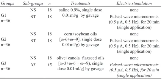
Groups Sub-groups n Treatments Electric stimulation
G1
n=36
NS 18 saline 0.9%, single dose 0.01ml/g by gavage
none
ST 18 Pulsed-wave microcurrents (0.5 µA, 0.5 Hz), for 20 min
(single application)
G2
n=36
NS 18 corn+soybean oils
[ω-6+ω--9], single dose
0.01ml/g) by gavage
none
ST 18 Pulsed-wave microcurrents (0.5 µA, 0.5 Hz), for 20 min
(single application)
G3 n=36
NS 18 olive+canola+laxseed oils
[ω-3+ω-6 + ω--9), single
dose 0.01ml/g) by gavage
none
ST 18 Pulsed-wave microcurrents
(0.5 µA, 0.5 Hz), for 20 min (single application)
TABLE 1 - Groups, number of animals, treatments and procedures.
ST=stimulated, NS=non-stimulated
Samples were collected from each subgroup on the 7th (n = 6), 14th (n = 6) and 21st day (n = 6) of the experi-ment. After sample collection, the animals were killed by an overdose of anesthetics (ketamine 500mg/kg + Xy -lazine250mg/kg)
Exsudates: 0=normal intensity, 1=mild; 2=moderate
and 3=intense
Epithelialization: 0=absent, 1=partial, 3=total Fibrosis: 0=absent, 1=mild, 2=moderate, 3=intense
Vascularization: 0=absent, 1=mild, 2=moderate,
3=intense
Immunohistochemistry analysis
The immunohistochemical reactions (Streptavidin-Biotin method) were performed at the laboratory of the Center for Studies in Microscopy Images and Procedures of the department of Morphology, Federal University of Ceara. Tissue sections
were deparafinized and rehydrated through xylene and graded
alcohols after antigen retrieval. Endogenous peroxidase was
blocked (15 min) with hydrogen peroxide and 3% in phosphate
buffered and washed in saline (PBS). Quantitative analysis of samples of epithelial cells in the incision areas were carried out with an Olympus Optical microscope - model DX41 (Olympus
Corporation, Tokyo, Japan) and a Digitimer Blood cell counter.
(Digitimer Ltd, Hertfordshire, England).
Statistical analysis
Statistical analysis was performed using Graphpad Prism 5.0 (GraphPad Software, San Diego California USA, www.graphpad. com). After testing all data for distribution, ANOVA/Bonferroni,
Kruskal-Wallis or Mann-Witney tests were used, as required. The signiicance level for rejecting the null hypothesis was 5% (p<0.05).
Results
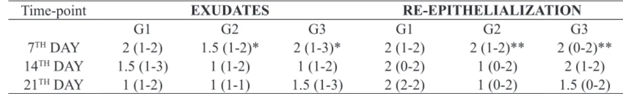
Effect of oil mixes (G2 and G3 groups) on exsudates and re-epithelialization of skin lesions in rats compared to negative control
Groups G2 and G3 rats (non-stimulated rats, treated with oil mixes) presented no signiicant differences when comparing
exudates and re-epithelialization variables with control groups,
in timepoints 14 and 21 days. However there was a signiicant
difference between rats on the 7th day regarding both the exudates
(p<0.05) and the re-epithelialization (p<0.01) (Table 2).
Time-point EXUDATES RE-EPITHELIALIZATION
G1 G2 G3 G1 G2 G3
7TH DAY 2 (1-2) 1.5 (1-2)* 2 (1-3)* 2 (1-2) 2 (1-2)** 2 (0-2)**
14TH DAY 1.5 (1-3) 1 (1-2) 1 (1-2) 2 (0-2) 1 (0-2) 2 (1-2)
21TH DAY 1 (1-2) 1 (1-1) 1.5 (1-3) 2 (2-2) 1 (0-2) 1.5 (0-2)
TABLE 2 - Effect of oil mixes (G2 and G3 groups) on exudates and re-epithelialization of skin lesions in rats compared to
negative controls.
Group G1, Negative Control (Saline); Group G2, Neutral control (ω-3+ω-6+ω-9); Group G3, test (ω-6+ω-9),. non-stimulated groups. The data represent Median+range (minimum-maximum) of microscopic scores. Tissue samples were collected on 7th, 14th and 21th days
after injury. Kruskal-Wallis/Dunn tests. Differences of G2, G3 groups (time points 14 and 21 days) compared to G1 are not signiicant. Differences on exsudates and re-epithelializaiton was identiied in G2 and, G3 rats (*p>0.05, **p<0,001 compared with G1)
Effect of oil mixes (G2 and G3 groups) on ibrosis
and vascularization of skin lesions in rats compared to negative control
Signiicant decrease in ibrosis and on day 14 in G2 and G3 and and in vascularization in G3 rats, compared with control
was identiied. Furthermore signiicant decreased vascularization
on the 7th post-operative day was observed in G2 rats compared
to G1 (Table 3).
Time-point FIBROSIS VASCULARIZATION
G1 G2 G3 G1 G2 G3
7TH DAY 2 (1-2) 1 (1-2) 2 (1-2) 2 (1-3) 0.5 (0-2) * 2 (1-3)
14TH DAY 2.5 (1-3) 1 (1-1) * 1 (1-1) * 2.5 (1-3) 1 (0-2) 0 (0-2) *
21TH DAY 1.5 (1-2) 1 (1-2) 1 (1-3) 1 (1-1) 1 (0-2) 0 (0-3)
TABLE 3 - Effect of oil mixes (G2 and G3 groups) on ibrosis and vascularization of skin lesions of rats compared to
negative control.
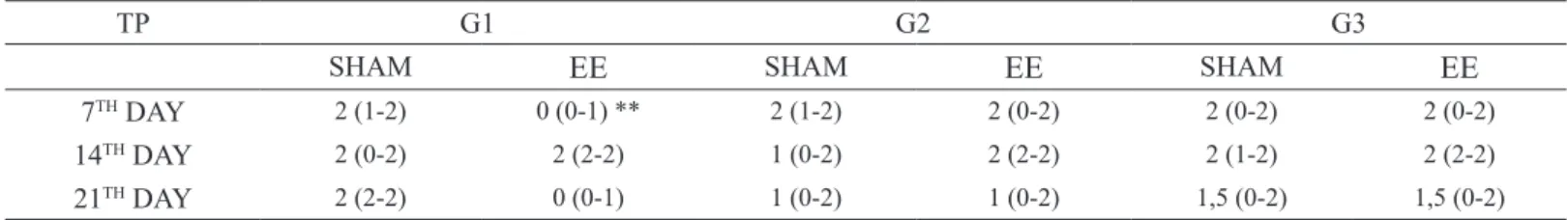
col-Effect of oil mixes (G2 and G3 groups) on exsudates of skin lesions in rats compared to sham subgroups
Signiicant increase (p<0.05) on exudates on day 7 was identiied when G1 rats were compared to sham subgroup
(Table 4).
Effect of oil mixes (G2 and G3 groups) on re-epithelization of skin lesions in rats compared to sham subgroups
Values were signiicantly different on day 7 (p<0.01) comparing stimulated vs. Sham groups. No signiicant differences
occurred on Day 14 and Day 21 timepoints (Table 5).
TP G1 G2 G3
SHAM EE SHAM EE SHAM EE
7TH DAY 2 (1-2) 3 (2-3)* 1.5 (1-2) 2 (1-3) 2 (1-3) 2 (1-3)
14TH DAY 1.5 (1-3) 2 (1-3) 1 (1-2) 2 (1-2) 1 (1-2) 1 (1-2)
21TH DAY 1 (1-2) 2 (1-3) 1 (1-1) 2 (1-3) 1.5 (1-3) 2 (1-3)
TABLE 4 - Effect of saline (G1) or oil mixes (G2 and G3 groups) on exudate of skin lesions of rats comparing Stimulated to
Sham subgroups.
TP = Time-point. Group G1, Negative Control (Saline);group G2, Neutral control ( ω-3+ω-6+ω-9); group G3, test (ω-6+ω-9),. non-stimulated (Sham) and stimulated (EE) groups. The data represent Median+range (minimum-maximum) of microscopic scores. Tissue samples were collected on 7th, 14th and 21th days after injury. Kruskal-Wallis/Dunn tests.
*p<0.05 compared to Sham
TP G1 G2 G3
SHAM EE SHAM EE SHAM EE
7TH DAY 2 (1-2) 0 (0-1) ** 2 (1-2) 2 (0-2) 2 (0-2) 2 (0-2)
14TH DAY 2 (0-2) 2 (2-2) 1 (0-2) 2 (2-2) 2 (1-2) 2 (2-2)
21TH DAY 2 (2-2) 0 (0-1) 1 (0-2) 1 (0-2) 1,5 (0-2) 1,5 (0-2)
TABLE 5 - Effect of saline (G1) or oil mixes (G2 and G3 groups) on re-epithelization of skin lesions of rats comparing
Stimulated to Sham subgroups.
Effect of oil mixes (G2 and G3 groups) on HSP27, nuclear transcription factor kappa b (NF-kB) and nitrotyrosine (NT) immunohistochemical results on the seventh day, in non-stimulated rats compared to negative control
HSP27 and NT expression levels were not signiicant different on the 7th day. However, NF-kB expression levels were signiicantly greater (p<0.05) in G2 rats compared to G1 at the
same timepoint (Table 6).
G1 G2 G3
HSP27 88.80 ± 7.01 76.60 ± 13.55 73.00 ± 17.12
NF-kB 23.50 (17 – 61) 87.50 (23 – 198)* 72.00 (14 – 110) NT 77.50 ± 15.21 76.20 ± 14.43 87.70 ± 16.08
TABLE 6 - Heat shock protein 27 (HSP27), nuclear
transcription factor kappa b (NF-kB) and nitrotyrosine (NT)
immunohistochemical results on the seventh day, in non-stimulated
rats treated with saline (G1) or oil mixes (G2 and G3 groups).
Histopathology
Figure 2 depicts histology indings of all groups.
Immunohistochemistry showed positive staining for epithelial
cells (black arrows) in stimulated G-3 rats, identiied by the intense
brown coloration (Figure 2).
FIGURE 2 - Histological section of rat skin from non-stimulated (A) and stimulated saline groups (B); corn+soybean oils non-stimulated (C) and stimulated (D) groups; olive+canola+laxseed oils non-stimulated (E) and stimulated (F) groups (x300).
Notes: A=Mono and polymorphonuclear cells, total reepithelializarion, moderate ibrosis; B=Mononuclear cells, total reepithelialization, mild vascularization; C=Mononuclear cells, total reepithelialization, mild vascularization; D=Exsudates, mononuclear cells, partial reepithelialization, moderate ibrosis; E=Mononuclear cells, partial reepithelialization, moderate ibrosis, no vascularization; F=Mononuclear cells, total reepithelialization, moderate ibrosis,. stained epithelial cells (black arrows).
Discussion
The relationship between endogenous electrical activity and wound healing has been investigated in several areas of clinical practice and has been well documented15-17. M i c r o c u r r e n t
electrical neuromuscular stimulation (MENS), applied for treating ulcers by venous impairment accelerates the healing process18. A
previous study16 demonstrated an increase of ATP generation in the skin of rats subjected to MENS, which makes evident the increase of
protein synthesis, as well as the increment of transportation by plasma
membrane. Considering that ATP is a key factor within the healing
calcium) transportation towards inside and outside the cell19. A recent
study investigated the effect of microcurrent stimulation (10µA) on wound healing in rats. The results have demonstrated that the treatment is effective in promoting tissue repair besides exerting positive effects
on the newly formed tissue area, number of ibroblasts, number of newly formed vessels, and epithelial thickness2.
Prior administration of nutraceutical blends for seven, 14 and 21 days before the surgical incision characterizes nutritional preconditioning. In our experiments, blends of oils containing polyunsaturated fatty acids were used: a mixture with nutraceutical
potential with ALA (canola+laxseed+olive oils) and an isolipidic
mixture without nutraceutical potential (corn+soybean oils), was used as a neutral control.
Tissue repair was studied in the different groups by
comparing inlammatory and proliferative processes and tissue
reorganization. For this purpose, samples were collected from the wound area seven, fourteen and 21 days after experimentally induced injury. Temporal differences in tissue repair were observed between the different treatments.
In the present study, the administration of oil mixes before surgery without associated electric stimulation had no
beneicial effects on the healing (exsudates and epithelialization
(Table 2) of wounds on the 14 and 21th day timepoints. However,
signiicant effects were observed on the 7th day timepoint
demonstrating a positive effect of the oil mixes. On the other hand
the absence of signiicant differences between oil+microcurrent
stimulation groups and control group (saline) concerning exudate and epithelialization variables opposes the result found in another study where the application of Jatropha curcas L. seed oil alone was not effective on experimental wound healing when compared to control, but microcurrent application alone or combined with
the oil exerted signiicant differences in the parameters studied. The use of ω-6+ω--9 oils (corn+soybean oils) promoted a signiicant decrease in vascularization on the 7th post-operative day and decreased ibrosis on 14th day (Table 3). When ω-3+ω-6+ω--9 mix (olive+canola+laxseed oils) was used the same effects
were observed at day 7 only. Therefore, when a single dose of corn+soybean oils is given, these effects last up to the 14th day.
Tissue expression of nitrotyrosine and HSP27 showed no statistical difference when comparing groups and timepoints. The results demonstrate that preconditioning with oil mixes as well as microcurrents do not promote and increase in nitrotyrosine
and HSP27 expressions in the skin of rats in this experimental model. However the increase in NF-kB expression (p<0.05) in G2 rats compared to G1 on the 7th day. shows the beneicial effect of corn+soybean oil mix during the irst phase of the healing process.
Conclusion
Preconditioning with microcurrents has
pro-inlammatory effects while oil mixes 1 and 2 decrease ibrosis and
vascularization in the proliferative phase of cicatrization, whereas preconditioning with microcurrents or mono and polyunsaturated oils induces no alterations in scar repair.
References
1. Agarwal PK, Singh A, Gaurav K, Goel S, Khanna HD, Goel RK. Evaluation of wound healing activity of extracts of plantain banana
(Musa sapientum var. paradisiaca) in rats. Indian J Exp Biol. 2009 Jan;47(1):32-40. PMID: 19317349.
2. Mendonça FA, Passarini Junior JR, Esquisatto MA, Mendonça JS,
Franchini CC, Santos GM. Effects of the application of Aloe vera (L.) and microcurrent on the healing of wounds surgically induced in Wistar rats. Acta Cir Bras. 2009 Mar-Apr;24(2):150-5. PMID:
19377785.
3. Sen R, Baltimore D. Multiple nuclear factors interact with the
immunoglobulin enhancer sequences. Cell. 1986 Aug
29;46(5):705-16. PMID: 3091258.
4. Siebenlist U. NF kappa B/I kappa B proteins. Their role in cell growth, differentiation and development. Madrid, Spain, July 7-10, 1996. Biochim Biophys Acta. 1997 Feb 22;1332(1):R7-13 PMID: 9061013.
5. Landry J, Huot J. Modulation of actin dynamics during stress and physiological stimulation by a signaling pathway involving p38 MAP kinase and heat-shock protein 27. Biochem Cell Biol. 1995 Sep-Oct;73(9-10):703-7. PMID: 8714691.
6. Okamoto CT. HSP27 and signaling to the actin cytoskeleton focus
on “HSP27 expression regulates CCK-induced changes of the actin
cytoskeleton in CHO-CCK-A cells”. Am J Physiol. 1999 Dec;277(6 Pt 1):C1029-31 PMID: 10600753.
7. Beckmann JS, Ye YZ, Anderson PG, Chen J, Accavitti MA, Tarpey
MM, White CR. Extensive nitration of protein tyrosines in human atherosclerosis detected by immunohistochemistry. Biol Chem
Hoppe Seyler. 1994 Feb;375(2):81-8. PMID: 8192861.
8. Levonen AL, Patel RP, Brookes P, Go YM, Jo H, Parthasarathy S,
Anderson PG, Darley-Usmar VM. Mechanisms of cell signaling by
nitric oxide and peroxynitrite: from mitochondria to MAP kinases. Antioxid Redox Signal. 2001 Apr;3(2):215-29. PMID: 11396477.
9. Padmaja S, Huie RE. The reaction of nitric oxide with organic
peroxyl radicals. Biochem Biophys Res Commun. 1993 Sep 15;195(2):539-44. PMID: 8373394.
10. Calder PC. Immunoregulatory and anti-inlammatory effects of n-3 polyunsaturated fatty acids. Braz J Med Biol Res. 1998 Abr;31(4):467-90.
11. Calder PC. Fatty acids and gene expression related to inlammation. Nestle Nutr Workshop Ser Clin Perform Programme. 2002;7:19-36. PMID: 12481693.
12. Santos VNS, Ferreira LM, Horibe EK, Duarte IS. Electric
microcurrent in the restoration of the skin undergone a trichloroacetic
acid peeling in rats. Acta Cir. Bras. 2004;19(5):466-70. doi: 10.1590/
S0102-86502004000500003.
13. Borba GC. Estimulação elétrica pré-incisional na cicatrização em
pele de rato (Dissertação). Universidade Federal de São Paulo; 2009.
wound healing in rats. Photomed Laser Surg. 2006 Aug;24(4):480-8. PMID: 1694242Aug;24(4):480-8.
15. Watson T. Current concepts in electrotherapy. Haemophilia. 2002
May;8(3):413-8. PMID: 12010443.
16. de G de Gaspi FO, Foglio MA, de Carvalho JE, Santos GM, Testa M, Passarini JR Jr, de Moraes CP, Esquisatto MA, Mendonça JS,
Mendonça FA. Effects of the topical application of hydroalcoholic
leaf extract of oncidium lexuosum sims (Orchidaceae) and
microcurrent on the healing of wounds surgically induced in Wistar
rats. Evid Based Complement Alternat Med. 2011;2011:950347. doi: 10.1155/2011/950347.
17. Castro FC, Magre A, Cherpinski R, Zelante PM, Neves LM,
Esquisatto MA, Mendonça FA, Santos GM. Effects of microcurrent application alone or in combination with topical Hypericum perforatum L. and Arnica montana L. on surgically induced wound
healing in Wistar rats. Homeopathy. 2012 Jul;101(3):147-53. doi:
10.1016/j.homp.2012.05.006.
18. Assimacopoulos D. Wound healing promotion by the use of negative
electric current. Am Surg. 1968 Jun;34(6):423-31. PMID: 5651495. 19. Cheng N, Van Hoof H, Bockx E, Hoogmartens MJ, Mulier JC, De
Dijcker FJ, Sansen WM, De Loecker W. The effects of electric currents
on ATP generation, protein synthesis, and membrane transport of rat
skin. Clin Orthop Relat Res. 1982 Nov-Dec;(171):264-72. PMID:
7140077.
Acknowledgements
To Mr. Osvaldo Fedell, Managing Director of HTM-Electronics (Amparo-SP, Brazil), who provided the Stimulus Face HTM equipment for this study; Mrs. Conceição da Silva Martins laboratory technician and Daniele Feijão de Souza nursing technician
for their valuable work during the development of this research.
Correspondence:
Prof. Dr. Paulo Roberto Leitão de Vasconcelos
Rua Prof. Costa Mendes, 1608/3º andar, Bloco Didático 60430-140 Fortaleza – CE Brasil
Tel.: (55 85)3366-8063
Received: Oct 15, 2014 Review: Dec 17, 2014
Accepted: Jan 12, 2015 Conlict of interest: none
Financial source: National Council for Scientiic and Technological De -velopment (CNPq)
1Research performed at Experimental Surgery Laboratory (LABCEX),