ANAlySIS oF DyNAmIC pulmoNARy hypeRINFlAtIoN (Dh)
FollowINg ACtIvItIeS oF DAIly lIvINg IN pAtIeNtS wIth ChRoNIC
oBStRuCtIve pulmoNARy DISeASe
S
clauSerP
eSSoaIMB
1, P
arreIraVF
2, l
orenzoVaP
3, r
eISMaS
4& c
oStaD
51 Department of physical therapy, pontifícia universidade Católica de minas gerais, Betim, mg - Brazil
2 Department of physical therapy, universidade Federal de minas gerais, Belo horizonte, mg - Brazil
3 Department of physical therapy, universidade Federal de São Carlos, São Carlos, Sp - Brazil
4 Department of medicine, Faculdade de Ciências médicas de Belo horizonte, Belo horizonte, mg - Brazil
5 graduate physical therapy program, Faculty of health Sciences, universidade metodista de piracicaba, piracicaba, Sp - Brazil
Correspondence to: Dirceu Costa, laboratório de Avaliação Funcional Respiratória, Faculdade de Ciências da Saúde, uNImep, Rod. do Açúcar, Km 156, taquaral, Cep 13400-911, piracicaba, Sp – Brasil, e-mail: [email protected]
Received: 12/02/2007 - Revised: 12/06/2007 - Accepted: 25/07/2007
AbstrAct
Introduction: Dynamic hyperinflation (Dh) is one of the ventilatory mechanisms that may contribute towards limiting the activities of daily living (ADls) in patients with chronic obstructive pulmonary disease (CopD). the objectives of this study were to evaluate the presence of Dh, by means of inspiratory capacity (IC), IC / total lung capacity (tlC) ratio and by the sensation of dyspnea, following an ADl performed using the upper limbs. method: the participants were 32 individuals aged 54 to 87 years (69.4 ± 7.4) who presented moderate-to-severe CopD. the patients selected underwent pulmonary function tests, spirometry and whole-body plethysmography. For the spirometric and pulmonary volume maneuvers, a conventional system was used (vmax22 Autobox). the IC was determined using a vmax229d ventilatory measurement system. the patients were asked to lift up pots weighing between 0.5 and 5.0 kg over a five-minute period, picking up the pots from a surface at waist level and putting them onto a shelf above head height. All the patients were evaluated regarding IC and using the Borg scale for dyspnea. the data were analyzed using Student’s t test for paired samples, pearson’s correlation and the wilcoxon test (p< 0.05). Results: there were reductions in IC and IC/tlC (p= 0.0001) following the ADl. the dyspnea increased after the exercise (p< 0.05). Conclusion: the ADl using the upper limbs caused Dh, as shown by the reductions in IC and IC/tlC and also by the increase in dyspnea.
Key words: dynamic hyperinflation; Chronic obstructive pulmonary Disease; activities of daily living.
resumo
Análise da hiperinsulação pulmonar dinâmica (hd) após atividade de vida diária em pacientes
com doença pulmonar obstrutiva crônica
introdução: A hiperinsuflação dinâmica (hD) é um dos mecanismos ventilatórios que podem contribuir para a limitação das atividades de vida diária (AvD) em pacientes com Doença pulmonar obstrutiva Crônica (DpoC). os objetivos deste trabalho foram avaliar a presença da hD, pela capacidade inspiratória (CI), e sua razão CI/Cpt (capacidade pulmonar total), e a sensação de dispnéia após uma AvD realizada com os membros superiores (mmSS). métodos: participaram 32 pacientes com DpoC de moderada a muito grave, com idades entre 54 a 87 anos (69,4 ± 7,4). os pacientes selecionados foram submetidos a testes de função pulmonar, es-pirometria e pletismografia de corpo inteiro. para as manobras espirométricas e dos volumes pulmonares, foi utilizado um sistema convencional (vmáx22 Autobox). A CI foi determinada usando um sistema de medidas ventilatórias (vmáx229d). Foi solicitado elevar potes com pesos de 0,5 a 5,0kg no tempo total de 5 minutos, pegando os potes em cima de uma superfície situada no nível da cintura pélvica e posicionando-os em uma prateleira localizada acima do nível da cabeça. em todos os pacientes, foram avali-ados a CI e a escala de Borg para dispnéia. para a análise dos davali-ados, foram utilizavali-ados o teste t de Student para amostras pareadas, a correlação de Pearson, e o teste de Wilcoxon (p< 0,05). Resultados: houve diminuição da CI e da CI/Cpt (p= 0,0001) após AvD. A dispnéia aumentou após o exercício (p< 0,05). Conclusão: A AvD com os mmSS resultou em hD evidenciada pela diminuição da CI e da razão CI/Cpt e, também, em aumento da dispnéia.
INtroductIoN
Chronic obstructive pulmonary Disease (CopD) is characterized by limited air flow and its most common symptom is dyspnea. Air flow limitation is caused by a combination of decrease in lung elastic recoil and increase in airway resistance. Dyspnea is the primary symptom of exercise limitation in patients at advanced stages of the disease and this often leads to limitation in activities resulting in peripheral muscle deconditioning1.
the complex interaction between peripheral, venti-latory and cardiovascular muscle abnormalities explain the intolerance to physical exercise, even during simple, trivial activities of daily living (ADl)2. Dynamic lung hyperinflation or dynamic hyperinflation (Dh) is con-sidered an important pulmonary-ventilatory mechanism that can contribute to the difficulty or inability to per-form physical exercise. During physical exercise, with the increase in ventilatory demand in patients with air flow limitation, the progressive increase of air entrap-ment becomes inevitable and, consequently, so does Dh above already elevated values2.
It is imperative to measure this hyperinflation in order to follow-up and constantly monitor these patients. however, conventional spirometric techniques, such as air flow measurements, do not always detect this hyperinfla-tion. therefore, the inspiratory capacity (IC) maneuver has been used to monitor exercise-induced Dh3-5. Since
total lung capacity (tlC) does not change with exercise in CopD patients, changes in IC reflect changes in func-tional residual capacity (FRC)6.
A recent study used inspiratory fraction (IF), which is IC divided by tlC (IC/tlC), explaining that it better represents the fraction of available volume for inspiration than the isolated IC measure, because a reduction in IF better relects the unhealthy combination of air entrapment and lung hyperinlation7,8.
Symptom-limited incremental tests on the treadmill and cycle ergometer are frequently used to determine hD in CopD patients2,9-12. unlike studies on exercises
using lower limbs, reports on the hD control during ADl with upper limbs are scarce13. CopD patients often
complain of difficulty performing ADl with upper limbs particularly when these limbs are raised and unsup-ported14-17. It is a known fact that lung hyperinflation,
present in most patients with moderate to acute CopD, greatly increases during physical exercise involving upper limbs due to changes in the breathing pattern, which has various sensorial and mechanic consequences for the respiratory system18,19. According to tangri and
wolf20, CopD patients display an irregular, superfi-cial and rapid breathing pattern, followed by dyspnea
during activities such as bathing, brushing their teeth, brushing their hair and tying their shoes. In the study by velloso et al.21, CopD patients that performed four ADl involving upper limbs showed an increase in the minute ventilation to maximum voluntary ventilation ratio (ve/mvv), justifying the intense dyspnea reported by these patients during the performance of simple daily activities.
In view of the scarcity of studies on the effects of ADl performed with upper limbs on lung volumes of CopD patients, it seems important to elucidate a venti-latory-pulmonary mechanism which results in functional limitation for these patients during their ADl. therefore, the objectives of this study were to verify the presence of Dh by analyzing IC and its IC/tlC ratio, as well as the feeling of dyspnea after ADl performed with the upper limbs.
mAterIALs ANd methods
Subjects
the present study was conducted in the lung function laboratory of hospital madre teresa (Belo horizonte-mg) where 32 CopD patients from the local community were assessed. they were recruited at a school clinic belonging to an academic institution. the sample had 22 patients, and the sample size calculation was based on a study by marin et al.9 whose methodology was similar to that of
the present study.
Inclusion criteria were patients with clinical and functional diagnosis of stage II to Iv CopD, according to criteria from the global Initiative for Chronic obstruc-tive lung Disease22,with a Forced expiratory volume in first second to Forced vital Capacity ratio - Fev1/FvC < 70% and Fev1 < 80%, with air entrapment (increase in residual volume-Rv and in Rv/tlC ratio >140% and >40% of predicted value respectively) and/or lung hyperinflation (tlC values>120% of predicted value)23 in stable
clini-cal conditions, with no lung infections in the four weeks preceding the tests, with a smoking history greater than 20 years per pack and with reported limitation during ADl with upper limbs due to breathing difficulty.
patients were excluded from the study if they dis-played asthma, heart failure, peripheral vascular diseases, orthopedic limitations in the shoulder girdle and recent surgeries.
Procedure
All selected patients were initially submitted to lung function tests, spirometry and full body plethysmog-raphy, according to recommendations by the American thoracic Society24. In order to evaluate lung function
under optimum ventilatory conditions, patients performed the lung function tests after 20 minutes inhaling 400 mg of albuterol via a metered dose inhaler7. For spiromet-ric and lung volume maneuvers, a conventional system was used (Vmax22 Autobox, Sensormedics Corporation, Yorba Linda, CA). Specific flow-volume curve software
was used to measure FvC, Fev1 and forced expiratory flows (FeF25% and FeF75%) during the spirometric test. thoracic gas volumes were measured with the patient seated in the plesthysmography chamber with constant volume and variable pressure25. the predicted values
were the same as in Knudson et al.26 in order to ac-commodate patients over 80 years of age and Neder et al.27, respectively.
Intervention
patients simulated the following ADl: lift pots weighing 0.5, 1.0, 2.0, 3.0, 4.0 and 5.0 kg, in a total of five minutes, with both arms extended, picking them up from a waist-high surface and positioning them on a shelf above head level21. the activity began by lifting the lightest pot and continued with the heavier pots, progressively, until the series was completed. the ac-tivity was repeated as many times as necessary during the total time frame of 5 minutes, and the number of completed activities was not taken into account. oxygen supply was given to three patients by nasal catheter in order to maintain Spo2 above 90% (portable wrist oxymeter, Nonim®). Before beginning and finishing the ADl, patients were asked to perform IC spirometric maneuvers and the dyspnea was evaluated according to the Borg modified scale28.
Inspiratory capacity analysis
IC was determined using a calibrated pneumotacho-graph, with the patient inhaling while seated. patients breathed through a mouth piece with a nasal clip. each respiratory cycle was measured and collected using an automated metabolic and ventilatory measurement system
Vmax229d Cardiopulmonary Exercise Testing Instrument (SensorMedics, Yorba Linda, CA) to analyze the IC
vari-able. the equipment was calibrated before the beginning of data collection. IC analysis is based on the study of inspiratory and expiratory loops in the flow-volume curve. FRC limitation, after the patient performs basic inspira-tions, is of the utmost importance to the establishment of current volume, inspiratory reserve volume (IRv), IC, expiratory reserve volume (eRv) and FRC in the
flow-volume curve. Dh is evidenced by the transfer of the inspiratory and expiratory loops to the left, reflecting the reduction in IC and the increase in FRC. Because tlC is not altered during exercise in CopD patients, the change in the abovementioned capacities confirmed the occur-rence of Dh.
Firstly, the patient performed the vCF spiromet-ric maneuvers, with one chosen maneuver, according to acceptability and reproducibility criteria (difference lower than 0.15l between the two larger values of Fev1 and FvC)24.
After four to six respiratory cycles, in the FRC level, patients were instructed to breathe in until tlC with the command “breathe in deep” and, at the end, return to basal breathing with the intention of establishing basal IC. the greater of the two reproducible IC maneuvers (≤10% or ≤150ml difference between the two) was selected for analysis before the beginning of ADl simulation2. After intervention, the patient was immediately asked to perform two IC maneuvers, following the same verbal command. the first maneuver was chosen for analysis because the end of the protocol was approaching.
Analysis of breathing dificulty
the modified Borg scale28 was used to evaluate the
effect of intervention on breathing discomfort, with a score of 0 representing the absence of respiratory fatigue and a score of 10 representing the greatest feeling of respira-tory fatigue.
Functional dyspnea was evaluated with the use of the modiied medical Research Council (mRC) scale29 that
in-cludes ive physical activity scenarios that cause dyspnea (0 – no dyspnea, except during strenuous exercise; 1 – dyspnea while running on a level surface or slight incline; 2 – due to dyspnea, you walk on level surface at a slower pace than others of the same age or have to stop to take a breath when walking on a level surface at your own pace; 3 – you stop to take a breath after walking about a 100 meters or after walking for a few minutes on level ground; 4 – dyspnea prevents you from leaving the house or it occurs while you dress or undress), with the intention of describing the sample.
Statistical analysis
the results were presented as mean ± standard de-viations (mean ± SD). the variables showed a normal distribution except for the Borg variable, according to the
Shapiro-Wilks and Lilliefors tests. Statistics were analyzed
with the use of the Pearson correlation test and the Student
t-test for paired samples. the Wilcoxon test was used to
Table 2. Inspiratory capacity (IC) and Inspiratory fraction (IC/ tlC) of CopD patient (n=32) at rest and after activities of daily living (ADl)
Data are presented as mean ± SD.CopD: Chronic obstructive pulmonary disease; *p<0.05.
resuLts
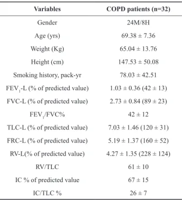
All 32 patients completed the protocol. table 1 shows all the patients’ data in relation to gender, age, body mass index (BmI), the pack-year ratio and the lung function test. patients displayed Fev1 between 22 and 64% of the predicted total with evidence of air entrap-ment (n= 32) and lung hyperinflation (n= 12). In the mRC scale, thirteen patients scored 4; fifteen scored 3 and four scored 2.
Both IC measures taken at rest before performing ADl displayed a strong correlation and signiicance (r= 0.90; p= 0.0000).
As can be seen from the results presented in table 2, there was a signiicant reduction in IC or dynamic lung hyperinlation after ADl, from 2.06 ± 0.60 before to 1.68 ± 0.59 after (p= 0.0001).
the results obtained from the inspiratory fraction (IC/tlC), presented in table 2, also reduced significantly with ADl, from 0.29 ± 0.07 before to 0.24 ± 0.07 after (p= 0.0001).
A strong correlation and signiicance was observed be-tween IC and IC/tlC when they were analyzed before (r= 0.78; p= 0.0000) and after (r= 0.85; p= 0.0000) the exercise.
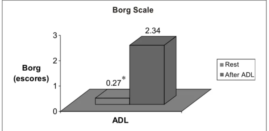
Figure 1 shows that the patients displayed a signiicant increase in dyspnea assessed with the Borg scale after the ADl simulation (p= 0.00000).
dIscussIoN
Dh has been the object of scientific investigation because it is an important aggravating factor in CopD patients, causing dyspnea and limiting their ability of perform physical exercise2,13 and simple daily routine
activities such as taking a shower, combing their hair and brushing their teeth20. yet the lack of studies that explore Dh during specific upper limb exercises motivated this investigation and, through the results, made it possible to confirm that physical exercise such as transferring pots with different weights from a lower shelf to a higher shelf, simulating daily living activities, caused moder-ate to serious Dh in CopD patients. In order to discuss the causes of the Dh seen after the performance of the ADl, it is very important to understand the ventilatory pattern during upper limb lifting. the difference between healthy subjects and CopD patients during exercises that involve lifting with the upper limbs is in the latter’s difficulty optimizing ve due to the increase in tv (tidal volume)30. these patients increase the tv marginally
during upper limb elevation because they already breathe at high lung volumes and, consequently, an additional volume increase results in disproportionate reduction in dynamic compliance, inspiratory muscle load increase and respiratory effort increase6.
therefore, the only way CopD patients can increase mv while lifting upper limbs is to increase respiratory frequency (RF), because tlC does not change with ex-ercise6. premature lack of expiration caused by rapid RF increases air entrapment even more and reduces IC19.
given this evidence, it would be interesting to conduct studies on the creation of therapeutic strategies geared toward the prevention and reduction of Dh during ADl Table 1. Anthropometric, demographic and lung function data of
CopD patients.
Data are presented as mean ± SD. CopD: Chronic obstructive pulmonary disease; Fev1 = Forced expiratory volume in one second ; FvC = Forced vital capacity; Fev1/CvF = tiffeneau index; tlC = total lung capacity ; FRC = Functional residual capacity ; Rv = Residual volume; IC = Inspiratory capacity; IC/tlC = Inspiratory fraction.
Variables COPD patients (n=32)
gender 24m/8h
Age (yrs) 69.38 ± 7.36
weight (Kg) 65.04 ± 13.76
height (cm) 147.53 ± 50.08
Smoking history, pack-yr 78.03 ± 42.51 Fev1-l (% of predicted value) 1.03 ± 0.36 (42 ± 13) FvC-l (% of predicted value) 2.73 ± 0.84 (89 ± 23)
Fev1/FvC% 42 ± 12
tlC-l (% of predicted value) 7.03 ± 1.46 (120 ± 31) FRC-l (% of predicted value) 5.19 ± 1.37 (160 ± 52) Rv-l(% of predicted value) 4.27 ± 1.35 (228 ± 124)
Rv/tlC 61 ± 10
IC % of predicted value 67 ± 15
IC/tlC % 26 ± 7
AdL At rest After ADL p
Ic 2.06 ± 0.60 1.69 ± 0.60 0.0001*
Figure 1. Data are presented as median.
* p<0.05; ADl: Activities of Daily living.
involving upper limb elevation, with non-invasive ven-tilation devices or pursed-lip breathing associated with ADl performance.
the patient’s basal IC was established after the per-formance of two maneuvers according to reproducibility criteria, and the highest basal IC value was selected for analysis1. when analyzing the Pearson correlation for these two basal IC values, we found evidence of a strong correlation. therefore, we recommend that future studies using the same methodology perform only one maneu-ver to establish basal IC, reducing patient exertion and collection time.
Dh was determined by IC reduction in absolute values and also by inspiratory fraction reduction after upper limb elevation. the supposed advantage of IC/ tlC analysis over isolated IC measurement may be re-lated to the fact that patients can display an absolute value similar to IC, however with a great difference in available maximum volume for lung expansion. In this context, the combination of a low IC/tlC ratio and the increase in tlC (evidenced by FRC increase) is possibly more important than the IC reduction because patients must breathe near tlC, with negative consequences to the elastic work of breathing8.
Albuquerque et al.7 veriied that the inspiratory frac-tion, measured at rest, after bronchial dilafrac-tion, was a better predictor of low maximum exercise capacity in CopD pa-tients than the isolated IC measure. this study did not aim to verify the best index (IC or IC/tlC) to predict CopD patient tolerance to exercise, but it was able to verify a strong correlation between these variables, which indicates that one or the other may be chosen to evaluate Dh occur-rence during upper limb elevation exercises.
the central theme of this study arose from the evi-dence that CopD patients complain about dyspnea while performing tasks using their arms20,21. this study did not aim to evaluate the feeling of dyspnea as an exercise
tolerance factor, given the complexity of the protocol. however, the dyspnea assessed by the Borg scale was used to observe respiratory fatigue after lifting pots with different weights, simulating an ADl. the study confirmed that upper limb elevation exercise causes dyspnea. the physiopathological mechanism of dyspnea and the ex-planation for the relationship between dyspnea and Dh have not been completely clarified, although there are many studies that confirm this relationship9,12,13. Dyspnea
during exercise can be explained by continuous afferent sensorial information (collected by specialized receptors located in the airways, lungs and rib cage) on the reduc-tion of strength generareduc-tion capacity of respiratory muscles to the higher cerebral centers. Functional weakness of respiratory muscles associated with increase in elastic and resistive work due to acute Dh increases afferent senso-rial information to the central motor command in order to maintain efficient ventilation and generate strength for the inspiratory muscles. that causes an imbalance in the relationship between the perception of breathing ef-fort and the anticipation of neurological center response. o’Donnell et al.12 gave this efferent – afferent dissociation the title of neuroventilatory dissociation.
this study’s findings are compatible with those of velloso et al.21, which used the Borg scale to evaluate the feeling of dyspnea in CopD patients during certain ADl simulations, such as sweeping the floor, wiping a blackboard, lifting pots with various weights and changing light bulbs. Among these activities, the authors found a high Borg score for the activity that involved lifting pots with different weights.
In conclusion, ADl performed with upper limbs, such as lifting pots with different weights, caused an increase in dynamic lung hyperinlation and dyspnea, with inspiratory capacity and inspiratory ratio being sensitive variables in the assessment and detection of dynamic hyperinlation in CopD patients.
0.27
2.34
0 1 2 3
Borg (escores)
ADL
Rest
After ADL Borg Scale
reFereNces
1. o’Donnell De. hyperinflation, dyspnea, and exercise intole-rance in Chronic obstructive pulmonary Disease. proc Am thorac Soc. 2006;3:180-4.
2. o’Donnell De, Revill Sm, webb KA. Dynamic hyperinflation and exercise intolerance in Chronic obstructive pulmonary Disease. Am J Respir Crit Care med. 2001;164:770-7. 3. Dolmage te, goldstein RS. Repeatability of inspiratory
capac-ity during incremental exercise in patients with severe CopD. Chest. 2002;121:708-14.
4. yan S, Kaminski D, Sliwinski p. Reliability of inspiratory capacity for estimating end-expiratory lung volume change during exercise in patients with Chronic obstructive pulmo-nary Disease. Am J Respir Crit Care med. 1997;156:55-9. 5. grimby g, Striksa J. Flow-volume curves and breathing
pat-terns during exercise in patients with obstructive lung disease. Scand J Clin lab Invest. 1970;25:303-13.
6. Stubbing Dg, pengelly lD, morse JlC, Jones Nl. pulmonary mechanics during exercise in subjects with chronic airflow obstruction. J Appl physiol. 1980;49:511-5.
7. Albuquerque Alp, Nery le, machado tyS, oliveira CC, paes At, Neder JA. Inpiratory fraction and exercise impair-ment in CopD patients golD stages II-III. eur Respir J. 2006;28:939-44.
8. Casanova C, Cote C, torres Jp, Aguirre-jaime A, marin Jm, pinto-plata v, et al. Inspiratory-to-total lung capacity ratio predicts mortality in patients with Chronic obstructive pulmo-nary Disease. Am J Respir Crit Care med. 2005;171:591-7. 9. marin Jm, Carrizo SJ, gascon m, Sanchez A, gallego B, Celli
BR. Inspiratory capacity, dynamic hyperinflation, breathless-ness, and exercise performance during the 6-minute walk test in Chronic obstructive pulmonary Disease. Am J Respir Crit Care med. 2001;163:1395-9.
10. maesto lp, pedro Jg, Abad ym, onã JmR, lorente D, Cubillo Jm. Dyspnea, ventilatory pattern, and changes in dynamic hyperinflation related to the intensity of constant work rate exercise in CopD. Chest. 2005;128:651-6.
11. o’Donnell De, lam m, webb KA. measurement of symptoms, lung hyperinflation, and endurance during exercise in Chronic obstructive pulmonary Disease. Am J Respir Crit Care med. 1998;158:1557-65.
12. o’Donnell De, webb KA. exertion breathlessness in pa-tients with chronic airflow obstruction. Am Rev Respir Dis. 1993;148:1351-7.
13. gigliotti F, Coli C, Bianchi R, grazzini m, Stendardi l, Cas-tellani C, et al. Arm exercise and hyperinflation in patients with CopD. Chest. 2005;128:1225-32.
14. Baarends em, Schols AmwJ, Slebos DJ, mostert R, Jans-sen pp, wouters eFm. metabolic and ventilatory response pattern to arm elevation in patients with CopD and healthy age-matched subjects. eur Respir J. 1995;8:1345-51.
15. mcKeough ZJ, Alison JA, Bye ptp. Arm exercise capacity and dyspnea ratings in subjects with chronic obstructive pulmonary disease. J Cardiopulm Rehabil. 2003;23:218-25.
16. Criner gJ, Celli BR. effects of unsupported arm exercise on ventilatory muscle recruitment in patients with severe chronic airflow obstruction. Am Rev Respir Dis. 1988;138:856-61. 17. epstein SK, Celli BR, martinez FJ, Couser JI, Roa J, pollock
m, et al. Arm training reduces the vo2 and ve cost of un-supported arm exercise and elevation in chronic obstructive pulmonary disease. J Cardiopulm Rehabil. 1997;17:171-7. 18. mcKeough ZJ, Alison JA, Bye ptp. Arm positioning alters
lung volumes in subjects with CopD and healthy subjects. Aust J physiother. 2003;49:133-7.
19. Dolmage te, maestro l, Avendano mA, goldstein RS. the ventilatory response to arm elevation of patients with Chronic obstructive pulmonary Disease. Chest. 1993;104:1097-100. 20. tangri S, wolf CR. the breathing pattern in Chronic
obstruc-tive lung Disease during the performance of some common daily activities. Chest. 1973;63:126-7.
21. velloso m, Stella Sg, Cendon S, Silva AC, Jardim JR. meta-bolic and ventilatory parameters of four activities of daily living accomplished with arms in CpoD patients. Chest. 2003;123:1047-53.
22. Rabe KF, hurd S, Anzueto A, Barnes pJ, Buist SA, Calver-ley p, et al. global strategy for the diagnosis, management, and prevention of chronic obstructive pulmonary disease: golD executive Summary. Am J Respir Crit Care med. 2007;176:532-55.
23. Ruppel gl. lung volumes and gas distribution tests. In: Ruppel gl, editor. manual of pulmonary Function testing. St. louis: mosby; 1998. p. 69-94.
24. American thoracic Society. lung function testing; se-lection of reference values and interpretative strategies: American thoracic Society Statement. Am Rev Respir Dis. 1991;144:1202-18.
25. Diretrizes para teste de função pulmonar. J pneumol. 2002;28: 95-100.
26. Knudson RJ, lebowitz mD, holberg CJ, Burrows B. Changes in the normal maximal expiratory flow-volume curve with growth and aging. Am Rev Resp Dis. 1983;127:525-734. 27. Neder JA, Nery le, Castelo A, Andreoni S, lerario mC, Sachs
A, et al. prediction of metabolic and cardiopulmonary responses to maximum cycle ergometry: a randomized study. eur Respir J. 1999;14:1304-13.
28. Borg gA. phychophysical bases of perceived exertion. med Sci Sports exerc.1982;14:377-81.
29. American thoracic Society. Surveillance for respiratory hazards in the occupational setting: AtS statement. Am Rev Respir Dis. 1982;126:952-6.