Journal of Evolution of Medical and Dental Sciences/ Volume 3/ Issue 05/February 03, 2014 Page 1097
A HISTOLOGICAL STUDY TO COMPARE CLINICALLY UNINVOLVED AND
INVOLVED SKIN IN LEPROSY
Prachi Saffar Aneja1, Savita Bansal2, Manish Aneja3, Kuldip Singh Sood4, Amit Kumar Saxena5, Ankur
Sharma6
HOW TO CITE THIS ARTICLE:
Prachi Saffar Aneja, Savita Bansal, Manish Aneja, Kuldip Singh Sood, Amit Kumar Saxena, Ankur Sharma. A Histological Study to compare Clinically Uninvolved and Involved Skin in Leprosy. Journal of Evolution of Medical and Dental Sciences 2014; Vol. 3, Issue 05, February 03; Page: 1097-1103,
DOI: 10.14260/jemds/2014/1952
ABSTRACT: BACKGROUND: Leprosy or Kustha roga is no less than a social stigma in our country. In
1983 our government started The National Leprosy Eradication Programme . But even after the turn of century the goal is far from achieved. AIMS: To histologically verify affliction of apparently normal looking skin in leprosy patients so as to establish skin biopsy as a screening tool for early detection of leprosy, especially in high risk groups such as personnel working in leprosy homes. METHOD: The study was conducted on thirty clinically defined cases of lepromatous leprosy. Two skin biopsies were taken from every patient. The histology of the lesion was compared with the apparently uninvolved skin under pre-defined parameters. RESULTS: In Group B - All the cases presented with focal atrophy of epidermis, mild to moderate cellular infiltration in upper and mid dermis and varying degree of perivascular and periappendageal infiltration.10% cases revealed infiltration of deep dermis. INTERPRETATION AND CONCLUSION: Varying degree of histological alteration in clinically uninvolved skin is present in all cases under present study. 60% cases showed moderate and 10% cases revealed marked cellular infiltration. Mid dermis was involved in 20% of the cases. The author feels that for high risk groups such as those working in leprosy homes, having low levels of immunity skin biopsy as a screening tool is beneficial.
KEY WORDS: Biopsy, Clinically involved skin, histology, leprosy, lesions, uninvolved skin.
INTRODUCTION: Leprosy is a multi-system infection with widespread hematogenous dissemination, caused by Mycobacterium leprae. It predominantly affects the skin and peripheral nerves. The mode of transmission of leprosy is unknown, but inhalation of bacilli which may be excreted from the nasal passage of a multibacillary patient is the most accepted theory. Direct person-to-person infection by means of the skin though rare may occur.
Leprosy is a pleomorphic granulomatous disease with a spectrum of lesions ranging from paucibacillary epithelioid cell granuloma in tuberculoid forms to lymphopenic lesions with bacilli laden foamy macrophages in lepromatous leprosy1. The histological hallmark of leprosy is the
involvement of appendages, nerves and erector pili muscle along with the presence of granuloma.
Granuloma is defined as a focal collection of inflammatory cells, lymphocytes, macrophages,
epithelioid cells, giant cells, plasma cells with connective tissue and blood vessels.
As hematogenous spread is very common the infecting organism is expected to reach anywhere throughout the skin with or without clinical expression in the form of skin lesions. What then prevents the appearance of lesions at some sites while simultaneous development of lesions at other in the same patient has intrigued many2
Reports on the study of uninvolved skin in leprosy are rather scanty. Fernandez3 observed
Journal of Evolution of Medical and Dental Sciences/ Volume 3/ Issue 05/February 03, 2014 Page 1098 morphologically granular. Findings in normal skin in leprosy patients led Bechelli et al4 to comment
that in the apparently healthy integument a certain condition exists which may be called potential corresponding more or less to the type of lesions present in the other parts of the body. Histopathological evidence of involvement of apparently normal skin in non-bacilliferous types of leprosy that is in tuberculoid and borderline tuberculoid leprosy was studied by Singh et al5. They
suggested sub-clinical involvement of normal skin in all cases under their study.
In view of above references and work done by eminent researchers the author undertook the present research work with the view to further our knowledge about the involvement or the lack of it; in clinically uninvolved skin in leprosy patients.
Leprosy is endemic in many tropical and subtropical countries including the Indian subcontinent. If clinically uninvolved skin shows a definite histological preponderance towards leprosy, a screening test by means of skin biopsy can be advocated and devised for early detection of leprosy especially in high risk population such as those working in leprosy homes. Such early detection help not only in substantially reducing the disability and anesthesia caused by the disease it will also go a long way in eradicating the disease.
This study thus aims to advocate skin biopsy as a screening tool for leprosy.
MATERIAL AND METHODS: The present study was conducted on thirty clinically defined cases of lepromatous leprosy who reported in leprosy clinic of Department of Dermatology, STD and Leprosy SMS Medical College and Hospital Jaipur, Rajasthan.
The patients were selected randomly irrespective of age, sex, and the duration of disease. Two skin biopsies were taken from every patient up to the thickness of subcutaneous tissue using punch biopsy technique and categorized as follows:
Group A: consist of skin biopsy from the active lesion of the patient
Group B: consist of skin biopsy from clinically uninvolved skin approximately 8cm away from the lesion; where sensation, surface and pigmentation were normal.
Paraffin sections of 5µ thickness were subjected to routine Haematoxylin and Eosin staining. Study was conducted under the low power light microscopy. The histology of the lesion was compared with the apparently uninvolved skin taking normal skin histology as control. The patient s details and histological parameters were recorded as per Proforma.
Inclusion criteria:
(i) Only patients reporting to the clinic for the first time were considered (ii)Patients with clinical diagnosis of lepromatous leprosy.
Exclusion criteria:
(i) Patients already under treatment or declared cured were excluded. (ii)Patients with clinical diagnosis other than lepromatous leprosy.
PROFORMA
Serial No. Leprosy Clinic No.
Name & Age & Sex Marital Status
Address Occupation
Journal of Evolution of Medical and Dental Sciences/ Volume 3/ Issue 05/February 03, 2014 Page 1099
Characteristics of lesions
(a) Site (b) Number
(c) Duration (d) Margin, Surface, Size, Color
(e) Tenderness (f) Sensation.
H/o past illness.
Date of Removal of tissues
Histological Parameters
(i) Epidermis
(ii)Subepidermal zone (iii)Dermis
a) Cellular infiltrate - Location - Degree - Type
(iv)Periappendicular infiltration (v) Perivascular infiltration
RESULTS: All patients considered for study were male between the age group of 20-50yrs.
Clinical features: Of the 30 cases of lepromatous leprosy, 24 patients gave history of having lesions for over one year while six patients reported the lesions to be six to 8 months old. 27 patients had macular lesions while 3 patients had nodular lesions. 24 patients with macular lesions had multiple and bilaterally symmetrical lesions while 3 patients had multiple lesions. Out of the 27 patients having macular lesions, 7 had erythematous lesions. Of the 3 patients having nodular lesions 2 had multiple lesions and one patient had multiple and bilaterally symmetrical lesions. All the patients had hypopigmented areas and some degree of hypoesthesia.
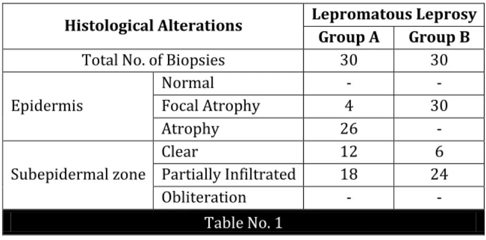
Table 1 and 2 depicts the Histological Parameters.
Histological Alterations Lepromatous Leprosy Group A Group B
Total No. of Biopsies 30 30
Epidermis
Normal - -
Focal Atrophy 4 30
Atrophy 26 -
Subepidermal zone
Clear 12 6
Partially Infiltrated 18 24
Obliteration - -
Journal of Evolution of Medical and Dental Sciences/ Volume 3/ Issue 05/February 03, 2014 Page 1100
Histological Alterations Lepromatous Leprosy Group A Group B
Total No. of Biopsies 30 30
Type of cells in dermis
Lymphocytes 30 30
Macrophages 28 25
Epithelioid Cells 25 19
Giant Cells 23 -
Degree of cellular infiltration
Absent/negligible - -
Mild - 8
Moderate 8 18
Marked 22 4
Dermis
(depth of infiltration)
Upper Dermis 30 30
Mid Dermis 21 7
Deep Dermis 8 -
Table No. 2
All the cases of Group A and Group B presented with perivascular and periappendageal infiltration though varying in degree.
DISCUSSION: Leprosy is not just another contagious disease; for the Indian population it is a social
stigma. A patient of leprosy or Kushtha roga is considered cursed, a person who is enduring the
wrath of God. Leprosy is an enigma. The causative organism Mycobacterium leprae has yet to be cultured and the last word about its pathogenesis and mode of spread has yet to be stated.
Leprosy exhibits a spectrum of manifestations, which correlates fairly well with the clinical spectrum but there is often great variability in the histological picture of different lesions in the same patient and even in different parts of the same lesion. Furthermore, the clinical and histological picture may not correlate at all.
Journal of Evolution of Medical and Dental Sciences/ Volume 3/ Issue 05/February 03, 2014 Page 1101 Reports on the study of histology of clinically uninvolved skin in leprosy are scanty. Khanolkar6 carried out studies on normal skin of asymptomatic contacts of leprosy patients with a
view to understanding the evolution of early lesions. Ramanujam and Ramu7 found bacilli in 17.9%
cases from the normal skin surrounding the borderline lesions within a distance of 0.5 to 2cm. Rea et al8 postulated that the uninvolved skin has sub-clinical involvement and the diffuse involvement may
precede the appearance of lesions. Bedi et al9 inspite of their inability to discern histological lesions in
normal skin in large majority of cases postulated the existence of sub-clinical involvement. The existence of even 10-25% foam cell granuloma and AFB from clinically normal looking skin in lepromatous leprosy patients became significant, because majority of the cases under their study were receiving treatment.
Katoch et al10 showed that clinically unaffected sites in lepromatous leprosy have lower
bacillary index and lesser bacterial load. Histologically the granulomas were smaller and biopsy index were lower in uninvolved areas. Sakuntala et al10 studied elliptical skin biopsies from actual
lesion as well as from apparently uninvolved area minimum of 8cm away from the actual lesion and reported that uninvolved skin showed infiltration around the skin appendages blood vessels, nerves and subepidermal region along with erosion of basal layer. They revealed presence of bacilli in LL patients.
Rea TH11 using the clinically normal skin from lepromatous leprosy subjects as a standard for
comparison studied the mean thickness of the nucleated epidermis and found it to be significantly increased in untreated lesions form borderline tuberculoid, erythema nodosum leprosum and reversal reaction patients but was unchanged in borderline lepromatous and lepromatous patients.
Suneetha S 12 concluded that apparently normal skin from the area of sensory changes
revealed microscopic evidence of nerve involvement. Clinical studies found that a proportion of patients develop visible skin lesions during follow-up.
Palaskar13 postulated that apparently normal areas of skin in lepromatous leprosy are the
probable sites of future extension of the pathological process and the events taking place in these sites might present the earlier stage of such an extension. Biopsies done on about twenty patients of apparently normal oral mucosa did not show granulation formation of acid-fast bacilli histologically. The only change present was mild inflammation.
El-Darouti MA et al14 carried out a study to document the microscopic affection of apparently
normal looking skin in different types of leprosy. They found that fifty two percent of the cases showed affection of which there was higher incidence towards the lepromatous end of the disease. Moreover the microscopic picture of indeterminate leprosy can be observed in normal looking skin of patients with tuberculoid leprosy or lepromatous leprosy.
Journal of Evolution of Medical and Dental Sciences/ Volume 3/ Issue 05/February 03, 2014 Page 1102 also a very crucial determining factor. Such contacts with low levels of immunity are thus more prone to the disease and hence more likely to present with sub-clinical histologic alterations.
The author is thus concluding on the note that at least for high-risk groups such as those working in leprosy homes, having low levels of immunity skin biopsy as a screening tool is beneficial. The study needs to be furthered and biopsies from actual healthy contacts should be studied. This will give an insight not only into the pathogenesis of the disease it might give a clue as to how to prevent the accumulation of bacilli and development of overt lesions.
REFERENCES:
1. Ridley DS and Jopling WH – classification of leprosy according to immunity a five group system. IJL 1966, 34: 255-273.
2. Katoch VM, Mukherjee A, Girdhar BK. A bacteriological and histopathological study of apparently normal skin in lepromatous leprosy. Leprosy in India, 1980 Oct, Volume 52, Issue 4, p508-12,
3. Fernandez J.M.M. Pathology of acute reactions in leprosy, Inter J Lepr. 1936, 4: 549 (Abstract). 4. Bechelli L.M., Da Silva E and Oliviera A.B. On the histopathological findings in biopsies of
apparently normal skin in cases of leprosy Inter. J. Lepr. 1945, 13: 175 (Abstract).
5. K. Iyenger, Singh B., and Singh R. Histopathological evidence of indeterminate leprosy in apparently uninvolved skin of entire spectrum of leprosy. Leprosy in India, 1983 Jul, Volume 55, Issue 3, p500-3,
6. Khanolkar V.R. Perspectives in pathology of leprosy. Ind. J. Med. Sci. 1955, 9: (Suppl 1) 13. 7. Ramanujam K and Ramu G. A study of borderline leprosy from the clinical, bacteriological and
immunological aspects. Lepr. India 1965, 37: 303.
8. Rea T.H., Gottlib B, and Levan N.E. Apparently normal skin in lepromatous leprosy. Arch Dermatol III, 1975: 1571.
9. Bedi T.R., Kumar B., Kaur S. Histopathological study of clinically normal appearing skin in lepromatous leprosy, Lepr. India. 1979, 51-178.
10.Sakuntala R, Pratap VK, Sharma NK, Dayal SS, Aggarwal SK. Histologic profile in apparently normal skin of leprosy patients. Lepr India. 1982; 54(1):40-7.
11.Thomas H. Rea. Frequency and extent of thickening of the nucleated epidermis in leprosy lesions. Int J Lepr Other Mycobact Dis. 2000 Dec; 68(4):410-6.
12.Suneetha S, Sigamoni A, Kurian N, Chacko CJ. The development of cutaneous lesions during follow-up of patients with primary neuritic leprosy. Int J Dermatol. 2005 Mar; 44(3):224-9. 13.Palaskar S. Histopathological study of apparently normal oral mucosa in lepromatous leprosy
Indian J. Dent Res, 2005 Jan-Mar; 16 (1): 12-4.
Journal of Evolution of Medical and Dental Sciences/ Volume 3/ Issue 05/February 03, 2014 Page 1103
AUTHORS:
1. Prachi Saffar Aneja 2. Savita Bansal 3. Manish Aneja 4. Kuldip Singh Sood 5. Amit Kumar Saxena 6. Ankur Sharma
PARTICULARS OF CONTRIBUTORS:
1. Assistant Professor, Department of Anatomy, SGT Medical College, Gurgaon.
2. Assistant Pathology, Department of Anatomy, Manav Rachna Dental College, Faridabad. 3. Attending Consultant, Department of
Anaesthesia, Medanda – The Medicity, Gurgaon.
4. Professor and Head, Department of Anatomy, SGT Medical Collge, Gurgaon.
5. Associate Professor, Department of Anatomy, SGT Medical College, Gurgaon.
6. Tutor, Department of Anatomy, SGT Medical College, Gurgaon
NAME ADDRESS EMAIL ID OF THE CORRESPONDING AUTHOR: Dr. Prachi Saffar Aneja, D-10/05, GF Ardee City, Sector – 52, Gurgaon, Haryana – 122011.
E-mail: [email protected]