Correspondence to: Lazar Velicki, Institute of Cardiovascular Diseases Vojvodina, Put doktora Goldmana 4, 21 204 Sremska Kamenica,
C U R R E N T T O P I C UDC: 616.12-08
DOI:10.2298/VSP121024007V
Long-term ventricular assist devices in current clinical practice
Dugotrajne ventrikularne pumpe u savremenoj praksi
Lazar Velicki*†, Frazier OH‡§
*Faculty of Medicine, University of Novi Sad, Novi Sad, Serbia; †Institute of Cardiovascular Diseases Vojvodina, Sremska Kamenica, Serbia; ‡Texas Heart Institute,
St. Luke's Episcopal Hospital, Houston, Texas, USA; §Baylor College of Medicine, Houston, Texas, USA
Key words:
heart failure; heart assist device; prognosis.
Kljuÿne reÿi:
srce, insuficijencija; srce, implantabilni mehaniÿki aparati; prognoza.
Introduction
Chronic heart failure (CHF) is a major healthcare issue associated with significant reduction of the quality of life, poor prognosis – high mortality rate, and still no adequate therapeutic approach available to the majority of the patients. Statistics indicate that survival rate for patients with CHF is only 50% after 5 years, and significantly less so for those with advanced heart disease, less than 50% after one year. Incidence and prevalence of CHF are also on the in-crease with more than 16 million diagnosed across Europe and United States – 2.5% of total population, resulting in frequent hospital admissions and long term costs of palliative support. Further still, numbers indicate significant increase of CHF in elderly patient group (65+ years of age), popula-tion here expected to double over the next 20 years 1–3.
Although, numerous advancements in medical therapy have improved patient outcomes in CHF, prognosis is still poor and the quality of life remains limited. For patients with end-stage heart failure, heart transplant (HTX) remains to be the only long-term satisfactory option. However, the in-creasing demand of donor organs is not equilibrated with limited number of available hearts. Current estimates indi-cate that up to 100,000 patients meet criteria for HTX in the United States 4. The scarcity of donor organs has fuelled the development of interim interventions such as different types of cardiac surgery operations aimed at restoration of left ventricle geometry and functionality, cardiac resynchroniza-tion therapy (CRT) and mechanical circulatory support (MCS). According to the Registry of the International Soci-ety for Heart and Lung Transplantation – the 25th Official Adult Heart Transplant Report – 2008, almost 29% of pa-tients at the time of transplant were on some type of MCS
modality (22% on left ventricular assist device-LVAD) 5. Technology advancements resulting in smaller MCS units offer alternative and permanent treatment option for many patients on the heart-transplant waiting list thus mitigating mortality rate 2.
aorta or pulmonary artery. Depending on the site of the pump, VADs can be classified as intracorporeal (inside the patient), extracorporeal (outside the body) or paracorporeal (immediately adjacent to the patient).
A pivotal randomized study that investigated the effect of mechanical assist devices as a DT on the outcome and quality of life in patients with CHF was the Randomized Evaluation of Mechanical Assistance for the Treatment of Congestive Heart Failure (REMATCH) trial 3. In this land-mark trial, a total of 129 patients with end-stage heart failure – New York Heart Association (NYHA) class IV – who were ineligible for cardiac transplantation were assigned to receive a LVAD (68 patients) or optimal medical management (61 patients). “The rates of survival at 1 year were 52% in the device group and 25% in the medical-therapy group (p = 0.002), and the rates at 2 years were 23% and 8% (p = 0.09), respectively” 3. The final conclusion of the trial was that the use of a LVAD in patients with advanced heart failure re-sulted in a prolonged survival period and an improved qual-ity of life and as such, LVADs may be considered as an ac-ceptable alternative therapy in selected patients who are not candidates for HTX. This trial was the first to establish the efficacy of device therapy for end-stage heart failure and set the standards for CHF treatment using VADs.
Depending on the level of ventricular reserve or resid-ual volume in the left ventricle to open the aortic valve and generate a pulse, LVAD may be added to support the circu-lation in two ways. With total unloading of the left ventricle, LVAD is connected to the systemic circulation in a serial manner and the aortic valve is closed all the time 8. If the failing myocardium has the capability of generating a pulse, the pump is added to the circulation in a parallel fashion – both the device and the native ventricle can pump blood into the ascending aorta. Aortic valve may open, usually during exercise when veins contract and produce increased venous return to the heart.
As a result of significant technological advancements with mechanical pumps in the last decade and their profound impact on the mainstream of our daily practice, guidelines for the CHF treatment need to be regularly updated. Guide-lines are expected to be published by all major bodies, in-cluding International Society for Heart and Lung Transplan-tation, American Heart Association and American College of Cardiology Task Force, and the Heart Failure Society of America 9. One may expect that these guidelines will rec-ommend that every patient with refractory end-stage heart failure should be considered and evaluated for some kind of MCS. The Centre for Medicare and Medicaid Services re-quires that patients exhibit NYHA class IV symptoms (opti-mal medical therapy refractory patients) to qualify for MCS therapy 10. Currently, MCS is recommended to those pa-tients facing imminent death due to heart failure still having sustained end-organ function. In other words, candidates for long-term assist devices are those with inadequate hemody-namics despite optimized drug therapy and/or intra-aortic balloon pump assistance 11. Hemodynamic parameters that may guide selection of patients suitable for device therapy include: pulmonary capillary wedge pressure (PCWP) of >20
mmHg, a cardiac index of 2 Lminí1mí2, and a systolic blood pressure 80 mmHg. However, interpretation of he-modynamic profiles may be very difficult in certain cases emphasizing the need for thorough clinical assessment and careful decision making.
A study from Holman et al. 12 included a total of 420 patients with 497 implanted assist devices (314 LVADs, 5 RVADs, and 77 BiVADs). The authors found that older age, ascites, increased bilirubin, and Interagency Registry for Mechanically Assisted Circulatory Support (INTERMACS) level 1 (cardiogenic shock) may be regarded as independent risk factors for mortality. A similar study using multivariate analysis on data from 47 patients receiving LVAD found that the preoperative total bilirubin value, age, and preoperative right heart dysfunction are independent predictors of unfa-vourable outcome and death 10. These findings underscore the importance of proper patient selection and early implan-tation of LVAD 13.
This paper briefly reviews currently available VAD systems for long-term support of the left ventricle (with the focus on HeartMate II and HeartWare), their role in today’s clinical practice, patient selection, observed complications, and further directions in this rapidly advancing field.
Historical notes
20,000 device implantations occurred worldwide since then (around 55% being HeartMate II).
An ideal VAD
An ideal VAD would be durable, capable of providing long-term reliable systemic flows, sufficient to meet meta-bolic needs over a substantial range of physical activity (self-adjusting operational mode), small in size, easy to implant (preferably intrapericardially) 18. The device should produce minimal immunobiological response; it would have to be re-sistant to infection, with minimal risk for complications (thrombosis, bleeding, and haemolysis), possibly not requir-ing permanent anticoagulation therapy, consumrequir-ing small amount of electrical power thus not requiring an external power source. The device should also be affordable and readily available, providing short learning curve for physi-cians as for the patients.
Currently, there are three generations of VADs avail-able for clinical application (Tavail-able 1). The classification of
the devices into three distinct generations does not only im-ply the order in which they appeared, but also functional properties, generated flow characteristics and mechanical de-sign among other things.
First generation VADs
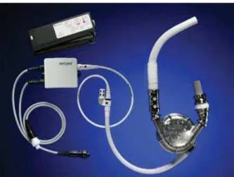
Initially, the first devices that were developed relied on the close imitation of the basic circulatory physiology and its inherent property – pulsatile flow. These devices (first gen-eration VADs) were developed during 1970s and 1980s and were characterized by the use of positive volume displace-ment and pulsatile flow. The first generation generally con-sists of pumps such as the Thoratec PVAD/IVAD, the HeartMate IP/VE/XVE (Figure 1), and the Novacor LVAS. Although provided satisfactory circulatory support allowing improved survival until HTX, the first generation pumps had many limitations, such as big size that required substantial surgical dissection for the placement of the device, noisy pump operation, presence of a large diameter driveline and, most importantly, limited mechanical durability due to its mechanical construction 2. The first generation pumps were also related to serious complications including bleeding, in-fections and thromboembolic events. It was the HeartMate XVE that was used in the REMATCH trial 3 to compare medical and circulatory assist device treatments for end-stage heart failure. Despite the obvious problems, a great
number of patients has been successfully supported with these devices which remain in use today for selected patients.
Fig. 1– Thoratec HeartMate XVE (with permission from Thoratec Corporation).
Second generation VADs
Growing waiting lists for HTX and long waiting times of up to 1 year have been urging the need for more reliable and smaller devices 19. Although the first generation pumps completely relied on the imitation of the physiologic prop-erty of the circulation – its pulsatile nature – introduction of the continuous flow pumps into everyday clinical practice was milestone concept that fundamentally changed the no-tion of human circulano-tion physiology. Pulse is not strictly necessary despite evolutionary adaptation of the human body to pulsatile circulation. That said, continuous flow VADs are able to mimic physiologic flow only to a certain extent – a special mode of operation (pulsatility index) that permits aortic valve opening during the systole. Pulsatility index is defined as the magnitude of flow pulse provoked by the pump through each cycle 20. Continuous flow pumps use electrical energy to rotate an axle on which a turbine or pro-peller system is mounted to pushing the blood through the body at a steady rate. These devices have now largely re-placed use of the first generation pulsatile, volume displace-ment pumps. Second generation pumps have no requiredisplace-ments for external venting – one of the reasons for their reduced size 18. Second generation rotary pumps are characterized by an axial blood flow path suspended by contact bearings and an internal rotor driven by an electromagnetic field 2. The ba-sic principle employed has been known for years –
Ar-Table 1 Major characteristics of different pump generations
Characteristics First generation pumps Second generation pumps Third generation pumps Operational
method
Pulsatile chamber with vol-ume displacement by external compression
Spinning rotor mounted on a central shaft
Hydrodynamic and/or elec-tromagnetic suspended spin-ning rotor
Type of flow Pulsatile Constant, nonpulsatile Constant, nonpulsatile Devices HeartMate XVE, Novacor,
Thoratec PVAD and IVAD, Abiomed 5000
HeartMate II, Jarvik 2000, MicroMed DeBakey
chimedes’ screw. Rotation of the rotor provides the driving force to propel the blood from the left ventricle through the pump and into the circulation. However, the system works on high rotational speeds 21, heat is generated, haemolysis with damage to the blood cells and thrombi may occur 22. Anaemia and platelet damage along with the activation of contact coagulation system may also ensue 8. Main advan-tages of these pumps are smaller size enabling easier im-plantation (even in small bodies), improved durability due to its design characteristics (only one moving part), less elec-tricity consumption, improved survival and quality of life, reduction of post-implantation adverse effect (bleeding, thrombosis, infection). The control systems and power deliv-ery mechanisms are easily portable and manageable by the patient.
Second-generation rotary pump LVADs were first in-troduced with the development of Hemopump. Researchers have applied its design to other circulatory assist devices (particularly HeartMate II). Subsequently, the Jarvik 2000 in 1999 and HeartMate II LVADs in 2000 have been used to support patients to HTX 14. Up to date, the HeartMate II is the most successful second-generation pump worldwide and Food and Drug Association (FDA) approved as BTT and DT 23–25. Eligibility criteria are essentially the same as those used to select patients for the pivotal clinical trial that in-cluded patients with shortness of breath and/or fatigue at rest or during minimal exertion despite treatment with optimal therapy for heart failure associated with a low ejection frac-tion (<25%) who were not candidates for HTX due to their age or comorbid conditions 26.
The physiologic response to the reduced arterial pulse or its absence during support with continuous flow pumps is not completely understood and it is unclear whether any ad-verse effects may surface in patients to be supported for many years 27. Clinical experience to-date indicates that no detrimental effect of these devices on end-organ function is to be expected in the long term 28, 29. There is a major shift among most VAD programs towards implantation of con-tinuous flow devices though discussion about the flow mo-dalities remains relevant 30.
HeartMate II
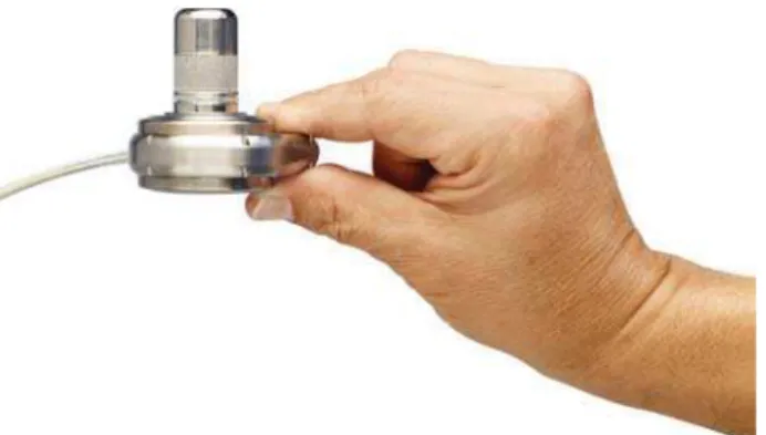
The HeartMate II (Thoratec Corp.) LVAD is an axial flow pump that had its origin in the early 1990s (Figure 2) and is intended for long-term support for BTT and DT in pa-tients with CHF31, 32. The HeartMate II contains a rotor (spinning impeller – the only moving part) capable of pro-ducing flow rates greater than 10 L/min at resolutions rang-ing from 6,000 to 15,000 rpm (Figure 3). HeartMate II is ap-proximately one seventh the size and one fourth the weight of the XVE pump. The pump can be implanted in a pre-peritoneal fashion or intra-abdominally, with the inflow can-nula connected to the left ventricle (the apex of the heart), and the outflow graft sutured to the ascending aorta. A driv-eline connected to the pump should be routed transcutane-ously, usually in the region of right upper quadrant of the ab-domen. Power is delivered by external power sources – re-chargeable batteries – that enable ambulance of the patient.
The system is operated at a fixed rotational speed set by the clinician with the aim of providing optimal circulatory sup-port for each patient. Although the internal surfaces of the device were designed to help resist the development of thrombi, anticoagulation is at present recommended to keep INR between 1.5 and 2.5.
Fig. 2 – Thoratec HeartMate II device (with permission from Thoratec Corporation).
Fig. 3 – Thoratec HeartMate II device – construction design (with permission from Thoratec Corporation).
Of all the continuous flow pumps currently in the use worldwide, only the Thoratec HeartMate II VAD has re-ceived approval from the US Food and Drug Administration (FDA) for use as BTT and DT in the United States 34.
The BTT pivotal multicentre, nonrandomised trial ini-tially enrolled 133 NYHA class IV patients who were listed for cardiac transplant and were at imminent risk of dying 35. The primary outcome of the BTT trial was survival to HTX or cardiac recovery, or being listed as United Network for Organ Sharing (UNOS) status 1A or 1B at 180 days of LVAD support. Of the 133 patients enrolled, 100 patients (75%) reached the primary end-point of HTX, cardiac recov-ery or survival at 180 days with ongoing mechanical support. The group of 100 patients included 56 patients who under-went HTX, 43 patients continued to receive LVAD support and were eligible for HTX, and 1 patient whose cardiac function recovered leading to LVAD explant. The overall rate of survival to HTX, recovery, or continued support was 75% at 180 days. The overall actuarial survival for patients continuing to receive HeartMate II device support was 89% at 1 month, 75% at 6 months and 68% at 12 months (the me-dian LVAD support was 126 days).
After being FDA approved in 2008, a multi-institutional study was carried out using the INTERMACS database com-prised of 169 consecutive BTT patients 36. The study com-pared the effectiveness of HeartMate II and previously ap-proved LVAD devices such as the HeartMate XVE and Thoratec IVAD. The 6 and 12-month survival of HeartMate II patients was 90% and 85% respectively whereas the con-trol group using pulsatile devices had 6 and 12-month sur-vival of 79% and 70% respectively. Significant reduction of device related complications was also observed for Heart-Mate II group (infection, neurological dysfunction, renal and respiratory dysfunction).
Another trial was carried out to evaluate effectiveness of HeartMate II device as DT in patients ineligible for HTX 25. The study was conducted in 38 centres in the USA. Patients were randomly assigned to undergo implantation of the HeartMate II continuous flow device or HeartMate XVE pulsatile device in a 2 : 1 ratio (a total of 200 patients – 134 in the continuous flow device arm and 66 in the pulsatile de-vice arm). The trial compared these dede-vices with established composite end-point of survival free from disabling stroke or reoperation to repair or replace the device for mechanical failure over a 2-year period (46% and 11% for HeartMate II and HeartMate XVE respectively). Overall 2-year survival was 58% and 24%, respectively; again with significant re-duction of adverse events in HeartMate II group. In total, 21 pump replacements (and 3 device explantations) were re-quired in the pulsatile LVAD cohort as opposed to only 13 pump replacements in the continuous flow device group (p < 0.001). After enrolment of the initial 200 patients in the DT trial, hundreds of additional patients have been enlisted part of continued access protocol 34.
Despite a dramatic improvement in survival with the HeartMate II, there still remains the burden of morbidity as-sociated with the device, including infection, bleeding, and thromboembolic events 36. According to the INTERMACS
annual report, the event rates (events per 100 patient months) during the first 12 months of HeartMate II therapy in BTT patients were 17.41 for bleeding, 11.8 for infection, and 1.93 for neurologic dysfunction 23.
Third generation VADs
During their extensive clinical use, it became apparent that implantation of second generation pumps in a form of BiVAD is extremely challenging if not impossible in cases of miniscule patients. An idea of developing even smaller devices that can be implanted within pericardial cavity emerged as an option to tackle the problem of BiVAD im-plantation and unsuitable patient’s anatomy.
Further advancement in design and construction of the continuous flow pumps has led to development of bearing-less devices, which in theory ought to be more durable than the previous generation pumps, and due to their smaller size allow intra-pericardial placement. Third generation VADs are continuous flow pumps broadly classified based on pump design into centrifugal and axial flow devices, and on whether the impeller is hydrodynamically or magnetically levitated. Levitation systems of third-generation rotary blood pumps suspend the moving impeller in pump thereby re-moving mechanical contact 2. Centrifugal flow pumps have cone-shaped or cylindrical rotors that drive the blood flow using the centrifugal force generated from the centre of the rotor to its circumference 37. One anticipated benefit is that the centrifugal design results in a flatter and more sensitive pressure flow curve at lower rounds-per-minute (RPM) com-pared to axial flow devices 17. Examples from this heteroge-neous group are the DuraHeart (Terumo Heart, Inc., Michi-gan), VentrAssist (Ventracor, Australia), CorAide (Arrow International, Pennsylvania), HeartWare HVAD (HeartWare, Inc., Massachusetts) and Levacor (World Heart, Inc., Oak-land, CA) systems.
HVAD
The HVAD (HeartWare Inc.) is a small third-generation continuous flow rotary pump with a centrifugal and bearing-less design (Figure 4). What distinguishes this pump from other of its generation is the size – the pump is small enough to be placed inside the pericardial cavity (no need for pump
pocket) at the apex of the heart or left ventricle inferior wall. As a consequence, surgical trauma to the surrounding tissue is significantly reduced while the implantation procedure is simplified – not requiring abdominal incision for implanta-tion. It usually operates at a speed of 2,400–3,500 RPMs and can provide up to 10L/min of flow 13. The impeller is sus-pended in place by a combination of passive magnetic and hy-drodynamic bearing systems, avoiding mechanical contact and wear (Figure 5) 18. Physical contact between the casing and the impeller is prevented by a thin blood film generated by the hy-drodynamic bearings 38–40. The device was implanted for the first time in humans in 2006 and, since then, it has been clini-cally evaluated in Europe (approved for BTT) and Australia with an ongoing bridge-to-transplantation trial in the US.
Fig. 5 – HVAD – internal design (with permission from Heartware Inc.).
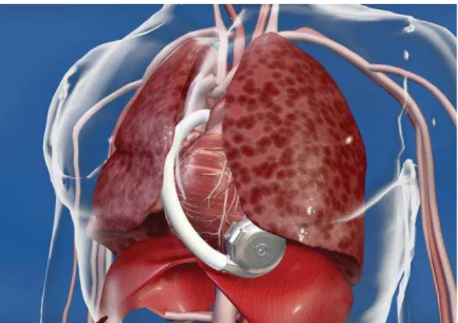
The HVAD is implanted through a median sternotomy with the assistance of cardiopulmonary bypass. An integrated inflow cannula is inserted into the left ventricle through the apex and is held in position by an adjustable sewing ring while pump is positioned in the pericardial cavity. After-wards, the 10 mm outflow graft is anastomosed to the as-cending aorta (Figure 6) 41.
Fig. 6 – HVAD position inside the chest (with permission from Heartware Inc.).
The HeartWare device has been evaluated in several clinical trials in terms of patient survival and quality of life. A recent multicentre trial in Europe and Australia found that
the actuarial survival after 6 and 12 months following HVAD implantation was 91% and 86%, respectively 42. An-other multicentre, non-randomized trial that included NYHA IV class patients, evaluated the safety of HVAD 43. Fifty pa-tients were included in the trial and they were supported us-ing HVAD for 180 days until HTX, myocardial recovery, device explantation or death with following actual survival at 6, 12, and 24 months: 90%, 84%, and 79% respectively. Death as an end-point was reached in 9 cases (18%) – 3 pa-tients died as a result of sepsis, 3 from multi-organ failure, and 3 from haemorrhagic stroke. The most frequent adverse events were infection and bleeding.
The ongoing Evaluate the HeartWare Ventricular Assist System (ENDURANCE) trial may demonstrate an important clinical advantage of this device.
Complications
Since growing number of patients are being supported with VADs for extended period of time, the interaction be-tween the body and the VAD during long-term or even life-long period and the management of complications have gained the interest of clinicians and biomedical engineers 44.
Thrombosis, thromboembolic events and the risk of bleeding
After the implantation of the device blood is exposed to a foreign surface requiring the use of systemic anticoagulation. Manufacturer guidelines for continuous flow devices recom-mend use of both antiplatelet and anticoagulation therapy in order to reduce the risk of pump thrombosis and consequent thromboembolism 37. Although the first recommended range of the international normalized ratio (INR) for the patients with HeartMate II was 2.5 to 3.5, the target INR has been re-cently decreased due to greater risk of bleeding 35, 45.
It has been noted that some patients may develop thrombosis of the aortic root and the ascending aorta – espe-cially when the outflow graft is implanted into the descend-ing aorta 44. In these settings, HTX should be performed on an emergency basis, if possible. In the case of pump throm-bosis, immediate pump exchange is mandatory. All other thromboembolic events should be dealt with in the usual manner.
haemolysis may occur after VAD implantation. This is usu-ally seen when the rotor speed in the continuous flow pumps is set to higher operation mode or as a result of postoperative complications (malposition of the apical cannula, pump thrombosis, outflow graft kinking) 44, 48, 49.
Infections
Infectious complications associated with VAD place-ment are often encountered, although the rate has improved with the new generation devices 50. Infection can involve any portion of a VAD – surgical site, driveline, pocket or the pump itself. Most infections involve the percutaneous driv-eline 51, 52. Length of device support was associated with more than 50% of 1-year survivors developing a driveline-related infection 53. The rate of driveline infections appears to have reduced after the introduction of newer generation pumps most likely due to smaller drivelines used by these pumps as well as the reduction in movement of the device within a surgically fashioned pocket 25. Another potential site of infection is the pump pocket usually originating from the driveline infection or secondary to surgical trauma or hema-toma formation 54. The infections is most likely to be caused by Staphyloccocus aureus, Corynebacterium or Pseudomo-nas aeruginosan 44.
Aortic valve pathology
Patients treated with long-term continuous flow devices are at higher risk of developing aortic insufficiency (AI) or some degree of aortic valve degeneration 37. It is believed that this may be the result of reduced (limited) or absent opening of the aortic valve mostly seen when the device is working in the serial fashion with continuous load of the left ventricle. The fusion of the aortic leaflets may be seen as early as 6 months following implantation. This complication
is encountered in approximately 25% patients, and several risk factors have been designated: aortic root diameter, fe-male sex, non-pulsatile flow 55. After 18 months on device support, up to 50% of patients present with moderate or se-vere AI 56.
Mechanical complications
Although rare, complications such as device malfunc-tion, inflow conduit rupture, and driveline break may occur. It is important to establish the correct cause of malfunction which, in most cases, requires device exchange. Despite modern technology and the use of high resistant materials, cable damage 57, due to kinking and twisting, or as a result of a suicide attempt occurs with an incidence of 5%–9% or up to 0.06 events per patient per year 27, 44, 58, 59.
Conclusion
Currently, long-term circulatory support with VADs of-fers viable choice for end-stage heart failure patients, either as BTT option or as DT. Survival rate along with the quality of life of these patients have been significantly improved. Patients supported with VADs continue to be affected by a variety of complications – the fact that only emphasizes the need to further improve this technology. Meticulous risk-benefit evaluation by a multidisciplinary team is mandatory for each patient in order to achieve optimal survival and minimize the risk of morbidity.
Grant support
The author (VL) worked on ventricular assist device sys-tems in the Texas Heart Institute in St. Luke's Hospital, Hous-ton, Texas, USA (2012–13) as a Fulbright Visiting Scholar.
R E F E R E N C E S
1. Gaddam KK, Ventura H. Developments in heart failure 2011. Congest Heart Fail 2012; 18(2): 112î26.
2. Garbade J, Bittner HB, Barten MJ, Mohr FW. Current trends in implantable left ventricular assist devices. Cardiol Res Pract 2011; 2011: 290561.
3. Rose EA, Gelijns AC, Moskowitz AJ, Heitjan DF, Stevenson LW, Dembitsky W, et al. Long-term use of a left ventricular assist device for end-stage heart failure. N Engl J Med 2001; 345(20): 1435î43. 4. Stevenson LW, Rose EA. Left ventricular assist devices: bridges
to transplantation, recovery, and destination for whom? Cir-culation 2003; 108(25): 3059î63.
5. Taylor DO, Edwards LB, Aurora P, Christie JD, Dobbels F, Kirk R, et al. Registry of the International Society for Heart and Lung Transplantation: twenty-fifth official adult heart transplant re-port--2008. J Heart Lung Transplant 2008; 27(9): 943î56. 6. Frazier OH, Rose EA, McCarthy P, Burton NA, Tector A, Levin H,
et al. Improved mortality and rehabilitation of transplant can-didates treated with a long-term implantable left ventricular as-sist system. Ann Surg 1995; 222(3): 327î36; discussion 336î8. 7. Frazier OH, Rose EA, Oz MC, Dembitsky W, McCarthy P, Rado-vancevic B, et al. Multicenter clinical evaluation of the Heart-Mate vented electric left ventricularassist system in patients awaiting heart transplantation. J Thorac Cardiovasc Surg 2001; 122(6): 1186î95.
8. Vural KM. Ventricular assist device applications. Anadolu Kardiyol Derg 2008; 8(Suppl 2): 117î30.
9. Moazami N, Feldman D. Rethinking the terminology of me-chanical circulatory support. J Thorac Cardiovasc Surg 2012; 144(1): 2î3.
10.Shiga T, Kinugawa K, Hatano M, Yao A, Nishimura T, Endo M, et al. Age and preoperative total bilirubin level can stratify prog-nosis after extracorporeal pulsatile left ventricular assist device implantation. Circ J 2011; 75(1): 121î8.
11.Frazier OH, Delgado RM. Mechanical circulatory support for advanced heart failure: where does it stand in 2003? Circula-tion 2003; 108(25): 3064î8.
12. Holman WL, Kormos RL, Naftel DC, Miller MA, Pagani FD, Blume E, et al. Predictors of death and transplant in patients with a me-chanical circulatory support device: a multi-institutional study. J Heart Lung Transplant 2009; 28(1): 44î50.
13.Neragi-Miandoab S. A ventricular assist device as a bridge to re-covery, decision making, or transplantation in patients with advanced cardiac failure. Surg Today 2012; 42(10): 917î26. 14.Clegg AJ, Scott DA, Loveman E, Colquitt J, Royle P, Bryant J.
15.Hall, CW, Liotta D, Henly WS, Crawford ES, DeBakey ME. De-velopment of artificial intrathoracic circulatory pumps. Am J Surg 1964; 108:685-92.
16.DeBakey ME. Left ventricular bypass pump for cardiac assis-tance. Clinical experience. Am J Cardiol 1971; 27(1): 3î11. 17.Tang DG, Oyer PE, Mallidi HR. Ventricular assist devices:
his-tory, patient selection, and timing of therapy. J Cardiovasc Transl Res 2009; 2(2): 159î67.
18.Krishnamani R, DeNofrio D, Konstam MA. Emerging ventricular assist devices for long-term cardiac support. Nat Rev Cardiol 2010; 7(2) :71î6.
19.Lahpor JR. State of the art: implantable ventricular assist de-vices. Curr Opin Organ Transplant 2009; 14(5): 554î9. 20.Wilson SR, Givertz MM, Stewart GC, Mudge GH Jr. Ventricular
assist devices the challenges of outpatient management. J Am Coll Cardiol 2009; 54(18):1647-1659.
21.Nose Y. Design and development strategy for the rotary blood pump. Artif Organs 1998; 22(6): 438î46.
22.Zhang Y, Zhan Z, Gui XM, Sun HS, Zhang H, Zheng Z, et al. De-sign optimization of an axial blood pump with computational fluid dynamics. ASAIO J 2008; 54(2): 150î5.
23.Kirklin JK, Naftel DC, Kormos RL, Stevenson LW, Pagani FD, Miller MA, et al. Second INTERMACS annual report: more than 1,000 primary left ventricular assist device implants. J Heart Lung Transplant 2010; 29(1): 1î10.
24.Rogers JG, Aaronson KD, Boyle AJ, Russell SD, Milano CA, Pagani FD, et al. Continuous flow left ventricular assist device im-proves functional capacity and quality of life of advanced heart failure patients. J Am Coll Cardiol 2010; 55(17): 1826î34. 25.Slaughter MS, Rogers JG, Milano CA, Russell SD, Conte JV,
Feld-man D, et al. Advanced heart failure treated with continuous-flow left ventricular assist device. N Engl J Med 2009; 361(23): 2241î51.
26.Rector TS, Taylor BC, Greer N, Rutks I, Wilt TJ. Use of Left Ventricular Assist Devices as Destination Therapy in End-Stage Congestive Heart Failure: A Systematic Review. Wash-ington (DC): Department of Veterans Affairs; 2012.
27.Slaughter MS. Long-term continuous flow left ventricular assist device support and end-organ function: prospects for destina-tion therapy. J Card Surg 2010; 25(4): 490î4.
28.Frazier OH, Benedict CR, Radovancevic B, Bick RJ, Capek P, Springer WE, et al. Improved left ventricular function after chronic left ventricular unloading. Ann Thorac Surg 1996; 62(3): 675î81.
29.Radovancevic B, Vrtovec B, de KE, Radovancevic R, Gregoric ID, Fra-zier OH. End-organ function in patients on long-term circula-tory support with continuous- or pulsatile-flow assist devices. J Heart Lung Transplant 2007; 26(8): 815î8.
30.Sansone F, Zingarelli E, Flocco R, Dato GM, Parisi F, Punta G, et al. Pulsed or continuous flow in long-term assist devices: a debated topic. Transplant Rev (Orlando) 2012; 26(4): 241î5.
31.Frazier OH, Myers TJ, Westaby S, Gregoric ID. Clinical experi-ence with an implantable, intracardiac, continuous flow cir-culatory support device: physiologic implications and their relationship to patient selection. Ann Thorac Surg 2004; 77(1): 133î42.
32.Frazier OH, Gemmato C, Myers TJ, Gregoric ID, Radovancevic B, Loyalka P, et al. Initial clinical experience with the HeartMate II axial-flow left ventricular assist device. Tex Heart Inst J 2007; 34(3): 275î81.
33.Sheikh FH, Russell SD. HeartMate(R) II continuous-flow left ventricular assist system. Expert Rev Med Devices 2011; 8(1): 11î21.
34.Milla F, Pinney SP, Anyanwu AC. Indications for heart trans-plantation in current era of left ventricular assist devices. Mt Sinai J Med 2012; 79(3): 305î16.
35.Miller LW, Pagani FD, Russell SD, John R, Boyle AJ, Aaronson KD, et al. Use of a continuous-flow device in patients awaiting heart transplantation. N Engl J Med 2007; 357(9): 885î96. 36.Starling RC, Naka Y, Boyle AJ, Gonzalez-Stawinski G, John R,
Jorde U, et al. Results of the post-U.S. Food and Drug Admini-stration-approval study with a continuous flow left ventricular assist device as a bridge to heart transplantation: a prospective study using the INTERMACS (Interagency Registry for Me-chanically Assisted Circulatory Support). J Am Coll Cardiol 2011; 57(19): 1890î8.
37.Patel CB, Rogers JG. Durable mechanical circulatory support devices. Prog Cardiovasc Dis 2011; 54(2):132î43.
38.Slaughter MS, Sobieski MA, Tamez D, Horrell T, Graham J, Pappas PS, et al. HeartWare miniature axial-flow ventricular assist de-vice: design and initial feasibility test. Tex Heart Inst J 2009; 36(1): 12î6.
39.Slaughter MS. Implantation of the HeartWare left ventricular assist device. Semin Thorac Cardiovasc Surg 2011; 23(3): 245î7.
40.Tuzun E, Roberts K, Cohn WE, Sargin M, Gemmato CJ, Radovance-vic B, et al. In vivo evaluation of the HeartWare centrifugal ventricular assist device. Tex Heart Inst J 2007; 34(4): 406î11. 41.Larose JA, Tamez D, Ashenuga M, Reyes C. Design concepts and
principle of operation of the HeartWare ventricular assist sys-tem. ASAIO J 2010; 56(4): 285î9.
42.Wieselthaler GM, Driscoll O, Jansz P, Khaghani A, Strueber M. Ini-tial clinical experience with a novel left ventricular assist device with a magnetically levitated rotor in a multi-institutional trial. J Heart Lung Transplant 2010; 29(11): 1218î25.
43.Strueber M, Meyer AL, Malehsa D, Haverich A. Successful use of the HeartWare HVAD rotary blood pump for biventricular support. J Thorac Cardiovasc Surg 2010; 140(4): 936î7. 44.Potapov EV, Stepanenko A, Krabatsch T, Hetzer R. Managing
long-term complications of left ventricular assist device ther-apy. Curr Opin Cardiol 2011; 26(3): 237î44.
45.Boyle AJ, Russell SD, Teuteberg JJ, Slaughter MS, Moazami N, Pa-gani FD, et al. Low thromboembolism and pump thrombosis with the HeartMate II left ventricular assist device: analysis of outpatient anti-coagulation. J Heart Lung Transplant 2009; 28(9): 881î7.
46.Slaughter MS, Pagani FD, Rogers JG, Miller LW, Sun B, Russell SD, et al. Clinical management of continuous-flow left ven-tricular assist devices in advanced heart failure. J Heart Lung Transplant 2010; 29(4 Suppl): S1î39.
47.John R, Kamdar F, Liao K, Colvin-Adams M, Boyle A, Joyce L. Im-proved survival and decreasing incidence of adverse events with the HeartMate II left ventricular assist device as bridge-to-transplant therapy. Ann Thorac Surg 2008; 86(4): 1227î34. 48.Bhamidipati CM, Ailawadi G, Bergin J, Kern JA. Early thrombus
in a HeartMate II left ventricular assist device: a potential cause of hemolysis and diagnostic dilemma. J Thorac Cardio-vasc Surg 2010; 140(1): e7î8.
49.Meyer AL, Kuehn C, Weidemann J, Malehsa D, Bara C, Fischer S, et al. Thrombus formation in a HeartMate II left ventricular as-sist device. J Thorac Cardiovasc Surg 2008; 135(1): 203î4. 50.Califano S, Pagani FD, Malani PN. Left ventricular assist
device-associated infections. Infect Dis Clin North Am 2012; 26(1): 77î87.
51.Argenziano M, Catanese KA, Moazami N, Gardocki MT, Weinberg AD, Clavenna MW, et al. The influence of infection on survival and successful transplantation in patients with left ventricular assist devices. J Heart Lung Transplant 1997; 16(8): 822î31. 52.Bentz B, Hupcey JE, Polomano RC, Boehmer JP. A retrospective
study of left ventricular assist device-related infections. J Car-diovasc Manag 2004; 15(1): 9î16.
continu-ous-flow left ventricular assist device: impact of duration of left ventricular assist device support and other variables. J Thorac Cardiovasc Surg 2010; 140(1): 174î81.
54.Akay MH, Gregoric I, Cohn WE, Frazier OH. HeartMate-II Left Ventricular Assist Device Infections Resulting from Gastro-intestinal-Tract Fistulas. J Card Surg 2012; 27(5): 643î5. 55.Cowger J, Pagani FD, Haft JW, Romano MA, Aaronson KD, Kolias
TJ. The development of aortic insufficiency in left ventricular assist device-supported patients. Circ Heart Fail 2010; 3(6): 668î74.
56.Mudd JO, Cuda JD, Halushka M, Soderlund KA, Conte JV, Russell SD. Fusion of aortic valve commissures in patients supported by a continuous axial flow left ventricular assist device. J Heart Lung Transplant 2008; 27(12): 1269î74.
57. Jafar M, Gregoric ID, Radovancevic R, Cohn WE, McGuire N, Frazier OH. Urgent exchange of a HeartMate II left ventricular assist de-vice after percutaneous lead fracture. ASAIO J 2009; 55(5): 523î4. 58.Birks EJ, Tansley PD, Yacoub MH, Bowles CT, Hipkin M, Hardy J, et al. Incidence and clinical management of life-threatening left ventricular assist device failure. J Heart Lung Transplant 2004; 23(8): 964î9.
59.Pelenghi S, Colombo T, Montorsi E, Newcomb A, Frigerio M, Marti-nelli L. Failure and off-pump replacement of Incor LVAD system. ASAIO J 2009; 55(1): 121î2.