Case 10268
Malignant melanoma of the vagina
C. Tentugal*, A. Félix**, T. M. Cunha**
Genital (Female) Imaging
Section:
2012, Sep. 24
Published:
63 year(s), female
Patient:
Authors' Institution
*Centro Hospitalar do Barlavento Algarvio, Portimão, PORTUGAL
**Instituto Português de Oncologia de Francisco Gentil de Lisboa, PORTUGAL.
Email: claudiatentugal@gmail.com
Clinical History
63-year-old woman presented with vaginal rash and a palpable nodule of the vulva. At physical examination an exophytic pigmented lesion was observed in the lower third of the vagina.
Imaging Findings
An abdominal and pelvic MRI with intravaginal contrast (ultrasound gel) was performed to further characterise and depict the extent of the vaginal lesion.
This study demonstrated an exophytic lesion with a lobulated contour, arising from the anterior and lateral wall of the lower third of the vagina. The lesion showed homogeneous intermediate signal intensity - slightly higher than muscle - and a small high signal intensity spot on T1-WI (Fig.1). On T2-WI the lesion showed homogeneous intermediate/high signal intensity - higher than muscle, lower than fat (Figs.2-4). On T2-WI it is also possible to observe invasion of the inferior segment of the urethra (Fig.4). After the administration of gadolinium the tumour shows homogeneous
An anterior pelvic exenteration was performed, removing the uterus, adnexa, bladder, urethra and vagina. The gross specimen showed a pigmented lesion (Fig.7).
Discussion
Primary vaginal malignant melanoma corresponds to less than 3% of all vaginal malignancies [1], being more common in the postmenopausal woman.
Primary vaginal melanoma may arise anywhere in the vagina but there is a predilection for the lower third and for the anterior and lateral walls [2].
The macroscopic characteristics usually suggest the diagnosis at physical examination. Malignant melanoma is usually pigmented but may be devoid of pigment (amelanotic melanoma) and can contain both pigmented and nonpigmented lesions in a zosteriform pattern [2], with ulceration and necrosis being usually present.
The melanotic type classically demonstrates intermediate to high signal intensity on T1-weighted imaging due to the paramagnetic effect of melanin and methemoglobin from intratumoral
haemorrhage, with corresponding low to intermediate signal intensity on T2-weighted imaging. Many primary malignant melanoma of the vagina do not present with these typical features on MRI, showing high to intermediate signal intensity on T1- and T2-weighted images.
Amelanotic melanomas show low signal intensity on T1-weighted images and intermediate to high signal intensity on T2-weighted images [2].
Therefore, the absence of high signal intensity on T1-weighted images should not exclude the diagnosis of malignant melanoma.
Final Diagnosis
Primary malignant melanoma of the vagina
Differential Diagnosis List
Squamous cell primary vaginal carcinoma, Vaginal metastases, Vaginal lymphoma, Vaginal leiomyosarcoma
Figures
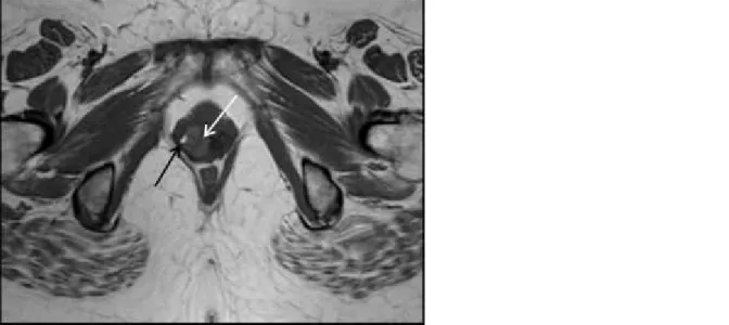
Axial T1-weighted image demonstrates the antero-lateral lesion of intermediate signal
intensity (white arrow) - slightly higher than muscle - with a hyperintense spot (black arrow)
that may correspond to melanin or haemorrhage (methemoglobin).
© Cunha TM, Department of Radiology, IPOFG - Lisboa, Portugal
Area of Interest: Genital / Reproductive system female;
Imaging Technique: MR;
Procedure: Staging;
Special Focus: Neoplasia;
Figure 2 Sagittal T2-weighted image
Sagittal T2-weighted image shows intermediate/high signal intensity lesion arising from the
anterior wall of the lower third of the vagina (arrow). Note that the intravaginal contrast
(ultrasound gel) helps to depict the lesion.
© Cunha TM, Department of Radiology, IPOFG - Lisboa, Portugal
Area of Interest: Genital / Reproductive system female;
Imaging Technique: MR;
Special Focus: Neoplasia;
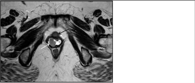
Figure 3 Axial T2-weighted image
Axial T2-weighted image shows that the vaginal lesion has slightly high signal intensity
(white arrow) and that the urethra wall at this level is preserved, with no signs of invasion
(black arrow).
© Cunha TM, Department of Radiology, IPOFG -Lisboa, Portugal
Area of Interest: Genital / Reproductive system female;
Imaging Technique: MR;
Procedure: Staging;
Special Focus: Neoplasia;
Figure 4 Axial T2-weighted image
Axial T2-weighted image demonstrates invasion of the urethra by the vaginal tumour at a
lower level than previous picture (white arrow).
© Cunha TM, Department of Radiology, IPOFG - Lisboa, Portugal
Procedure: Staging;
Special Focus: Neoplasia;
Figure 5 Sagittal fat-suppressed T1- weighted image + gadolinium
Sagittal fat-suppressed T1- weighted image after the administration of gadolinium shows the
homogeneous enhancement of the vaginal lesion (arrow).
© Cunha TM, Department of Radiology, IPOFG - Lisboa, Portugal
Area of Interest: Genital / Reproductive system female;
Imaging Technique: MR;
Procedure: Staging;
Special Focus: Neoplasia;
Axial diffusion-weighted image (b = 600 sec/mm2) shows the mass (arrow) with bright
signal intensity.
© Cunha TM, Department of Radiology, IPOFG - Lisboa, Portugal
Area of Interest: Genital / Reproductive system female;
Imaging Technique: MR;
Procedure: Staging;
Special Focus: Neoplasia;
Figure 7 Sagittal sections of the gross specimen
Two sagittal sections of the gross specimen of the lower third of the vagina. A polypoid
pigmented tumour (arrow) is observed in the anterior wall of the vagina.
© Félix A, Department of Pathology, IPOFG - Lisboa, Portugal
Area of Interest: Genital / Reproductive system female;
Imaging Technique: PACS;
Special Focus: Neoplasia;
MeSH
[A05.360.319.779]
Vagina
The genital canal in the female, extending from the uterus to the vulva. (Stedman, 25th ed)
References
[1] Chung AF, Casey MJ, Flannery JT, Woodruff JM, Lewis JL Jr. (1980) Malignant melanoma of the vagina: report of 19 cases Obstet Gynecol 55(6):720-727
[2] Parikh JH, Barton DP, Ind TE, Sohaib SA (2008) MR Imaging features of Vaginal Malignancies Radiographics 28(1):49-63; quiz 322
Citation
C. Tentugal*, A. Félix**, T. M. Cunha** (2012, Sep. 24)
Malignant melanoma of the vagina {Online}