DISTRIBUTION
Abstract
The Currais Islands is a group of a few small rocky islands in the state of Paraná, in southern Brazil, which provides an interesting location for the study of ascidians. Subtidal diversity is very high and the islands have recently been proposed as a Conservation Unit. A field study was established on the largest island to understand ascidian distributions on spatial and temporal scales. Transects, sampled three times during 2002 and 2003, were established on northern and southern locations of the island as well as at three depths. Twenty species were recorded; the most common were
Didemnum rodriguesi, Didemnum speciosum and Didemnum granulatum. Three species are possibly new and will be
described elsewhere. An additional nine are new records for the state of Paraná: Perophora regina, Didemnum speciosum, Trididemnum orbiculatum, Eudistoma carolinense, Aplidium pentatrema, Molgula phytophila, Botryllus tuberatus, Symplegma brakenhielmi and Polyandrocarpa anguinea. While all these species are distributed between 6 and 15 m, there
is a tendency to reduction of abundance towards 15 m in several species. Some species appear to prefer the north side of the island. Several species show temporal changes in abundance, with some being most common in the spring and others in the summer, but we can not affirm yet that there is a seasonal pattern between years.
Key words: Ascidiacea, Tunicata, taxonomy, spatial distribution, temporal distribution.
Resumo
As Ilhas Currais são formadas por um grupo de três pequenas formações rochosas localizadas no Estado do Paraná, sul do Brasil, sendo um local de interesse para o estudo do grupo Ascidiacea. A diversidade da região sublitoral local é bastante alta, e Currais foi recentemente proposto como local a ser transformado em Unidade de Conservação. Foi desenvolvido um estudo de campo na maior das três ilhas, com o intuito de se conhecer a distribuição espacial e temporal de ascídias. Foram realizadas três amostragens entre 2002 e 2003, nos lados norte e sul da ilha, em três diferentes profundidades. Vinte espécies foram registradas, sendo as mais comuns Didemnum rodriguesi, Didemnum speciosum e Didemnum granulatum. Três são possíveis espécies novas e serão descritas futuramente. Nove espécies representam novos registros
para o Estado do Paraná: Perophora regina, Didemnum speciosum, Trididemnum orbiculatum, Eudistoma carolinense, Aplidium pentatrema,Molgula phytophila, Botryllus tuberatus, Symplegma brakenhielmi e Polyandrocarpa anguinea.
As espécies encontram-se distribuídas principalmente entre a faixa dos seis aos 15 m, tendo sido observada uma redução da abundância em profundidades maiores de 15 m. Algumas espécies parecem preferir o lado norte da ilha. Algumas espécies apresentam variações temporais de abundância, sendo mais comuns ou na primavera ou no verão, mas não é possível ainda afirmar que exista um padrão sazonal entre anos.
Palavras-chave: Ascidiacea, Tunicata, taxonomia, distribuição espacial, distribuição temporal.
Biota Neotropica v5 (n2) – http://www.biotaneotropica.org.br/v5n2/pt/abstract?article+BN03105022005 Rosana Moreira da Rocha1 & Suzana Barros de Faria2
1 Departamento de Zoologia, Universidade Federal do Paraná, Caixa postal 19020, 81531-980, Brazil. Corresponding author: E-mail: rmrocha@ufpr.br
2 Graduate student, Programa de Pós-graduação em Zoologia, Universidade Federal do Paraná, Brazil. E-mail: suzanabf@ufpr.br
Date Received: 09/03/2004
Introduction
The coastline of the state of Paraná (98 km) includes two important estuarine systems. Rocky substrates are re-stricted to several small islands near the coast and SCUBA diving in these areas has been limited because of the poor visibility of water. Together, these conditions have resulted in little exploration and knowledge of the fauna in this region, resulting in only two studies of ascidians of Paraná. Didemnum candidum Savigny, 1816 (probably a mixture of white
didemnids), Polysyncraton amethysteum (Van Name, 1902)
and Styela plicata (Lesueur, 1823) were collected mainly at
Mel Island (Moure et al. 1954). Didemnum candidum has not
been found again in Brazil and its report was probably an error. An additional 11 species were found by Rocha & Nasser (1998): Ascidia curvata (Traustedt, 1882), Ascidia sydneiensis
Stimpson, 1855, Clavelina oblonga Herdman, 1880, Distaplia bermudensis Van Name, 1902, Didemnum granulatum
Tokioka, 1954, Didemnum lutarium Van Name, 1910, Didemnum rodriguesi Rocha & Monniot, 1993, Diplosoma listerianum (Milne-Edwards, 1841), Cystodytes dellechiajei
(Della Valle, 1877), Symplegma rubra Monniot, 1972, and Microcosmus exasperatus Heller, 1978.
Since faunal surveys are far from comprehensive, the distribution patterns of this fauna are still unknown. Due to their sessile and filter-feeding habit, ascidians have limited distribution on soft bottoms because of the lack of sub-strate for attachment and the clogging effect of the sus-pended particles. Also, delicate species may be damaged by sand abrasion (Millar 1971, Monniot et al. 1991). There-fore, lower diversity and abundance of ascidians is expected near soft bottoms compared with rocky substrates farther from the bottom. Light may also be an important selection force resulting in specific depth distribution, due to the ten-dency of ascidian larvae to attach to shadowed substrates (Young & Chia 1984, Hurlbut 1993). Even species symbiotic with photosynthetic prokaryotes initially settle in crevices or other shadowed places and move into the light later in their life cycles (Olson 1983). Thus, vertical distributions of ascidian species may be strongly influenced by light condi-tions, while some species show depth preferences that are not clearly associated with light per se (Oren & Benayahu
1998, Hurlbut 1991). Colonial ascidians are more abundant in shallow water, usually found on vertical and horizontal surfaces, while solitary forms are more abundant in deeper water, inside crevices and underneath rocks (Turon 1990).
This study of the ascidians at Currais Islands in south-ern Brazil was undertaken to record the species composi-tion of the ascidian community, to describe the species in-cluded in this study, and to examine the spatial and tempo-ral distributions of the species forming that community.
Material and Methods 1. Study site
The Currais Islands are three small rocky outcrops located 12 km from the coast of the state of Paraná (25S44’; 48W22’) in the municipality of Pontal do Paraná. Guapirá Island was the one used in this study; it is the easternmost island and the largest with 54,700 m2. Large granite blocks
of varying size comprise the sublittoral zone. Rocky sub-strates are found to a depth of 15 -- 18 m where sandy bottoms start. Shallow substrates (down to 6 m) are cov-ered by macroalgae, coralline algae and large colonies of zoanthids. Encrusting communities of deeper substrate are dominated by algae, tunicates, sponges, oysters, hydroids and other anthozoan cnidarians (Silva 2001).
Waves may reach 2.4 m (monthly mean maximum) in September and October. Winds from the east and south prevail (Silva 2001), therefore the island is the most exposed to waves, winds and strong currents on its southern and eastern sides. It is also more exposed during the winter because of cold fronts coming from the south. Diving con-ditions are variable and water visibility varies from 1 m to rarely 15 m (usually 3 - 5 m). Water temperature ranges from 15oC during the winter to 24oC during the summer.
2. Sampling
Ascidians were sampled by SCUBA diving on the southeast and north sides of the largest island during July 2002 (winter), October 2002 (spring), and January 2003 (sum-mer). During those dives, specimens were counted, col-lected for identification and quantified within small quad-rats (5 x 5 cm2) that were placed uniformly along a transect
line at a fixed depth. Only vertical surfaces were sampled to minimize variability due to light, since light is an important recruitment cue for many ascidians (Olson 1983, Young & Chia 1984, Hurlbut 1993). The presence of ascidian species inside each quadrat was registered.
For identification, specimens collected both within and outside the quadrats were anesthetized with menthol crystals for 90 minutes, then transferred to 10% seawater formalin. Voucher specimens were deposited in the collec-tion of the Zoology Department of the Federal University of Paraná (DZUFPR) and at the Museum of Zoology of the University of São Paulo (MZUSP).
Results
Fifteen species were encountered in the samples with an additional five species collected outside the quadrats, to complete the ascidian survey. The most frequent species in the samples were Didemnum rodriguesi Rocha & Monniot,
1993, Didemnum speciosum (Herdman, 1886) and Didemnum granulatum Tokioka, 1954. One new species (Eudistoma
sp.) was found among the species at Currais Islands with possibly two more new species (Didemnum sp., Lissoclinum
sp.), while ten species are reported here for the first time from the state of Paraná: Perophora regina Goodbody &
Cole, 1987, Didemnum speciosum, Diplosoma sp., Trididemnum orbiculatum (Van Name, 1902), Eudistoma carolinense Van Name, 1945, Aplidium pentatrema
(Monniot, 1972), Molgula phytophila Monniot, 1969-70, Botryllus tuberatus Ritter & Forsyth, 1917, Symplegma brakenhielmi (Michaelsen, 1904) and Polyandrocarpa anguinea (Sluiter, 1898).
Systematics
Order ENTEROGONA Perrier, 1898 Suborder PHLEBOBRANCHIA Lahille, 1887
Family PEROPHORIDAE Giard, 1872
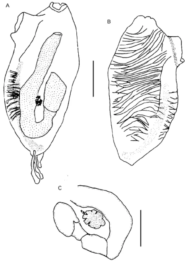
Genus Perophora Wiegmann, 1835 Perophora regina Goodbody & Cole, 1987
(Figure 1A-C)
Examined material
DZUFPR - PEROPH 013: 1 colony, Currais Islands, south side, 12 m, PR, 19 July 2002.
DZUFPR - PEROPH 014: 1 colony, Currais Islands, north side, PR, 31 January 2003.
DZUFPR - PEROPH 011: 1 colony, Saco d’Água, 10 m, Arvoredo Island, Marine Biological Reserve of Arvoredo, SC, 27 January 2000.
DZUFPR - PERO 016: 1 colony, Alcatrazes Island, north side, SP, 03 May 2002.
The colony comprises ramified stolons from which zoo-ids arise vertically. Zoozoo-ids are crowded and completely cover
the substrate around the stolons. The tunic is transparent and colorless except for two oval white spots between the siphons, one on each side of the neural ganglion, which is covered by a yellow line. The orange stomach can also be seen through the transparent body. Zooids are 2.5 - 3.5 mm long. The tubular or conical oral siphon has a delicate fringe of six to eight rounded lobes. In anesthetized individuals the margin is nearly smooth, sometimes folding over the siphon and exposing the oral tentacles. The apical, tubular atrial si-phon has six to eight rounded lobes. About 22 simple oral tentacles of three sizes are common; one zooid had 35. The circular siphonal muscles are more superficial and conspicu-ous than are the longitudinal muscles. Oblique muscles on the left side of the thorax are short and do not cover the gut, while on the right side the muscles are remarkably developed. The pharynx has four rows of stigmata, which are posterior to the non-perforated anterior area. The first three pharyngeal rows have about 35 stigmata while the fourth row has about 45 per side. Trifurcating papillae arise from each of the three transversal bars (nine to ten papillae in each half row), some of which form complete longitudinal vessels. Three languets arise from the dorsal lamina. There is a simple ciliated ring and the dorsal tubercle form a small oval aperture. The esophagus is short wider in its proximal end; the stomach is conical or globular without folds; a short duodenal region enters to the tubular posterior stomach. The intestinal loop is straight, not twisted, and the bilobed anus ends between the second and the third row of stigmata. Three stoloniferous vessels arise posteriorly: the middle vessel is short and wide while the other two are thin and long and descend into the peduncle. The gonads are inside the intestinal loop; the testis has 14 or fewer round or pyriform follicles and are found in the central part of the gut loop; the ovary contains four immature oocytes at the base of the sperm duct, just dorsal to the testis. Gonads were not found in the colony collected in July (winter) and an ovary was present only in the colony from Alcatrazes Island (May – fall). Larvae were not seen.
Remarks
Perophora with four rows of stigmata that occur in
the western Atlantic are P. viridis Verrill, 1871 and P. regina
Goodbody & Cole, 1987 (Goodbody 1994). The material described here has all the characters described for P. regina
Suborder APLOUSOBRANCHIA Lahille, 1887 Family CLAVELINIDAE Forbes & Hanley, 1848
Genus Clavelina Savigny, 1816 Clavelina oblonga Herdman, 1880
Examined material
DZUFPR - POLY 049: 1 colony, Currais Islands, PR, south side, 8 m, 19 July 2002.
DZUFPR - POLY 057: 1 colony, Currais Islands, PR, north side, 04 October 2002.
DZUFPR - POLY 061: 1 colony, Currais Islands, PR, 31 January 2003.
DZUFPR - POLY 062: 2 colonies, Currais Islands, PR, north side, 8 and 12 m, 31 January 2003.
DZUFPR - POLY 063: 1 colony, Currais Islands, PR, south side, 15 m, 31 January 2003.
Zooids are linked witch each other only at the base and are about 2 cm long. They are very transparent, some-times with a white line on the prepharyngeal grove and on the endostyle. Zooids can be densely packed in the colony or not. The two apertures are circular without lobes. There are 13 oral tentacles of three sizes and 12 - 13 rows of stig-mata. The pyriform follicles of the testis lay on the gut loop, around the central ovary. Larvae were present in summer.
Remarks
There are four species of Clavelina in the tropical
Atlantic: C. oblonga Herdman, 1880, C. picta (Verrill, 1900), C. brasiliensis (Millar, 1977) and C. puertosecensis Millar &
Goodbody, 1974. The first three have been reported along the Brazilian coast, but although C. picta was recorded from
São Sebastião, SP, (Van Name 1945) it has not since been found there. Millar (1977) collected some specimens in the northern Brazilian coast, which he identified as C. picta.
Compared with C. oblonga, C. picta has larger colonies
with up to 1,000 zooids, the endostyle, dorsal lamina and peripharyngeal bands are purple colored and the larvae have four rows of stigmata (Van Name 1945). Clavelina brasiliensis has long zooids with a cylindrical stalk of firm
consistency, the position of oral and atrial openings is sub-terminal, and the larvae present modified adhesive papillae (Millar 1977), characters not observed in our specimens.
Family POLYCITORIDAE Michaelsen, 1904 Genus Eudistoma Caullery, 1909 Eudistoma carolinense Van Name, 1945
Examined material
DZUFPR - POLY 055: 1 colony, Currais Islands, PR, north side, 8 m, 04 October 2002.
DZUFPR - POLY 056: 1 colony, Currais Islands, PR, north side, 04 October 2002.
DZUFPR - POLY 065: 1 colony, Currais Islands, PR, south side, 12 m, 31 January 2003.
DZUFPR - POLY 066: 1 colony, Currais Islands, PR, north side, 31 January 2003.
Colonies are lobed and completely covered with sand. Lobes are 2 to 5 cm long and compressed laterally. The zooids reach 5.5 mm in length with three rows of stigmata and 14 - 15 stigmata are found in each half-row. The gonad is formed by the central ovary surrounded by seven to 14 testicular follicles, all of which forms a compact mass. The larval body is 0.3 mm long with three adhesive papillae and four pairs of ectodermal ampullae.
Remarks
This species has been reported in northeastern Bra-zilian coastal waters (Millar 1977) and less than 50 km south of Currais Islands in the state of Santa Catarina (Rocha & Moreno 2000), even though it has never been reported from the intervening areas (Lotufo 2002). It is possible that it has been introduced to the southern coast of Brazil through ship transportation.
Family POLYCITORIDAE Michaelsen, 1904 Genus Eudistoma Caullery, 1909
Eudistoma sp.
Examined material
DZUFPR - POLY 058: 1 colony, Currais Islands, PR, south side, 04 October 2002.
DZUFPR - POLY 059: 1 colony, Currais Islands, PR, north side, 8 m, 04 October 2002.
The colony is white, with grayish round spots on the surface of each zooid. Colonies are small (1.5 x 2.0 cm and 4 mm thick) and oval-shaped. The colorless tunic is gelati-nous but offers some resistance to tear; the white zooids may be seen through the tunic. Colonies were always found on encrusting coralline algae.
Remarks
Gonads and larvae were present. This is a new spe-cies to be described elsewhere.
Family POLYCLINIDAE Milne-Edwards, 1842
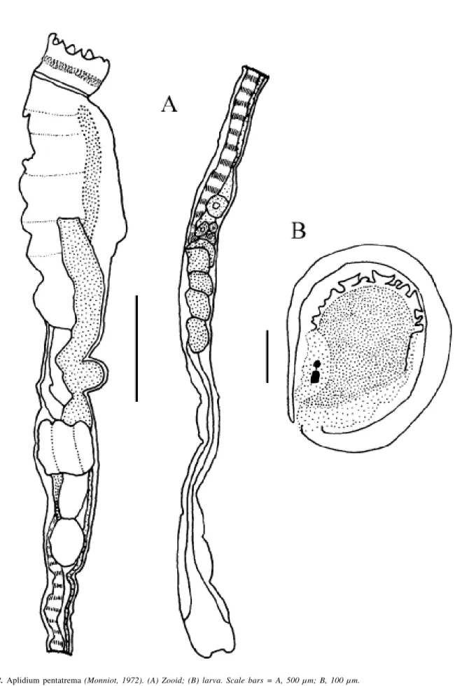
Genus Aplidium Savigny, 1816 Aplidium pentatrema (Monniot, 1972)
Examined material
DZUFPR - POCL 029: 1 colony, Currais Islands, PR, north side, 8 m, 04 October 2002.
Colonies were brown, encrusting and cushion-like, of 2 x 3 cm in area and 8 mm thick. The tunic is firm, cartilagi-nous and packed with sand. When alive, zooids form sys-tems that are difficult to observe in preserved specimens. Zooids are 1.2 cm long with the post-abdomen portion be-ing twice the length of the rest of the body. The apical oral siphon has eight lobes and circular muscles that form a sphincter, while the atrial siphon is lateral, small and circu-lar. A long dorsal languet divides into three points with the middle point longer than the others.
The pharynx has five rows of stigmata with at least 10 stigmata in the first half-row. No more than 10 thin longitudi-nal muscles are found on each side of the thorax continuing to the end of the post-abdomen. The esophagus is long, the stomach has six folds, the intestine has two constrictions and the bilobed anus is between the third and fourth rows of stig-mata. Up to seven embryos were found in the atrial cavity. The oval larvae appear to be immature and are only 0.33 mm long, with the tail wrapping entirely around the trunk on the left side. Three adhesive papillae form a line with short and thin peduncles. A concentration of epidermal vesicles is found in the anterior region of the larvae. The ocellus and statocyte are visible inside the sensory vesicle.
Remarks
This species was described from Bermuda under the genus Sidnyum by Monniot (1972). The only difference
between the genera Aplidium and Sidnyum is the number
of lobes on the oral siphon. Therefore, in agreement with Kott (1992), we suggest that these genera are synonymous and the name Aplidium has precedence. The species
de-scribed here is very similar to the original description of A. pentatrema, the main difference is that the size of the larvae
of that species is twice that of the species described herein. This could be explained by the immature aspect of the lar-vae from Currais. Aplidium appendiculatum (Michaelsen,
1923) from the Azores also have small larvae and eight lobes on the oral siphon, while the thorax is longer with 10 to 12 rows of stigmata (Monniot 1974).
Family HOLOZOIDAE Berrill, 1950 Genus Distaplia Della Valle, 1881 Distaplia bermudensis Van Name, 1902
Examined material
DZUFPR - POLY 064: 1 colony, Currais Islands, PR, north side, 12 m, 31 January 2003.
The colony is encrusting, approximately 3 mm thick, with a surface area of 1 x 1 cm. The preserved tunic is greenish, transparent and soft. Zooids are 1.8 mm long. The internal characteristics of the zooids correspond to the de-scription in the literature (Van Name 1945).
Remarks
Gonads were present inside the intestinal loop but larvae were not found.
Family DIDEMNIDAE Verrill, 1871 Genus Diplosoma MacDonald, 1859
Diplosoma listerianum (Milne-Edwards, 1841)
Examined material
DZUFPR - DID 159: 1 colony, Currais Islands, PR, south side, 12 m, 19 July 2002.
DZUFPR - DID 162: many colonies, Currais Islands, PR, south side 04 October 2002.
DZUFPR - DID 163: many colonies, Currais Islands, PR, north side, 04 October 2002.
The encrusting colony is very thin, transparent and grayish. Dark pigment is found over the abdomen and some-times the endostyle and oral siphon. Zooids are 0.8 mm long and the thorax is longer than the abdomen. The margin of the oral siphon is formed by six triangular lobes while the open atrial siphon lacks a dorsal languet. The thorax is transparent, with four rows of stigmata, the first two rows with 14 stigmata, the third with 13 and the last with 12 in each side. A very delicate muscular process is present. The esophagus is short, the stomach is rounded and smooth walled and the anus is not lobed. The testis has two fol-licles connected to a straight sperm duct. The ovary has four oocytes alongside the testis. A large embryo may be visible inside the abdomen but fully developed larvae are inside the tunic. The larvae are oval and 0.55 mm in length. The tail extends along the ventral side of the body to the anterior end. There are three adhesive papillae in a line, close together, with long peduncles, and two ectodermal ampullae in each side. The ocellus and statocyte are visible inside the sensory vesicle. Larvae are gemmiparous, with the oozoid and one blastozooid, being present only in colo-nies collected in October (spring).
Remarks
Diplosoma macdonaldi Herdman, 1886 was originally
described from the state of Bahia in Brazil and is a common species in shallow waters of the West Atlantic (Van Name 1945). Despite the fact that Rowe (1966) considered D. macdonaldi and D. listerianum synonyms, Lafargue (1968)
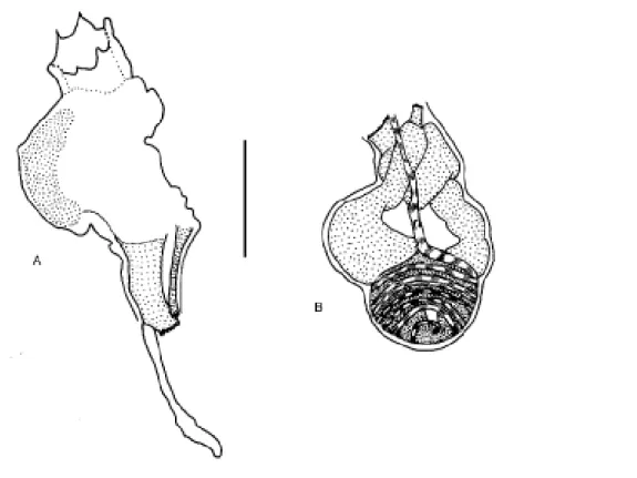
Figure 3.Diplosoma sp. (A) Zooid; (B) larva. Scale bars = A, 200 µm; B, 500 µm.
the latter. Kott (2001) recently suggested that the Atlantic specimens should be considered a different species from D. listerianum, but her only argument is the geographical
dis-tance between Pacific and Atlantic localities. We did not find any consistent morphological characteristics to justify this position and we therefore maintain the south Brazilian specimens as D. listerianum. This is a cosmopolitan
spe-cies, usually included in lists of nonindigenous species (Lam-bert 2001), previously found in southern localities along the Brazilian coast (Rocha & Nasser 1998).
Family DIDEMNIDAE Verrill, 1871 Genus Diplosoma MacDonald, 1859
Diplosoma sp.
(Figure 3A, B)
Examined material
MZUSP-15.779: many colonies, Currais Islands, PR, south side, 04 October 2002, and permanent slide.
DZUFPR - DID 177: many colonies, Currais Islands, PR, north side, 04 October 2002.
The colony and tunic are very similar to D. listerianum, but the black pigment over the abdomen,
Remarks
Diplosoma listerianum (Herdman, 1886) is the only
species known in Brazil, and normally has two pairs of larval ectodermal ampullae. Six other species are known from the Atlantic Ocean and differ from this species as follows:
Diplosoma glandulosum Monniot, 1983 from Guadalupe
and the Bermudas has a larger zooid (1.75 mm), glandular tissue on the side of the testis, and two pairs of larval ecto-dermal ampullae; Diplosoma tritestis Monniot, 1984, also
from Guadalupe, has a smaller larva (0.3 mm), testis with three follicles, and only two pairs of ectodermal ampullae;
Diplosoma lafargueae Vazquez, 1993 from the Atlantic coast
of the Iberian Peninsula, has orange colonies with large zooids (1.7 mm) without a muscular process; Diplosoma singulare Lafargue, 1968 also from Glenan Archipelago, has
an orange colony with larger zooids (2 mm), without a mus-cular process, and in spite of the large number of ampullae, has fewer ampullae on each side of the larva compared with our specimen (Lafargue 1968, Lafargue & Wahl 1987); and finally Diplosoma spongiforme (Giard, 1872) from Glenan
Archipelago. This last one is the most similar to the present species but Lafargue & Wahl (1987) report the following diagnostic characteristics: a long subendostylar thoracic appendix (longer than the abdomen – Lafargue 1968) which was not seen in our material; three to eight adhesive papil-lae (all the larvae we found had three), and two to seven pairs of ectodermal ampullae in the larvae (we found only four to six). Furthermore, our larvae are smaller than the one described by Lafargue (1968) with a length of 0.8 mm, but they could have been juvenile. Since Diplosoma is known
to contain invasive species (Lambert 2001), it is possible that the colonies found at Currais Islands are a population of D. spongiforme, but only further genetic investigations
would solve the problem.
Among the species not from the Atlantic, the only
Diplosoma with asimilar larva is Diplosoma ata Monniot &
Monniot, 1987 from French Polynesia and New Caledonia. This larva is 0.55 to 0.8 mm long and presents two to six pairs of ectodermal ampullae. The zooids also have a small muscular process, but the testis has only one follicle and the oral siphon has long leaf-like lobes (Monniot & Monniot 1987, Monniot 1994).
Family DIDEMNIDAE Verrill, 1871 Genus Lissoclinum Verrill, 1871
Lissoclinum sp.
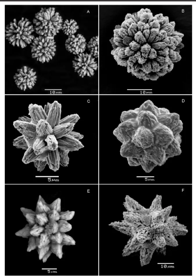
(Figure 4 A)
Examined material
DZUFPR - LIS 002: 1 colony, Currais Islands, PR, north side, 6 m, 31 January 2003.
The white encrusting colony becomes salmon-col-ored upon fixation. The tunic is soft, yet resistant and has a smooth surface. In live animals, the cloacae are large and slightly raised with a smooth margin. In the medium layer the tunic is restricted to the contour of the body of the zooids due to the wide cloacal cavities, giving great flexibil-ity to the colony. Spherical spicules, 10 - -20 ìm, with many cylindrical rays and truncated tips, are densely packed in the surface and base of the colony. The zooids were studied from decalcified cross sections of the tunic. Zooids are or-ange, 0.9 mm in length with the thorax larger than the abdo-men. The thorax is transparent with abundant yellow pig-ment in each margin of the branchial stigmata. The atrial opening is wide, exposing part of the pharynx, without a dorsal languet. The muscular process is absent. There are four rows of stigmata and in one bud six, six, five, four stig-mata were found in each half row. It was not possible to count the stigmata in mature thoraces. The abdomen is opaque and yellowish. The esophageal-rectal peduncle is horizontal and the abdomen makes a right angle with the thorax. Gonads are located in the intestinal loop with the testicle anterior to the ovary. The testicle has two follicles and a straight sperm duct. Larvae, 0.35 mm in length, were poorly developed in the material.
Remarks
Only two species in the genus Lissoclinum are
re-ported from Brazil: L. fragile (Van Name, 1902) and L. perforatum (Giard, 1872) (Lotufo 2002). Lissoclinum fragile
is characterized by the whitish color of the colony, even in preserved specimens, and no zooid pigmentation (Rodrigues et al. 1998). Also, the zooid is 1.5 mm long and the colony has low spicule density (Monniot 1983). On the other hand, the spicules of the present species are very similar to the spicules of L. fragile.Lissoclinum perforatum has orange
zooids of 2.0 mm in length and a unique type of spicule, which is larger (500 ìm), in low density, and restricted to the superficial layer of the tunic (Monniot 1983). In the Carib-bean there are also Lissoclinum abdominale Monniot, 1983
and Lissoclinum verrilli (Van Name, 1902), each with a single
testicular follicle. The species described here is apparently new, but because in didemnids the larva is usually crucial to differentiate species, we prefer not to name the species at present since larvae were not well developed.
Family DIDEMNIDAE Verrill, 1871 Genus Polysyncraton Nott, 1892 Polysyncraton amethysteum (Van Name, 1902)
(Figure 4B)
Examined material
The living colony is encrusting and bright purple, becoming orange after fixation. Several large, circular cloa-cae are distributed on the colony surface. The colony is transparent due to the low surface spicule density and the absence of spicules in the rest of the tunic. Spicules are only found around the openings of the oral siphons. Zoo-ids are 1-1.3 mm in length. A long dorsal languet narrows towards the base but expands into four finger-like projec-tions distally. The round thoracic organ is located between the endostyle and the border of the mantle and between the first and second rows of stigmata. Gonads were immature and larvae were absent.
Remarks
This species is a very common in intertidal and shal-low subtidal regions along the coast of southeastern and southern Brazil and was recently found in northeastern Bra-zil (Lotufo 2002).
Family DIDEMNIDAE Verrill, 1871 Genus Didemnum Savigny, 1816 Didemnum rodriguesi Rocha & Monniot, 1993
(Figure 4C)
Examined material
DZUFPR - DID 154: 1 colony, Currais Islands, PR, north side, 12 m, 19 July 2002.
DZUFPR - DID 155: 1 colony, Currais Islands, PR, south side, 12 m, 19 July 2002.
DZUFPR - DID 161: 4 colonies, Currais Islands, PR, south side 11-12 m, 04 October 2002.
DZUFPR - DID 172: 3 colonies, Currais Islands, PR, south side 11 m, 04 October 2002.
Small encrusting colonies may form large aggregates that cover relatively large areas of the substrate. The colony is red or bright orange, with a cream-colored ring bordering the oral siphon, becoming yellowish after fixation. The tu-nic is firm, brittle and packed with spicules. Zooids resemble the original description (Rocha & Monniot 1993) with the exception that the total length surpasses the 1.2 mm previ-ously described, measuring up to 1.5 mm in the examined material. No larvae were present.
Remarks
This species has been found from Ceará to Santa Catarina (Lotufo 2002).
Family DIDEMNIDAE Verrill, 1871 Genus Didemnum Savigny, 1816 Didemnum cf. granulatum Tokioka, 1954
Examined material
DZUFPR - DID 151: 1 colony, Currais Islands, PR, south side, 12 m, 19 July 2002.
DZUFPR - DID 152: 2 colonies, Currais Islands, PR, north side, 12 m, 19 July 2002.
DZUFPR - DID 153: 1 colony, Currais Islands, PR, south side, 12 m, 19 July 2002.
DZUFPR - DID 166: 3 colonies, Currais Islands, PR, south side, 9-12 m, 10 October 2002.
DZUFPR - DID 167: 1 colony, Currais Islands, PR, south side, 12 m, 04 October 2002.
DZUFPR - DID 168: 1 colony, Currais Islands, PR, north side, 9 m, 04 October 2002.
Colonies are large, encrusting, cream-colored, becom-ing white after fixation. The surface of the tunic is rough and granulated due to the presence of papillae with a high spicule density. Spicules are similar to the ones figured by Rocha & Monniot (1995) and Kott (2001). Cloacae are coni-cal with toothed borders. The zooid mantle is transparent and whitish. Thoraces were too contracted to permit count-ing the number of stigmata. The thoracic organ is circular, placed between the third and fourth rows of stigmata on the border of the body wall. The muscular process is attached to the esophageal-rectal peduncle. Neither gonads nor lar-vae were found in the material.
Remarks
Mature gonads and larvae were not found in this material, so we cannot identify this species with certainty. The cream color of the colony differs from that first de-scribed (orange, salmon or brown) for the Atlantic Ocean (Rocha & Monniot 1995). Also, cloacae do not have smooth margins. The color was always described as orange or brick in previous descriptions of this species in Brazil (Rocha & Nasser 1998, Lotufo 2002) while Tokioka (1954) described the species as small white colonies. Nishikawa (1990) also characterizes D.granulatum with white colonies and
zoo-ids with clear orange mantle. Omuro & Rocha (2000)
ob-served the presence of white colonies of Didemnum granulatum in the Marine Biological Reserve of Arvoredo
-SC, which indicates that colonies of clear coloration occur in the south of Brazil.
Family DIDEMNIDAE Verrill, 1871 Genus Didemnum Savigny, 1816 Didemnum speciosum (Herdman,1886)
(Figure 4D)
Examined material
DZUFPR - DID 174: 2 colonies, Currais Islands, PR, south side, 04 October 2002.
DZUFPR - DID 175: 2 colonies, Currais Islands, PR, south side, 8-2 m, 19 July 2002.
DZUFPR - DID 180: 2 colonies, Currais Islands, PR, south side, 15 m, 31 January 2003.
DZUFPR - DID 181: 1 colony, Currais Islands, PR, north side, 31 January 2003.
DZUFPR - DID 182: 1 colony, Currais Islands, PR, south side, 12 m, 31 January 2003.
The colony is encrusting and white. Star-shaped oral openings occur in small elevations on the colony surface. The tunic is firm and brittle with spicules densely packed. Spicules have several conical rays with rounded tips and are distributed evenly throughout the tunic. Zooids are small, approximately 0.6 mm. The muscular process is short arising from the esophageal-rectal peduncle. The thoracic organ is salient, but visible in only some individuals. Larvae were present only in the material collected in 31 January 2003 (sum-mer). Larval trunk is triangular and only 0.3 mm long even in mature larvae. The tail makes a complete turn around the trunk, on the left side of the larva. The three adhesive papillae have short and wide stalks, are distant from each other and form a line. Four digitiform ampullae are found on each side of the trunk. The ocellus and statocyte are present.
Remarks
Beyond the original locality in Bahia (NE) (Herdman 1886), this species has already been found in São Paulo (Rocha & Monniot 1995) and north and northeastern Brazil (Millar 1977).
Family DIDEMNIDAE Verrill, 1871 Genus Didemnum Savigny, 1816
Didemnum sp.
(Figures 4E; 5A, B)
Examined material
DZUFPR - DID 156: 2 colonies, Currais Islands, PR, south side, 12 m, 19 July 2002.
DZUFPR - DID 157: 1 colony, Currais Islands, PR, north side, 8 m, 19 July 2002.
DZUFPR - DID 164: 3 colonies, Currais Islands, PR, south side, 9-12 m, 04 October 2002.
DZUFPR - DID 165: 1 colony, Currais Islands, PR, north side, 04 October 2002.
DZUFPR - DID 178: 1 colony, Currais Islands, PR, south side, 12 m, 31 January 2003.
DZUFPR - DID 179: 1 colony, Currais Islands, PR, north side, 8 m, 31 January 2003.
The colony is encrusting and marbled due to the dark pigment (black or dark purple) on the cloacal channels and around the cloacae, contrasting with the beige color of the rest of the tunic. After fixation the colony becomes choco-late-brown and beige. The tunic is firm and brittle. Cloacae are conical with oval openings. Spicules are uniformly and densely distributed in the surface of the tunic, but in low density in the rest of the colony. Spicules are small (20 –30 µm), globular, with a small number of conical and short rays, sometimes with rounded tips. Zooids are 1.0 mm long, with the thorax and abdomen of equal length. The body wall is dark brown. The long, tubular, oral siphon has six triangu-lar lobes and conspicuous circutriangu-lar musculature. The atrial opening is lateral and wide, without a dorsal languet. The muscular process is longer than the abdomen, arising from the esophageal-rectal peduncle. The thoracic organ is sa-lient in shell form, but not observed in all individuals. The pharynx has four rows of stigmata. In one bud seven stig-mata were found in one half of the anterior row while six stigmata were found in the others. The esophagus is short and the esophageal-rectal peduncle presents a very evi-dent constriction. The stomach is round and smooth and the duodenum is as long as the stomach. The single testicle is surrounded by six or seven turns of the sperm duct, and located posterior in the intestinal loop. The ovary has two oocytes, anterior to the testis, and the sperm duct usually covers the larger oocyte. There were no larvae.
Remarks
The absence of larvae in the examined material hin-dered comparisons with other didemnid species, although the appearance of the colony is quite different from any species described from Brazil and the Atlantic. This suggests that the present material may represent a new species.
Family DIDEMNIDAE Verrill, 1871 Genus Trididemnum Della Valle, 1881 Trididemnum orbiculatum (Van Name, 1902)
(Figure 4F)
Examined material
DZUFPR - DID 160: 1 colony, Currais Islands, PR, north side, 8 m, 19 July 2002.
DZUFPR - DID 170: 1 colony, Currais Islands, PR, south side, 04 October 2002.
The colony is encrusting and transparent with black spots caused by the accumulation of pigment on the oral siphons of the zooids. The tunic is mucous, with low spicule density at the surface and absence of spicules in the base and medium layer. The muscular process is shorter than the abdo-men, arising from the esophageal-rectal peduncle. The tho-racic organ is round and salient, positioned between the
en-dostyle and the border of the body wall. There are three rows of stigmata in the pharynx. The atrial siphon is tubular and horizontal. The testis has one follicle surrounded by 5-6 turns of the sperm duct and the ovary with two small oocytes. There were no larvae in the examined material.
Remarks
The genus Trididemnum in Brazil is represented by T. orbiculatum (T. thetidis and T. orbiculatum are
synony-mous) (Van Name 1945, Rodrigues & Rocha 1993) and T. maragogi (Rocha, 2002). Recently T. solidum (Van Name,
1902)and Trididemnum sp. have been reported from the
Brazilian coast (Lotufo 2002). The examined material was identified as T. orbiculatum mainly because of the shape,
quantity and distribution of the spicules and the color of the colony. There were no larvae in the examined material.
Order PLEUROGONA Perrier, 1898 Suborder STOLIDOBRANCHIA Lahille, 1887
Family STYELIDAE Sluiter, 1895 Genus Botryllus Gaertner, 1774 Botryllus tuberatus Ritter & Forsyth, 1917
Examined material
DZUFPR - DID 151: 1 small colony on Didemnum granulatum, Currais Islands, PR, south side, 12 m, 19
July 2002.
The colony is encrusting and 4 mm in diameter. The tunic is transparent, thin and delicate. Zooids are sac-like, 0.6 mm. The tubular oral siphon has a smooth margin; the atrial opening is wide, with a long and pointed dorsal languet. The body wall is translucent and strongly pigmented brown, thus hiding the internal structures. The pharynx has four rows of stigmata.
Remarks
Several species of Botryllus have been reported in
Brazil: B.giganteum Aron & Sole-Cava, 1991, B. tabori
Rodrigues, 1962and B. tuberatus Ritter & Forsyth, 1917
occur in the coast of the state of São Paulo (Rodrigues et al. 1998), B.planus (Van Name, 1902) and B. schlosseri (Pallas,
1766) occur on the tropical Brazilian coast (Lotufo 2002). Among them, only B.tuberatus has four rows of stigmata.
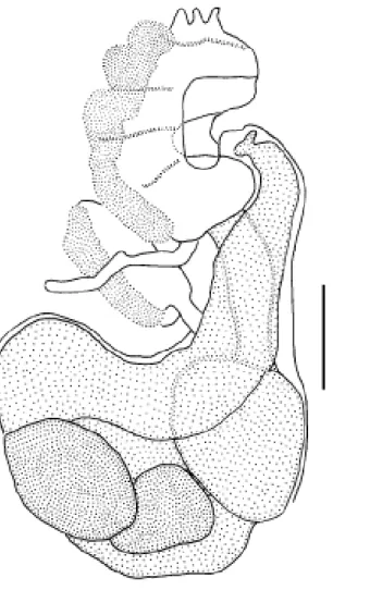
Family STYELIDAE Sluiter, 1895 Genus Polyandrocarpa Michaelsen, 1904 Polyandrocarpa anguinea (Sluiter, 1898)
Examined material
The colony is 5.5 cm long and 3 cm thick, comprising a compact mass of zooids, with siphons projecting from the colony surface. The tunic is firm and wrinkled with encrus-tations. Zooids are 1.5 cm long, yellowish with a very deli-cate body wall, usually tearing during removal from the tu-nic. Approximately 22 thin oral tentacles, of two sizes. The neural gland opening is “C” shaped with the ends of the “C” curved inward. The pharynx comprises four folds on each side. The longitudinal vessels were arranged in the following manner in one example: right E 7 12 7 16 4 13 5 11
2 DL 4 14 4 10 7 12 8 12 7 E left. The abdomen is formed by
a globular stomach, with 13 longitudinal folds, without a caecum. The intestinal loop contains no endocarp, and the anus is fringed by many lobes. The gonads are sac like, loosely connected to the body wall. In one example we counted 15 in the right side and 10 in the left side of the body; in another 15 in the right and two in the left.
Remarks
Only two species of Polyandrocarpa have been
found in Brazil: Polyandrocarpa anguinea (Sluiter, 1898)
and P. zorritensis (Van Name 1931). P. zorritensis has smaller
zooids each in its tunic and attached to each other only by branching and root-like stolons. The colony may be cov-ered by sand (Van Name 1945).
Family STYELIDAE Sluiter, 1895 Genus Symplegma Herdman, 1886 Symplegma brakenhielmi (Michaelsen, 1904)
Examined material
DZUFPR - STY 096: 1 colony, Currais Islands, PR, south side, 04 October 2002.
The colony has a very thin tunic and protuberant zooids. In formalin, the colony is uniformly brown and the tunic is transparent and delicate. The color comes from the zooids. The body is flattened dorso-ventrally, measuring up to 4 mm in length. There are six long, and a variable number of smaller, oral tentacles, all of which have a large base and pointed tip. The dorsal tubercle forms a swollen region around the small circular aperture. The pharynx has ten rows of stigmata. The first two longitudinal vessels of the left side are not parallel with the others, turning towards, and contacting the dorsal lamina at the fourth and sixth rows of stigmata. The caecum of the stomach has two con-nections with the intestinal loop, the first arising from the base of the caecum and the second, from the curve of the caecum. The testis has one or two anterior lobes and two or three posterior lobes, and the ovary has up to eight oocytes in each side. No larvae were found.
Remarks
In addition to S. brakenhielmi, the species reported
from Brazil are S. rubra Monniot, 1972 and S. viride Herdman,
1886. However, in a recent revision of specimens from the state of São Paulo, no S. viride were found (Couto 2003). Symplegma brakenhielmi was diagnosed by the color of
the body wall, a discontinuous prepharyngeal groove with a swollen region around the dorsal tubercle, weakly lobed testicular follicles and the presence of two tissue connec-tions between the caecum and the intestine. Symplegma rubra, on the other hand, has a continuous prepharyngeal
groove that usually covers the small opening of the dorsal tubercle, has very lobed testicular follicles and three tissue connections between the caecum and the intestine.
Family PYURIDAE Hartmeyer, 1908 Genus Microcosmus Heller, 1878 Microcosmus exasperatus Heller, 1878
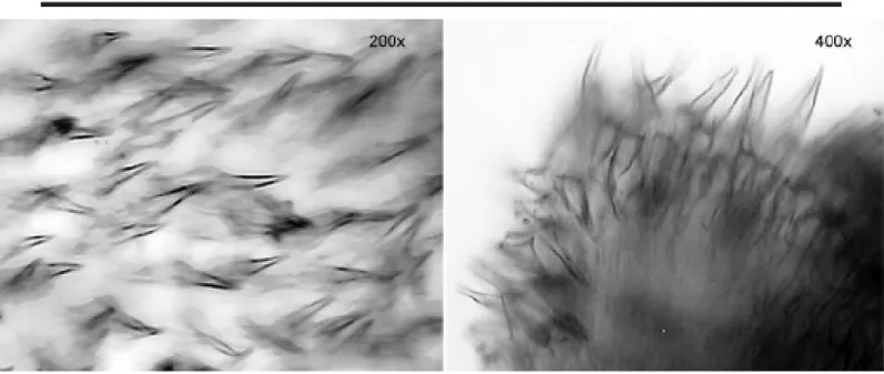
(Figure 6)
Examined material
DZUFPR - PYU 042: 2 individuals, Currais Islands, PR, 31 January 2003.
DZUFPR - PYU 043: 1 individual, Currais Islands, PR, 31 January 2003.
The globular body is enclosed within a leathery and wrinkled tunic. The animals were about 2.5 cm in height and 3 cm in width (other animals more or less the same size). The tunic is orange or purple, maintaining color in formalin, and contains some sand and encrusting organisms on the sur-face. Both siphons are lobed with four triangular lobes. There are 12 large and 18 smaller branched oral tentacles arranged on a muscular ring. The prepharyngeal groove is double, the neural gland opening U-shaped with very inrolled horns. The pharynx has 8 folds on each side, but the ventral fold on the right side is incomplete. The longitu-dinal vessels were disposed in the following manner in one exemplar: right E 2 12 4 17 3 18 4 20 5 17 5 20 4 15 5 18 3 DL
4 21 4 14 4 18 6 16 7 17 5 16 5 13 3 8 2 E left. One gonad on
each side of the body is formed by 3 - -5 masses of testis follicles surrounding a tubular ovary.
Remarks
Two species of the genus Microcosmus have been
reported from the Brazilian coast: Microcosmus exasperatus
Heller, 1878 and Microcosmus helleri Herdman, 1881. Microcosmus helleri differs from M. exasperatus by the
smaller number of branchial folds, the gut loop is less bent upwards at the anterior end and there is a more compacted gonad, not divided into segments (Van Name 1945).
Microcosmus squamiger has the gonads also divided into
three lobes and has been confused with M. exasperatus,
Figure 6.Microcosmus exasperatus Heller, 1878. Siphonal spines.
shape and spiny rims (Monniot et al. 2001). Microcosmus exasperatus is widely distributed in all oceans and, in the
western Atlantic Ocean, its southern distributional limit is Santa Catarina (Van Name 1945).
Family MOLGULIDAE Lacaze-Duthiers, 1877 Genus Molgula Forbes, 1848 Molgula phytophila Monniot, 1969-70
Examined material
DZUFPR - MOL 014: 2 individuals, Currais Islands, north side, 8-9 m, 31 January 2003.
Individuals are small, approximately 5 mm in length. The tunic is thin, delicate and encrusted with sand. The body is 3 mm long without the tunic, slightly flattened dor-sal-ventrally. The body wall is also transparent and deli-cate. Both siphons have 12 pointed or triangular lobes and conspicuous musculature, both circular and longitudinal. The longitudinal muscles form wide bands that extend ob-liquely up to 1/3 of the body. There are 10 long, branched oral tentacles. The prepharyngeal groove is double-walled. The dorsal lamina has a smooth margin. The pharynx has seven folds on each side, and each fold has two longitudi-nal vessels. The stigmata are spiral-shaped, but infundibula were not visible in the examined material. The abdomen includes a short esophagus, a tubular stomach overlaid by the lamellar digestive gland. The gonads are elongate and compact, the right one accompanies the renal sac dorsally, and the left lies inside the secondary intestinal loop. Sev-eral embryos were in the atrial cavity.
Remarks
Molgula phytophila has already been found on the
coast of Santa Catarina, to the south of the Currais Islands (Rocha & Moreno 2000). All the diagnostic characters (Monniot 1969-70) were found in these specimens: seven folds on each side of the pharynx, presence of two longitu-dinal vessels in each fold, the form and disposition of the gonads, and the presence of brooded embryos.
Ecological Section
The genus Didemnum (species D. rodriguesi, D. granulatum and D. speciosum) was the most common. Didemnum granulatum and D. speciosum, because they
were difficult to distinguish in the field, were treated as one species (white didemnids) in the first field sample period (winter, Table 1). During subsequent samplings, both D. granulatum and D. speciosum were identified in the field
and showed contrasting vertical patterns in the summer: D. granulatum was increasingly common from deep to shallow
water (G = 20.18; P < 0.05) while D. speciosum showed the
reverse trend (G = 13.1; P < 0.05; Table 1). Both species were very uncommon in winter (Table 2). Didemnum rodriguesi
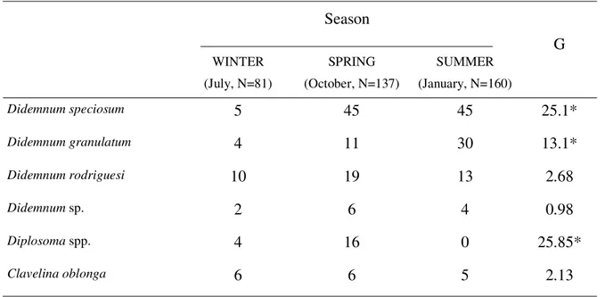
Table 2.
Seasonal occurrence of ascidian species, at Currais Islands.
Season
WINTER (July, N=81)
SPRING (October, N=137)
SUMMER (January, N=160)
G
Didemnum speciosum
5 45 45
25.1*
Didemnum granulatum
4 11 30
13.1*
Didemnum rodriguesi
10 19 13
2.68
Didemnum sp.
2 6 4
0.98
Diplosoma spp.
4 16 0
25.85*
Clavelina oblonga
6 6 5
2.13
G, Log-likelihood ratio test. *, P < 0.05.
Table 1.
Comparison of occurrence frequencies for selected ascidian species between
north and south sides, and at three depths, at Currais Islands.
Orientation Depth (m)
WINTER (July, N=81) N S
G
<10 1012 >12
Ga
White didemnids 18 16 0.29 20 14 - 1.6
Diplosoma spp. 3 1 1.15 3 1 - 1.05
SPRING (October, N=137)
D. granulatum 5 6 0.02 5 6 - 1.01
D. speciosum 28 17 5.90* 22 23 - 3.32
Diplosoma spp. 3 13 6.45* 16 0 - 17.83*
SUMMER (January, N=160)
D. granulatum 12 18 1.49 17 13 0 20.18*
D. speciosum 19 26 1.52 10 15 20 13.1*
NON-SEASONAL SPECIES
D. rodriguesi 30 12 9.81* 4/8b 29/5 -/0 22.82*/8.64*
Didemnum sp. 6 6 0.01 4/3 4/1 -/0 0.14/2.8
C. oblonga 15 2 12.3* 9/3 3/1 -/1 1.89/3.6
a
G, Log-likelihood ratio test. *, P < 0.05.
b
In Summer three depths were sampled. The numerator therefore is a comparison between
Diplosoma spp. includes both D. listerianum and D.
sp., which could not be separated in the field. Uncommon in winter and absent in summer (Table 2), Diplosoma was
more often encountered in shallow transects (G = 17.83; P < 0.05) and on the south side of the island (G = 6.45; P < 0.05) in spring (Table 1). Didemnum sp. occurred in all samples
with low frequency (Table 2) and without differences be-tween location and depths (Table 1).
Clavelina oblonga occurred in all seasons (Table
2), more commonly on the north side of the island (G = 12.3; P < 0.05) (Table 1). Additionally, Trididemnum orbiculatum, Distaplia bermudensis, Eudistoma carolinense,Eudistoma
sp., Aplidium pentatrema, Molgula phytophila, Botryllus tuberatus and Polyandrocarpa anguinea were rare with
only one or two occurrences (Table 3).
At this island, ascidians in general showed no verti-cal distribution patterns, perhaps due to the great seasonal variation in species abundance. Four species did show variation in occurrence by depth: D. rodriguesi was more
frequent in medium depth in spring and in shallow water in summer, Diplosoma spp. was more frequent in shallow
wa-ter in spring, D. granulatum was more frequent in shallow
water in summer, while D. speciosum was more frequent in
deep water in summer (Table 1).
Some species also showed geographical tendencies related to the side of the island. Didemnumrodriguesi and Clavelina oblonga were most frequent on the north side of
the island, Didemnum speciosum was also most frequent
on the north side in spring, and Diplosoma spp. were most
frequent in the south in spring (Table 1). Also, Trididemnum orbiculatum, Distaplia bermudensis, Eudistoma sp., Aplidium pentatrema, Molgula phytophila and Polyandrocarpa anguinea occurred exclusively on the
north side of the island and Botryllus tuberatus on the south
side (Table 3), but were uncommon. Further studies will be required to determine whether these species indeed have geographical tendencies or whether this restricted distribu-tion was due to sampling methods.
Discussion
Fifteen ascidian species were recorded in the transects with an additional five species encountered in the area while gathering the data. One of these is a new species for the Currais Islands: Eudistoma sp. (being
de-scribed elsewhere). An additional two species may also be new, pending further study: Didemnum sp. and Lissoclinum
sp., (see taxonomic session). Aplidium pentatrema is a
new record for Brazil, and another nine species were found for the first time in the state of Paraná: Perophora regina, Didemnum speciosum, Diplosoma sp., Trididemnum orbiculatum, Eudistoma carolinense, Botryllus tuberatus, Symplegma brakenhielmi, Polyandrocarpa anguinea and Molgula phytophila.
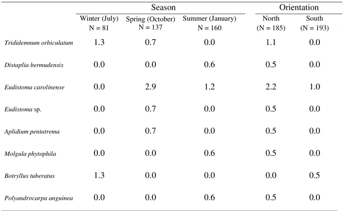
Table 3.
Percentage of 25 cm
2quadrats with rare ascidian species at Currais Islands.
Season
Orientation
Winter (July) N = 81
Spring (October) N = 137
Summer (January) N = 160
North (N = 185)
South (N = 193)
Trididemnum orbiculatum
1.3 0.7 0.0
1.1
0.0
Distaplia bermudensis
0.0 0.0 0.6
0.5
0.0
Eudistoma carolinense
0.0 2.9 1.2
2.2
1.0
Eudistoma sp.
0.0 0.7 0.0
0.5
0.0
Aplidium pentatrema
0.0 0.7 0.0
0.5
0.0
Molgula phytophila
0.0 0.0 0.6
0.5
0.0
Botryllus tuberatus
1.3 0.0 0.0
0.0
0.5
The presence of Aplidium pentatrema in the
Currais Islands could be explained by the existence of a major port receiving approximately 1,500 large ships each year, thus potentially being a source of introduction of this normally warm-water species. Aplidium pentatrema,
described by Monniot (1972) from the Bermudas, until now was only known from the type locality and Guadalupe (Monniot 1983), so it is surprising to find it here in colder subtropical waters. However, our knowl-edge of Brazilian ascidians is still very incomplete.
Eudistoma carolinense has also a very intriguing
distri-bution: Florida (Van Name 1945), northern Brazil (Millar 1977) and southern Brazil (Rocha & Moreno 2000, and in the present study). It has not been reported in the inter-vening states within Brazil (Lotufo 2002, Rodrigues et al. 1998). This distributional gap does not simply reflect the lack of sampling effort because this species occurs in intertidal zones and is easy to collect. The species was first collected in the south in 1994 but no ascidians were collected here prior to this date. Many species of ascidians have been recently reported as invasive, and currently have wide geographical distributions due to dispersal by ships (Lambert 2001).
For Symplegma brakenhielmi and Microcosmus exasperatus, the coast of the state of Paraná seems to
be their southern distributional limit in the West Atlan-tic. The other species have already been recorded fur-ther south in Brazil: Didemnum rodriguesi, D. granulatum, D. speciosum, Diplosoma listerianum, Clavelina oblonga, Distaplia bermudensis, Botryllus tuberatus, Polyandrocarpa anguinea and Molgula phytophila (Rocha et al. 1999, Rocha & Moreno 2000).
A vertical pattern of distribution was not generally observed. However, Didemnum rodriguesi,D. granulatum, andDiplosoma spp. were more frequent in shallow water,
while D. speciosum was more frequent in deep water, but
none of these species showed the same pattern in all sea-sons. The range in depths in this study was narrow be-cause the maximum depth around the island is 18 m, and most of the species were equally distributed between 6 and 15 m. However, abundance and diversity were much re-duced at 15 m, and this could possibly indicate an important shift in habitat conditions for these animals. Even in this shallow region, a marked thermocline forms during the sum-mer when the Central South Atlantic waters penetrate be-neath the warmer surface waters (Brandini 1990inSilva 2001). Temperatures near the bottom may reach 13oC (personal
observation), and low temperature can retard growth or kill ascidians (Grosholz 2001). Another possible explanation for low ascidian abundance at 18 m is the proximity to the bottom, and its associated sand, continually re-suspended by water currents. A population of Ascidia sp. was
de-stroyed after a disturbance of the sea-bed and the resulting increased turbidity (Monniot 1965). Thus, further studies, perhaps experimental, will be required to test which factors influence the abundance of ascidians in deeper waters.
Other studies have analyzed vertical distribution pat-terns for ascidians within the same depths as this study.
Microcosmus sabatieri, for instance, has different depth
distributions depending on the amount of illumination re-ceived and this varies with the inclination of the substrate (the upper limit of M. sabatieri is 15 m on horizontal
sur-faces and 0.5 m on under sursur-faces) and its orientation (the upper limit is 7-8 m on substrates facing south and 3-4 m on substrates facing north) (Monniot 1965). Light intensity seems to be the most important factor limiting spatial distri-bution of Didemnum granulatum at Eilat, Red Sea. Oren &
Benayahu 1998 reported that this species recruited faster and more intensively on experimental plates at 31 m than on shallower plates, but where the maximum depth was 17 m it also recruited more intensively on the deepest 17 m plates. Transplanted cuttings to shallower vertical and the top of horizontal plates resulted in complete mortality. Didemnum granulatum at the Currais Islands showed a different
pat-tern, being more abundant above 12 m. Future studies may determine whether this is a consequence of very poor water transparency in Currais Islands or the result of interspecific interactions after recruitment.
Depth zonation of ascidians was also observed on Deer Island of the Bay of Fundy, Canada, where photo-transects up to 140 m depth showed that some species oc-curred only at < 20 m and others reached peak densities between 60 and 90 m (Hatfield et al. 1992). Since maximum light penetration is only 14 m in that region, light is prob-ably not the factor limiting ascidian distribution. Strong tidal currents may be the most important environmental limita-tion in that study.
The south side of Currais Islands is more exposed to waves and currents, while the northern face is more pro-tected. Since intense hydrodynamics can limit the occur-rence of more sensitive ascidian species because of turbu-lence (Monniot 1965, Hatfield et al. 1992), we anticipated differences in species composition or abundance between the two sides of the island. Indeed, Didemnum rodriguesi, Clavelina oblonga and Didemnum speciosum were either
more common on the north side of the island, or equally common on both sides, depending upon the season.
Trididemnum orbiculatum, Distaplia bermudensis, Eudistoma sp., Aplidium pentatrema, Molgula phytophila
and Polyandrocarpa anguinea occurred exclusively on the
northern face, but because of their uncommonness, this pattern may be spurious. Botryllus tuberatus was found
only on the south and may prefer an exposed substrate; Van Name (1945) lists its California distribution occuring on kelp fronds and rocks in exposed areas. These consider-ations suggest that the north side of the island (more protected) could be richer in ascidians.
and abundance of species were greater than in other sea-sons. In summer, only D. speciosoum and D. granulatum
had an abundance peak, and Diplosoma spp., which was
very common in the spring, was absent. Colony growth and reproduction peaked in spring in four of five species studied in the Mediterranean, and summer was the period of colony regression for many which was attributed to ex-cessive heat (Turon & Becerro 1992). A slightly different situation has been observed in Phallusia nigra Savigny,
1816 at São Sebastião, 400 km to the north of Currais Is-lands: the peak of abundance occurred in the fall (recruit-ment) and the peak of growth in spring and summer (Rocha et al. 1999).
In conclusion, an abundant and species-rich colonial ascidian community is present at the Currais Islands, with
Didemnum rodriguesi, D. speciosum and D. granulatum
being the most abundant. Twenty species were recorded, of which one (and possibly another two) are new species. Ascidians are distributed between 6 and 15 m, with no strik-ing patterns of vertical distribution. There are some sug-gestions that the north side of the island may be richer in ascidians than the south side, but future work will be re-quired to test that hypothesis. A clear temporal variation in abundance was observed, with the greatest abundance of species and individuals in the spring, but we do not know if this pattern in repeatable between years.
Acknowlwdgements
We would like to thank the ECOPLAN Institute that generously provided us with the transport and diving lo-gistics during field trips, Dr James Roper for statistical ad-vice and revision of the English text, Carlos W. Hackradt, Rafael Metri and Igor S. de Oliveira for assistance in field work, and CNPq for a research grant to RMR. This paper is the contribution 1424 of Departamento de Zoologia, Universidade Federal do Paraná.
References
COUTO, A.C.F. 2003. Estudo taxonômico das ascídias do gênero Symplegma (Tunicata, Ascidiacea, Styelidae) encontradas no canal de São Sebastião, SP. Dissertação, Universidade de São Paulo, São Paulo, Brazil.
GOODBODY, I. & COLE, L. 1987. A new species of
Perophora (Ascidiacea) from the Western Atlantic,
in-cluding observations on muscle action in related spe-cies. Bull. Mar. Sci. 40:246--254.
GOODBODY, I. 1994. The tropical Western Atlantic Perophoridae (Ascidiacea): I. The genus Perophora.
Bull. Mar. Sci. 55:176--192.
GROSHOLZ, E. 2001. Small spatial-scale differentiation among populations of an introduced colonial inverte-brate. Oecologia 129:58--64.
HATFIELD, C., LOGAN, A. & THOMAS, M.L.H. 1992. As-cidian depth zonation on sublittoral hard substrates off Deer Island, New Brunswick, Canada. Estuar. Coast. Shelf Sci. 34:197--202.
HERDMAN, W.A. 1886. Report on the Tunicata collected during the years 1873-1876. Part 2, Ascidiae compositae Zool. Chall. Exp. 14:1-425.
HURLBUT, C.J. 1991. The effects of larval abundance, settle-ment and juvenile mortality on the depth distribution of a colonial ascidian. J. Exp. Mar. Biol. Ecol. 150:183--202. HURLBUT, C.J. 1993. The adaptive value of larval behavior
of a colonial ascidian. Marine Biology 115:253--262.
KOTT, P. 1992. The Australian Ascidiacea part 3, Aplousobranchia (2). Mem. Queensl. Mus. 32:375--620. KOTT, P. 2001. The Australian Ascidiacea Part 4, Aplousobranchia (3), Didemnidae. Mem. Queensl. Mus. 47:1--410.
LAFARGUE, F. 1968. Les peuplements sessiles de l’Archipel de Glénan. II. Les Didemnidae – Systématique – Écologie. Vie Milieu19:353--446.
LAFARGUE, F. & WAHL, M. 1987. The didemnid ascidian fauna of France. Ann. Inst. Oceanogr. Paris, 63:1--32. LAMBERT, G. 2001. A global overview of ascidian
introduc-tions and their possible impact on the endemic fauna. In The Biology of Ascidians (H. Sawada, H. Yokosawa and C.C. Lambert, eds.). Springer Verlag, Tokyo, p.249-257. LOTUFO, T.M.C. 2002. Ascidiacea (Chordata:
Tunicata) do litoral tropical brasileiro. PhD thesis. Instituto de Biociências da Universidade de São Paulo, São Paulo, Brazil.
MILLAR, R.H. 1971. The biology of ascidians. Adv. Mar. Biol.9:1--100.
MILLAR, R.H., 1977. Ascidians (Tunicata: Ascidiacea) from the northern and north-eastern Brazilian shelf. J. Nat. Hist.11:169--223.
MONNIOT, C. 1965. Étude systématique et évolutive de la Famille des Pyuridae (Ascidiacea). Mem. Mus. natl Hist. Nat., Paris, sér. A,36:1--203.
MONNIOT, C. 1969-70. Campagne de la Calypso au large des cotes Atlantiques de l’Amérique du Sud (1961-1962). Première partie (suite). 17 Ascidies Phlébobranches et Stolidobranches. Ann. Inst. Oceanogr. Paris, 36:33--59. MONNIOT, C. & MONNIOT, F. 1987. Les ascidies de
Polynésie française. Mem. Mus. natl Hist. Nat., Paris,
sér.A, Zoologie, 136:1--155.
MONNIOT, C., MONNIOT, F. & LABOUTE, P. 1991.Coral Reef Ascidians of New Caledonia. Orstom Editions, Paris.
MONNIOT, F. 1972. Ascidies aplousobranches des Bermudes. Polyclinidae et Polycitoridae. Bull. Mus. Hist. Nat., Paris, 3e sér. (82), Zoologie, 61: 949--962.
MONNIOT, F. 1974. Ascidies littorales et bathyales recoltées au cours de la campagne Biacores: Aplousobranches. Bull. Mus. Hist. Nat., Paris, 3e sér. (251), Zoologie, 173: 1287-1325.
MONNIOT, F. 1983. Ascidies littorales de Guadeloupe. I. Didemnidae. Bull. Mus. Hist. Nat., Paris,4e sér., section A, 5: 5--49.
MONNIOT, F. 1983. Ascidies littorales de Guadeloupe. III. Polyclinidae. Bull. Mus. Hist. Nat., Paris,4e sér., section A, 5: 413--422.
MONNIOT, F. 1994. Ascidies de Nouvelle-Calédonie. XIV. Le genre Diplosoma (Didemnidae). Bull. Mus. Hist. Nat.,
Paris, 4e sér., section A, 16: 3--11.
MOURE, J.S., BJORNBERG, T.K.S. & LOUREIRO, T.ST. 1954. Protochordata ocorrentes na entrada da Baía de Paranaguá. Dusenia 5: 233--242.
NISHIKAWA, T. 1990. The ascidians of the Japan Sea. I. Publ. Seto Mar. Biol. Lab. 34: 73--148.
OLSON, R.R. 1983. Ascidian-Prochloron symbiosis: The role of larval photo-adaptations in midday larval release and settlement. Biol. Bull. 165: 221--240.
OMURO, J.Y. & ROCHA, R.M. 2000. Distribuição e abundância de ascídias na Reserva Biológica Marinha do Arvoredo, Santa Catarina, Brasil. In Anais da XIII Semana Nacional de Oceanografia, Universidade do Vale do Itajaí, Santa Catarina, Brasil, p. 706--708.
OREN, U. & BENAYAHU, Y. 1998. Didemnid ascidians: rapid colonizers of artificial reefs in Eilat (Red Sea). Bull. Mar. Sci. 63: 199--206.
ROCHA, R. M. 2002. Trididemnum maragogi sp. nov.
(Ascidiacea, Didemnidae) from Alagoas, Northeastern Brazil. Revta Bras. Zool. 19: 1105-1110.
ROCHA, R. M. & MONNIOT, F. 1993. Didemnum rodriguesi
sp. nov. - a new didemnid ascidian common to south-eastern Brazil and New Caledonia Ann. Inst. Oceanogr., Paris, 69: 261-265.
ROCHA, R.M. & MONNIOT, F. 1995. Taxonomic and eco-logical notes on some Didemnum species (Ascidiacea, Didemnidae) from São Sebastião Channel, South – East-ern Brazil. Revta Bras. Biol. 55: 639--649.
ROCHA, R.M. & MORENO, T.R. 2000. Ascidians associated with Eudistoma carolinense Van Name, 1945. With
de-scription of a new species of Polycarpa. Ophelia 52: 9--16.
ROCHA, R.M. & NASSER, C.M. 1998. Some ascidians (Tunicata, Ascidiacea) from Paraná State, Southern Bra-zil. Revta Bras. Zool. 15: 633--642.
ROCHA, R.M., LOTUFO, T.M.C. & RODRIGUES, S.A. 1999. The biology of Phallusia nigra Savigny, 1816 (Tunicata,
Ascidiacea) in southern Brazil: spatial distribution and reproductive cycle. Bull. Mar. Sci. 64: 77--87.
ROCHA, R.M., MORENO, T.R. & METRI, R. 1999. Ascídias da Reserva Biológica Marinha do Arvoredo, Santa Catarina, Brasil. In Livro de Resumenes Ampliados del VIII Congresso Latinoamericano sobre Ciencias del Mar (A.E.T. Aguilar & Z.G.C. Malpica, eds.), Trujillo, Peru, p. 432--433. RODRIGUES, S.A. & ROCHA, R.M. 1993. Littoral compound ascidians (Tunicata) from São Sebastião, Estado de São Paulo, Brazil. Proc. Biol. Soc. Wash. 106: 728--739. RODRIGUES, S.A., ROCHA, R.M. & LOTUFO, T.M.C. 1998.
Guia Ilustrado para Identificação das Ascídias do Estado de São Paulo. FAPESP, Editora Parma Ltda, São Paulo. ROWE, F.W.E. 1966. A review of the genus Diplosoma
Macdonald, 1859 with a description of the proposed new type Diplosoma listerianum. Ann. Mag. Nat.
Hist. 13: 457--467.
SILVA, A.S. 2001. Estrutura e dinâmica de comunidades epilíticas de habitats artificiais e suas relações com os fatores ambientais na plataforma rasa do estado do Paraná. Tese de doutorado, Universidade Federal do Paraná, Paraná, Brasil.
TOKIOKA, T. 1954. Invertebrate fauna of the intertidal zone of the Tokara Island. Publ. Seto Mar. Biol. Lab. 3: 239--264. TURON, X. 1990. Distribution and abundance of ascidians from a locality on the Northeast Coast of Spain. Mar. Ecol. 11: 291--308.
TURON, X. & BECERRO, M.A. 1992. Growth and survival of several ascidian species from the northwestern Medi-terranean. Mar. Ecol. Prog. Ser. 82: 235--247.
VAN NAME, W. G. 1945. The North and South American ascidians. Bull. Am. Mus. Nat. Hist. 84: 1--476.
YOUNG, C.M & CHIA, F.S. 1984. Microhabitat-associated variability in survival and growth of subtidal solitary ascidians during the first 21 days after settlement. Mar. Biol. 8: 61--68.
Title: Ascidians at Currais Islands, Paraná, Brazil: Taxonomy and Distribution
Authors: Rosana Moreira da Rocha & Suzana Barros de Faria
Biota Neotropica, Vol. 5 ( number 2): 2005
h t t p : / / w w w. b i o t a n e o t r o p i c a . o rg . b r / v 5 n 2 / p t / abstract?article+BN03105022005
Date Received: 09/03/2004 - Revised: 09/26/2005 Accepted: 10/10/2005