Protein-mediated surface structuring
in biomembranes
Departamento de Química Biológica, CIQUIBIC, Facultad de Ciencias Químicas, Universidad Nacional de Córdoba, CONICET, Córdoba, Argentina
B. Maggio, C.M. Rosetti*, G.A. Borioli*, M.L. Fanani and M. Del Boca
Abstract
The lipids and proteins of biomembranes exhibit highly dissimilar conformations, geometrical shapes, amphipathicity, and thermody-namic properties which constrain their two-dimensional molecular packing, electrostatics, and interaction preferences. This causes inevi-table development of large local tensions that frequently relax into phase or compositional immiscibility along lateral and transverse planes of the membrane. On the other hand, these effects constitute the very codes that mediate molecular and structural changes determining and controlling the possibilities for enzymatic activity, apposition and recombination in biomembranes. The presence of proteins constitutes a major perturbing factor for the membrane sculpturing both in terms of its surface topography and dynamics. We will focus on some results from our group within this context and summarize some recent evidence for the active involvement of extrinsic (myelin basic pro-tein), integral (Folch-Lees proteolipid protein) and amphitropic (c-Fos and c-Jun) proteins, as well as a membrane-active amphitropic phosphohydrolytic enzyme (neutral sphingomyelinase), in the pro-cess of lateral segregation and dynamics of phase domains, sculptur-ing of the surface topography, and the bi-directional modulation of the membrane biochemical reactivity.
Correspondence
B. Maggio
Departamento de Química Biológica CIQUIBIC
Facultad de Ciencias Químicas Universidad Nacional de Córdoba Ciudad Universitaria
5000 Córdoba Argentina
Fax: +54-351-433-4074
E-mail: [email protected].
*These authors contributed equally to this study.
Presented at the XXXIII Annual Meeting of the Sociedade Brasileira de Bioquímica e Biologia Molecular, Caxambu, MG, Brazil, May 15-18, 2004.
Research supported by Secretaria de Ciencia y Técnica-Universidad Nacional de Córdoba, Consejo Nacional de Investigaciones Científicas y Técnicas (CONICET), Fondo para la Investigación Científica y Tecnológica, and Fundación Antorchas. B. Maggio and M.L. Fanani are Career Investigators and C.M. Rosetti and M. Del Boca are recipients of fellowships from CONICET, Argentina.
Received May 18, 2005 Accepted July 7, 2005
Key words
•Lipid monolayers •Sphingomyelinase •c-Fos
•Segregated lipid domains •Lipid-protein interaction •Amphitropic proteins
Introduction
The plasma membrane of eukaryotic cells has a formidable compositional heterogeneity in that it contains a wide variety of lipid and protein species. These are thermodynamically constrained to coexist within a lateral and transverse narrowly restricted anisotropic (vec-torial) structure whose fundamental topology was conceived on the basis of the fluid lipid bilayer paradigm (1). However, the lipids and proteins forming the membrane exhibit highly dissimilar conformations, geometrical shapes,
ef-fects constitute the molecular and structural codes that mediate changes of phase state, domain segregation determining lateral and transverse topography, surface electrostatics, membrane curvature and non-bilayer phases, as well as membrane-membrane recognition and/or recombination (2-6).
The above phenomena can also occur in protein-free lipid bilayers due to lipid phase segregation brought about by polar head group, hydrocarbon chain or packing geom-etry incompatibilities leading to immiscibil-ity (2,7). On the other hand, the presence of proteins constitutes a major perturbing fac-tor for the membrane sculpturing both in terms of its surface topography and dynam-ics (8,9). In turn, the compensating struc-tural features generated by their presence and activity concomitantly affect and regu-late protein function (10). Moreover, some membrane active proteins and enzymes do not remain permanently integrated to the membrane but are extrinsically adsorbed or exhibit amphitropic behavior whereby they can become associated or not with the biointerface depending on composition, in-teractions, or dynamic changes of membrane topology (11). Although the latter properties have been less popularized for lipids, sev-eral species can be readily incorporated, re-leased or rapidly exchanged between mem-branes depending on the membrane compo-sition and structural dynamics (12).
As a general overall concept, it is ther-modynamically inevitable that at least one way of relieving lateral and transverse ten-sions is by segregation of immiscible com-ponents into separate domains of different composition and/or phase state. However, it has been much more difficult to describe the local molecular properties and defined inter-actions representing the critical thresholds driving the intermolecular immiscibility pro-cesses leading to domain formation and sur-face microheterogeneity at the mesoscopic level (3,13). Although most details still re-main obscure, it has long been known to
membrane biophysicists that immiscible do-mains exist even in very simple binary and ternary systems (14,15), let alone in the com-positional complexity of whole cell mem-branes (3), and that both the lipid and protein components are likely to participate in es-tablishing the phenomena of membrane phase separation. The importance of this fact was rediscovered relatively recently in the fields of membrane biology and biochemistry (16, 17). Research in this area gained a long postponed and deserved impetus raising some hopes of recognizing “lipidomics” or “mem-branomics” as a formidable problem to be tackled to allow the understanding of one of the more complex condensed states of mat-ter, whose surface, metaphorically, we are only beginning “to scratch”.
In this paper we will focus on some re-cent results from our group within this con-text and summarize some evidence for the involvement of some extrinsic, integral or amphitropic proteins and a membrane-ac-tive phosphohydrolytic enzyme in the pro-cess of lateral segregation of phase domains, sculpturing of the surface topography, and the bi-directional modulation of its biochemi-cal and structural reactivity. We do not at-tempt to cover all the literature and our description will be restricted to some surface effects of myelin basic protein (MBP), a neutral sphingomyelinase, the amphitropic transcription factors c-Fos and c-Jun, and the Folch-Lees proteolipid protein (PLP).
Thermodynamic domains induced by myelin basic protein in lipid mixtures of myelin glycosphingolipids
biosynthetic pathways (7). In more complex systems other factors may override the in-trinsic thermodynamic tendencies of glyco-sphingolipids to undergo mixing or demixing processes. Among several physical factors, long-range tensions related to curvature and topological stress cause lateral and trans-verse reorganization through thermodynam-ic-geometric compensation (4,19-21) which may translate to alterations of the activity of membrane-related enzymes (22). Regarding composition, a major factor is the existence of differential interactions of particular gly-cosphingolipids with some membrane pro-teins such as MBP (23,24). As a result, defined glycosphingolipids may be laterally sequestered or segregated into different com-positional domains as recently described in whole myelin monolayers (25).
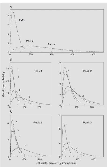
Apart from preferential interactions with negatively charged interfaces containing sulfatides and gangliosides, MBP causes lat-eral condensation (23) and affects differen-tially the thermotropic behavior of single glycosphingolipids (24). In ternary systems of MBP in bilayers of defined composition constituted by dipalmitoyl phosphatidylcho-line (dpPC) and different glycosphingolipids, the protein causes phase separation and can induce membrane-membrane interactions and recombination depending on the oligo-saccharide chain of the glycosphingolipid (4). The protein affects the thermotropic be-havior. Figure 1A shows that in mixtures of dpPC with the neutral galactosylceramide the protein causes an asymmetric distortion of the phase transition; this indicates that MBP preferably partitions into the liquid-crystalline lipid phase in which the high temperature asymmetry can be resolved into a broad low-cooperativity peak. Statistical thermodynamic calculations (26) of the clus-ter size distribution functions reveal that the most probable size (number of thermody-namically correlated molecules undergoing the transition at T1/2) of the high temperature
clusters induced by MBP is below 40 and is
Figure 1. Myelin basic protein induces phase separation in mixtures of dipalmitoyl phospha-tidylcholine (dpPC) and glycosphingolipids. Excess heat capacity-temperature scans (0.5ºC/ min) of mixtures of: Column A, galactosylceramide-dpPC (1:3) with a proportion of myelin basic protein (MBP) of 0 (a), 0.6 (b), 2.3 (c), and 4.3 (d) mol%; Column B, mixtures of sulfatide-dpPC (1:3) with a proportion of MBP of 0 (a), 0.3 (b), 1.1 (c), and 4.4 (d) mol%; Column C, mixtures of ganglioside GM1-dpPC (1:5) with a proportion of MBP of 0 (a), 0.1 (b), 1.2 (c), and 4.5 (d) mol%. Coexisting phase transitions are identified by numbers on the corresponding peaks.
narrowly distributed. On the other hand, the cluster size and distribution of the segre-gated domains undergoing the phase transi-tion at the T1/2 corresponding to the
protein-free mixture remain practically unchanged (Figure 2A).
Pre-vious publications have reported the complete temperature-composition phase diagrams for binary mixtures of several glycosphingolipids
with dpPC. These studies revealed that for protein-free mixtures containing sulfatide and gangliosides the phase diagrams were quite broad and, depending on composition, phase coexistence of phospholipid domains exclud-ing glycosphexclud-ingolipids into enriched clusters was present (18). This can be seen in Figure 1Ba and 1Ca where two overlapping calori-metric transitions are found in the protein-free mixtures. The presence of MBP in increasing amounts facilitates formation of high-temper-ature segregated glycosphingolipid-enriched domains (Figure 1B, peak 2, Figure 1C, peak 3). The statistical thermodynamic analysis (Fig-ure 1B,C) reveals that the most probable clus-ter size of the high-temperature transition com-ponent (glycosphingolipid-enriched domains) becomes larger and more broadly distributed with increasing proportions of MBP (Figure 2B, peak 2, Figure 2C, peak 3; see also Figure 1B,C). On the other hand, the most probable clusters of the segregated low-temperature component whose transition temperature mostly remains unaltered in the presence of MBP become very small and more narrowly distributed (mixtures with sulfatide; Figure 2B, peak 1) while the opposite occurs in mix-tures with GM1 (Figure 2C, peak 2). This points to the existence of a long-range struc-tural influence and intercommunication among the coexisting phase domains on the thermo-dynamic level whereby the transition features of one type of segregated cluster become in-fluenced by the presence of another type.
Lipid-protein surface reorganization induced by the transcription factors c-Fos and c-Jun
Two of the most studied transcription factors are the immediate early gene protein products c-Fos and c-Jun. Their amounts in the cell are very rapidly and transiently in-creased in response to stimuli, culminating with the activation of an array of target genes involved in normal cellular processes such as growth, differentiation and
eration (27). Imbalance of their highly regu-lated expression and or activity leads to on-cogenic or apoptotic processes (28,29). The transcriptional activity of c-Fos and c-Jun is conditioned to their entering the nucleus as a heterodimeric complex, the activator-pro-tein 1 (AP-1), whose formation depends on the relative amounts of both proteins, their presence in a same cellular compartment, and their post-translational state (30,31). These in turn depend on the cell type and its microenvironment. Phosphorylation, among the modifications that modulate AP-1 for-mation (32), may be seen as a mechanism involving competition between the associa-tion of its components with membranes and their dimerization. c-Fos association with membranes was first suggested by its re-cently reported capacity to specifically regu-late phospholipid metabolism (33). This is one of the normal c-Fos functions, occurring when the protein is expressed very rapidly after a cell or tissue receives stimuli of vari-ous kinds and associates with the endoplas-mic reticulum (34). The other normal, more studied, function of the protein is to tran-scriptionally regulate target genes of AP-1 at longer times (usually about one hour) after the stimulus, when located in the nucleus.
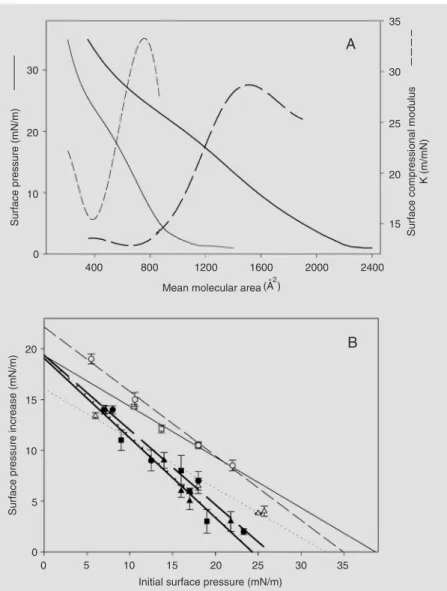
A few but important lines of evidence have opened new insights into the molecular function of some gene-regulatory proteins; among the most unexpected was the impres-sive membrane activity of some of them, like the DnaA protein (35). Within this con-text, the finding that c-Fos is strongly amphitropic and that it interacts differen-tially with phospholipids (Ref. 36 and Fig-ure 3) was a first step in the way to under-standing the nature of its association with membranes. This was followed by demon-stration of its capacity to finely modulate phospholipase activity against organized biointerfaces (37). On the other hand, c-Jun, the partner of c-Fos in the AP-1 complex, is also amphitropic but its interaction with phos-pholipids is not specific (Ref. 38 and Figure
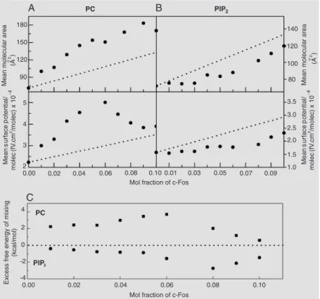
3). A further step in the characterization of the c-Fos association with membranes indi-cated more clearly the differential quality of the protein/lipid interaction: interestingly, c-Fos elicits opposite lipid packing and elec-trostatic effects in dilauroyl phosphatidyl-choline (dlPC) and phosphatidylinositol di-phosphate (PIP2), inducing expansion and
hyperpolarization of the former and conden-sation and depolarization of the latter
ure 4). Moreover, its effects on the organiza-tion of dlPC explain its capacity to modulate phospholipase A2 (PLA2) and PLC activity
on this phospholipid (37,39). The contrast-ing effects of the intermolecular interactions of c-Fos with dlPC and PIP2 are supported
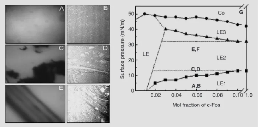
by the thermodynamic drive, as assessed by the excess free energy of mixing, regulating the composition-dependent specific lipid-protein interactions (Figure 4C). The two-dimensional phase diagram for the mixture of c-Fos with PIP2 also shows regions of
protein-induced surface immiscibility (40). This is illustrated by epifluorescence and Brewster angle microscopy images of the mixtures (Figure 5).
The phenomena underlying the interaction of c-Fos and c-Jun with phospholipids may
eventually affect their dimerization tendency, which leads to nuclear translocations and fur-ther transcriptional regulation. With respect to AP-1 heterodimers, their operational forma-tion and involvement in cellular responses have been ascertained (28). However, although association of c-Fos and c-Jun has been de-scribed (27-30), we have not found publica-tions describing the actual isolation of this complex as a stable chemical entity and we were not successful in achieving its formation in solution under a variety of conditions (38). On the other hand, we and others (27,38) obtained direct evidence indicating that inter-face interactions are essential to drive AP-1 formation. In this regard, not only both pro-teins can interact with each other at the inter-face, as they do separately with phospholipids, but their equimolecular complex is also strongly stabilized by the favorable thermody-namics of their association (Ref. 38 and Figure 6). The interplay and balance between the two sets of interaction forces are likely to deter-mine the fate and possible reversible location of these transcription factors in subcellular compartments, thus accomplishing the fine regulation of their function in the cell.
Bi-directional information transduction between surface domain structuring and membrane biocatalysis
A “dynamic” type of protein-induced surface structuring is mediated by the action of phospholipases (10,41). All phosphohy-drolytic enzymes, usually a kind of amphi-tropic proteins producing lipid second mes-sengers, are currently thought to be involved in some sort of membrane signal transduc-tion process (42,43). With respect to sphin-golipid signaling, ceramide is a pivotal com-pound that links the metabolism of phospho, sphingo-, and glycosphingolipids, all of which are important biomodulators that con-trol membrane topology and phospholipase activity. Besides the concept that ceramide
is an important second messenger derived from the sphingomyelin cycle (43), there are direct metabolic, structural and functional consequences of the sphingomyelinase-driven conversion of sphingomyelin to cera-mide in biomembranes (43,44).
Studies on the molecular and structural codes that underlay the enzyme specificities and the kinetics of the complex surface reac-tions are beginning to redefine the concept of membrane signaling by including pro-cesses taking place over length and time domains spanning several orders of magni-tude (21). This involves bi-directional trans-duction through a “bottom-up” flow of in-formation from the molecular level to the mesoscopic level, thus redefining local in-teractions on a wider range topological scale; conversely, the membrane topology and de-rived tensions at that level generate a “top-down” transduction to the local properties of the individual membrane molecules with a regulatory power on their function (10,45).
It is well documented that the activity and kinetics of lipases and phospholipases (mostly those of the A2 and C types) are
correlated with the existence of lateral mem-brane defects and structural domain micro-heterogeneity (41,46,47), coexistence of bi-layer and non-bibi-layer phases (48,49) and
Figure 5. Surface pressure- and composition-induced phase transitions by c-Fos of mixed films with phosphatidylinositol biphosphate (PIP2). The domain segregation is shown in the left panels and the respective phase diagrams in the right panels. Epifluorescence (panels A, C and E) and Brewster angle microscopy (BAM, panels B, D, and F). Microscopy images of a film with a 0.06-mol fraction of c-Fos at 3 (A,B), 15 (C,D) and 27 (E,F) mN/m. The vertical side of the BAM images corresponds to 4.8 mm. The magnification of the epifluo-rescence images is ten times that of the BAM images. Two-dimensional partial phase diagram of mixtures of PIP2 (G) with different proportions of c-Fos showing surface pressure-dependent presence of different liquid expanded (LE, LEn with N = 1-3) phases; Co represents the collapsed phase. The circles correspond to the collapse point, and the squares and triangles to the first and second molecular reorganization of c-Fos, respective-ly. AB, CD, and EF indicate the region of the phase diagram illustrated by the correspond-ing surface patterns on the left panels.
Figure 6. Interactions between c-Fos and c-Jun. A, Mean molecular area (open circles, left scale) and dipole potential density (filled squares, right scale) as a function of composition for Jun-Fos mixtures; the straight line corresponds to ideally mixed films. B, Excess free energy of mixing as a function of film composition.
45,49). A common structural element in all of these factors is the existence of lateral and/or transverse packing defects and inter-face tensions introduced by changes of lipid composition, anisotropic interactions, phase coexistence and connectivity of the domain lattice (10,21,41,45,49). Within this con-text, critical amounts of non-substrate lipids or proteins that may induce or disrupt specif-ic surface super-structuring (10,45,47) over ranges that exceed the local intermolecular interactions become important membrane-regulating factors, and some of the topologi-cal variations may be induced by the phos-phohydrolytic enzymes themselves.
The formation by a membrane-associated enzyme of a lipid product with markedly dif-ferent surface properties and interactions with the parent substrate generates a major thermo-dynamic problem in a local micro-region (7,10, 21). This is because of the emergence of local tensions due to variations of the relative com-position in lipid species having different mis-cibility properties. These tensions can only be resolved through long-range variations of the membrane topography involving compo-sitional and/or phase domain segregation, su-per-lattice structuring and curvature alterations (3,4). For sphingomyelinase, we could dem-onstrate that the enzyme not only actively modifies the surface topography but also that the dynamic features and shape variations of the latter finely regulate the time-course and extent of the enzymatic reaction (10,45).
Sphingomyelinase preferably degrades sphingomyelin in the liquid-expanded state, both in monolayers and bilayer vesicles (21, 49). The steady-state reaction is preceded by a pre-catalytic latency period that involves enzyme adsorption and a rate-limiting inter-face activation step (21). We were able to show the real-time dynamic sculpturing of the surface topography by sphingomyeli-nase through the organized formation, mor-phological evolution, and super-struc-turing of ceramide-enriched domains under conditions of controlled and known
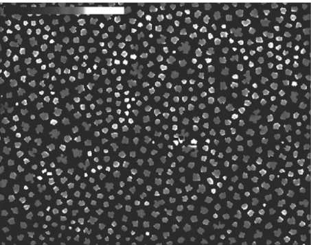
inter-molecular organization of the substrate (45). A succession of discrete shape transitions and lateral distribution of domains at de-fined times during the reaction underlay the topography generated by the activity of the enzyme, that is not found in enzyme-free mixtures of sphingomyelin and ceramide of the same composition (10,45). Advanced image processing routines in combination with time-resolved epifluorescence micros-copy (45) on Langmuir monolayers revealed: i) spontaneous nucleation and circular growth of ceramide-enriched domains after injec-tion of sphingomyelinase into the subphase of the sphingomyelin monolayer, ii) domain-intrinsic discrete shape transitions from cir-cular to periodically undulating shapes fol-lowed by a second transition towards in-creasingly branched morphologies, iii) lat-eral superstructure organization into pre-dominantly hexagonal domain lattices, iv) formation of super-superstructures by the hexagonal lattices, and v) rotationally and laterally coupled domain movement prior to domain border contact (Figure 7).
hex-agonal lattices and super-structuring over the long range (Figure 8). Lateral enzyme-specif-ic out-of-equilibrium organization of lipid do-mains represents a new level of signal trans-duction (45) from local (nm) to long-range (µm) scales, which controls and regulates in-formation exchange among various hierarchi-cal levels of the membrane function. This includes the variations in composition due to insertion of regulatory lipids and proteins that inherently modify the structure (4,21,39). This can be correlated with alterations of the mem-brane surface in terms of domain segregation and topological restructuring (10,41,45). In this manner, local and/or supramolecular in-formation generated by one phosphohydro-lytic pathway may be transduced to a non-biochemical regulation of other enzymatic pathways that do not even share common substrates or products. This was specifically shown for the cross-talk between the PLA2
-and sphingomyelinase-driven reactions where-by they can become mutually amplified or
dampened by fluctuations of the lateral sur-face pressure controlling the lateral lipid pack-ing and dipolar organization (21). Recently, it was shown that the lateral packing and in-plane elasticity of the lipid-protein interface can also regulate the activity of the GPI-an-chored protein alkaline phosphatase and modu-late information exchange between the inter-face and the aqueous environment (50).
Surface topography of whole myelin monolayers and influence of
Folch-Lees proteolipid protein on domain structuring and dynamics
In contrast to the commonly used binary or ternary lipid and lipid-protein model mem-brane systems, multicomponent systems such as natural membranes can establish a wide diversity of interactions among their lipid and protein components giving rise to an increasing number of ways to reduce local tensions derived from molecular
tries. The resultant loss of cooperativity, as the system becomes more complex, induces an apparent gradual (diffuse) response to changes in temperature, pressure and com-positional changes (4,25,51). Nevertheless, although some responses become buffered in multicomponent systems, marked rear-rangements transducing local stress over wider range scales occur under the effects of some perturbing factors that introduce large tensions in membranes. Examples of the latter are HII phase-forming molecules (19-22), cholesterol (52,53), and proteins (54). Cholesterol is known to induce, above a certain threshold, the coexistence of immis-cible liquid phases (52). This effect has been recognized as the basis for the surface het-erogeneity of monolayers prepared from some natural lipid membranes (55), and is postulated as an important factor in the ori-gin of the generically called “raft domains” (17). Membrane proteins embedded in mem-branes can behave as strong perturbing agents acting at different levels of organization.
They not only affect the immediate lipid environment but also influence the long-range thermotropic behavior of lipids ac-cording to their tendency to undergo aggre-gation or partitioning into particular lipid phases. Furthermore, peripheral and integral proteins have been shown to induce phase domain segregation in model lipid mem-branes consisting of few components (4,8, 9,24). Solvent-solubilized whole myelin membranes can form monomolecular layers at the air-water interface (23,56). We first reported (57) that the surface topography of a compositionally complex lipid-protein natural membrane, under precisely controlled conditions of intermolecular organization, is characterized by the coexistence of at least two liquid phases at low- and high-lateral surface pressures with a transition from round-border domains to fractal domains occurring during compression (Figure 9a-d). Prior to our studies, the presence of liq-uid-liquid surface immiscibility was de-scribed in complex monolayers containing cholesterol prepared with lipids from red blood cell membranes (55). In agreement with our observations on the complex natu-ral myelin interface (57), fluid-phase coex-istence was subsequently found in bilayer vesicles made from natural kidney brush border membranes (58) and was also re-cently reported in bilayer vesicles prepared with a natural mixture of lipids and pulmo-nary surfactant protein (59). In whole my-elin monolayers we identified the contribu-tion of some protein and lipid components to the topographic organization. A segregation of the protein components excluded from domains containing condensed lipids such as cholesterol and galactosylceramide at all surface pressures pointed to compositional immiscibility as an important factor for the domain phase coexistence (25).
The protein-depleted mixture of myelin lipids at low-surface pressures shows coex-istence of cholesterol-enriched and choles-terol-depleted liquid phases organized as
rounded domains of homogenous size dis-tributed in rather regular lattices (Figure 9i,j) but fails to undergo the topographic changes involving the formation of fractal domains under increasing compression that is charac-teristic of whole myelin monolayers (com-pare Figure 9k,l with Figure 9c,d). On the other hand, one of the major protein compo-nents of myelin membranes, the Folch-Lees PLP (about 50% by weight of the protein fraction), when mixed with myelin lipids in the absence of all the other proteins (51) is capable of reproducing the topographic or-ganization of the whole myelin monolayer in a concentration-dependent manner (Fig-ure 9e-g).
The addition of PLP to myelin lipids preserves the liquid character of the coexist-ing phases at low-surface pressures but modi-fies the size and shape distribution of do-mains (compare Figure 9i,j with Figure 9e,f). At high-surface pressures the PLP overrides the tendency of the lipids to merge, and the surface aggregation of PLP-enriched fractal domains provides a topographic explanation for the surface heterogeneity of the mono-layer. The fractal nature of the structure
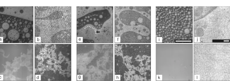
Figure 9. Surface topography of whole myelin and myelin lipid monolayers. Representative images of whole myelin extract monolayers (a, b, c, and d), lower phase extract monolayers (e, f, g, and h) and myelin lipid monolayers, except gangliosides (i, j, k, and l) containing 0.5 mol% of egg-Rho-PE as seen by epifluorescence microscopy (a, c, e, g, i, and k) and Brewster angle microscopy (b, d, f, h, j, and l). The lower phase extract contains only proteolipid protein as the protein component. The upper row of images corresponds to surface pressures between 1 and 1.5 mN/m during the second compression. The arrow in image “e” is pointing to the border of the gray phase (outlined with a dotted line) that is surrounded by a rim of bright phase. The images in the bottom row were taken between 35 and 37 mN/m. The reference bars in epifluorescence and Brewster angle microscopy images represent 150 µm.
microheterogeneity in the pattern and dy-namics of the surface topography, thus con-veying molecular to supramolecular differ-ential responses over to variations of the molecular packing (and surface pressure). Simultaneously with the directionality of out-of-equilibrium processes, the protein-induced effects described indicate the ca-pacity of membrane-associated proteins to store and vectorially convey information with respect to membrane perturbations and dis-sipation of surface tensions. This cannot be overlooked within the context of the extraor-dinary capability of membranes to act as sensing, transducing, and modulatory mani-fold elements for cell function as bio-electro-mechano-chemical devices.
Figure 10. Dynamic structuring of the fractal pattern in mixtures with proteolipid protein (PLP). Representative Brewster angle microscopy images of a film of myelin lipids (except gangliosides) containing 5.3 x 10⁻⁴ mol fraction of PLP. The monolayer was compressed up to 37 mN/m and was left at the same surface pressure for 100 min. Images 1a, 1b and 1c are representative of the topography of the films at 2, 50, and 100 min, respectively, after the target surface pressure was reached during the first compression. Images 2a and 2b show the relaxation pattern after 10 and 70 min elapsed from the time when the film was brought to 37 mN/m during the second compression. The scale bar represents 300 µm.
one of the fundamental structuring factors capable of determining the pattern and dy-namics of the surface topography in a man-ner indistinguishable from that observed in the whole myelin membrane monolayers that contain all the protein and lipid components (57).
Differing from the view of proteins as passively partitioning according to the fea-tures of pre-assembled lipid domains in cell membranes (8), our results directly demon-strate that some proteins such as PLP can have an active role of paramount importance and should be considered as key sculpturing factors determining membrane structuring over the long range. In addition, the pres-ence of the protein brings about features of
References
1. Edidin M (2003). Lipids on the frontier: a century of cell-membrane bilayers. Nature Reviews. Molecular Cell Biology, 4: 414-418. 2. Cevc G & Marsh D (1987). Phospholipid Bilayers. Wiley-Interscience
Publication, New York.
3. Vereb G, Szollosi J, Matko J et al. (2003). Dynamic, yet structured: The cell membrane three decades after the Singer-Nicolson model. Proceedings of the National Academy of Sciences, USA, 100: 8053-8058.
4. Maggio B (1994). The surface behavior of glycosphingolipids in
biomembranes: a new frontier of molecular ecology. Progress in Biophysics and Molecular Biology, 62: 55-117.
5. Siegel DP (1999). The modified stalk mechanism of lamellar/in-verted phase transitions and its implications for membrane fusion. Biophysical Journal, 76: 291-313.
6. Seddon JM (1990). Structure of the inverted hexagonal (HII) phase, and non-lamellar phase transitions of lipids. Biochimica et Biophy-sica Acta, 1031: 1-69.
ceramide, neutral glycosphingolipids and gangliosides in mixed monolayers. Chemistry and Physics of Lipids, 132: 209-224. 8. Gil T, Ipsen JH, Mouritsen OG et al. (1998). Theoretical analysis of
protein organization in lipid membranes. Biochimica et Biophysica Acta, 1376: 245-266.
9. Epand RM (2004). Do proteins facilitate the formation of cholesterol-rich domains? Biochimica et Biophysica Acta, 1666: 227-238. 10. Fanani ML, Hartel S, Oliveira RG et al. (2002). Bidirectional control
of sphingomyelinase activity and surface topography in lipid mono-layers. Biophysical Journal, 83: 3416-3424.
11. Fanani ML, Topham MK, Walsh JP et al. (2004). Lipid modulation of the activity of diacylglycerol kinase alpha- and zeta-isoforms: activa-tion by phosphatidylethanolamine and cholesterol. Biochemistry, 43: 14767-14777.
12. Malinina L, Malakhova ML, Teplov A et al. (2004). Structural basis for glycosphingolipid transfer specificity. Nature, 430: 1048-1053. 13. Edidin M (1997). Lipid microdomains in cell surface membranes.
Current Opinion in Structural Biology, 7: 528-532.
14. Phillips MC, Ladbrooke BD & Chapman D (1970). Molecular interac-tions in mixed lecithin systems. Biochimica et Biophysica Acta, 196: 35-44.
15. Jost PC & Griffith OH (1980). The lipid-protein interface in biological membranes. Annals of the New York Academy of Sciences, 348: 391-407.
16. Munro S (2003). Lipid rafts: elusive or illusive? Cell, 115: 377-388. 17. Simons K & Ikonen E (1997). Functional rafts in cell membranes.
Nature, 387: 569-572.
18. Maggio B, Ariga T, Sturtevant JM et al. (1985). Thermotropic behav-ior of binary mixtures of dipalmitoylphosphatidylcholine and glyco-sphingolipids in aqueous dispersions. Biochimica et Biophysica Acta, 818: 1-12.
19. Perillo MA, Scarsdale NJ, Yu RK et al. (1994). Modulation by gan-gliosides of the lamellar-inverted micelle (hexagonal II) phase tran-sition in mixtures containing phosphatidylethanolamine and dio-leoylglycerol. Proceedings of the National Academy of Sciences, USA, 91: 10019-10023.
20. Carrer DC & Maggio B (2001). Transduction to self-assembly of molecular geometry and local interactions in mixtures of ceramides and ganglioside GM1. Biochimica et Biophysica Acta, 1514: 87-99. 21. Maggio B, Carrer DC, Fanani ML et al. (2004). Interfacial behavior of glycosphingolipids and chemically related sphingolipids. Current Opinion in Colloid and Interface Science, 8: 448-458.
22. Basanez G, Fidelio GD, Goni FM et al. (1996). Dual inhibitory effect of gangliosides on phospholipase C-promoted fusion of lipidic vesicles. Biochemistry, 35: 7506-7513.
23. Fidelio GD, Maggio B & Cumar FA (1984). Interaction of myelin basic protein, melittin and bovine serum albumin with gangliosides, sulphatide and neutral glycosphingolipids in mixed monolayers. Chemistry and Physics of Lipids, 35: 231-245.
24. Maggio B, Sturtevant JM & Yu RK (1987). Effect of myelin basic protein on the thermotropic behavior of aqueous dispersions of neutral and anionic glycosphingolipids and their mixtures with dipal-mitoylphosphatidylcholine. Journal of Biological Chemistry, 262: 2652-2659.
25. Oliveira RG & Maggio B (2002). Compositional domain immiscibility in whole myelin monolayers at the air-water interface and Langmuir-Blodgett films. Biochimica et Biophysica Acta, 1561: 238-250. 26. Freire E & Biltonen R (1978). Estimation of molecular averages and
equilibrium fluctuations in lipid bilayer systems from the excess heat capacity function. Biochimica et Biophysica Acta, 514: 54-68. 27. Angel P & Karin M (1991). The role of Jun, Fos and the AP-1
complex in cell-proliferation and transformation. Biochimica et Bio-physica Acta, 1072: 129-157.
28. Shaulian E & Karin M (2002). AP-1 as a regulator of cell life and death. Nature Cell Biology, 4: 131-136.
29. Eferl R & Wagner EF (2003). AP-1: a double-edged sword in tumori-genesis. Nature Reviews. Cancer, 3: 859-868.
30. Hess J, Angel P & Schorpp-Kistner M (2004). AP-1 subunits: quarrel and harmony among siblings. Journal of Cell Science, 117: 5965-5973.
31. Horisawa K, Tateyama S, Ishizaka M et al. (2004). In vitro selection of Jun-associated proteins using mRNA display. Nucleic Acids Re-search, 32: e169.
32. Karin M & Hunter T (1995). Transcriptional control by protein phos-phorylation: signal transmission from the cell surface to the nucleus. Current Biology, 5: 747-757.
33. Bussolino DF, Guido ME, Gil GA et al. (2001). c-Fos associates with the endoplasmic reticulum and activates phospholipid metabolism. FASEB Journal, 15: 556-558.
34. Gil GA, Bussolino DF, Portal MM et al. (2004). c-Fos activated phospholipid synthesis is required for neurite elongation in differen-tiating PC12 cells. Molecular Biology of the Cell, 15: 1881-1894. 35. Castuma CE, Crooke E & Kornberg A (1993). Fluid membranes with
acidic domains activate DnaA, the initiator protein of replication in Escherichia coli. Journal of Biological Chemistry, 268: 24665-24668. 36. Borioli GA, Caputto BL & Maggio B (2001). c-Fos is surface active and interacts differentially with phospholipid monolayers. Biochemi-cal and BiophysiBiochemi-cal Research Communications, 280: 9-13. 37. Borioli GA, Fanani ML, Caputto BL et al. (2002). c-Fos is a surface
pressure-dependent diverter of phospholipase activity. Biochemical and Biophysical Research Communications, 295: 964-969. 38. Del Boca M, Caputto BL, Maggio B et al. (2005). c-Jun interacts with
phospholipids and c-Fos at the interface. Journal of Colloid and Interface Science, 287: 80-84.
39. Borioli GA, Caputto BL & Maggio B (2004). Phospholipase activity is modulated by c-Fos through substrate expansion and hyperpolar-ization. Federation of European Biochemical Societies Letters, 570: 82-86.
40. Borioli GA, Caputto BL & Maggio B (2005). c-Fos and phosphatidyl-inositol-4,5-bisphosphate reciprocally reorganize in mixed monolay-ers. Biochimica et Biophysica Acta, 1668: 41-52.
41. Grainger DW, Reichert A, Ringsdorf H et al. (1990). Hydrolytic action of phospholipase A2 in monolayers in the phase transition region: direct observation of enzyme domain formation using fluo-rescence microscopy. Biochimica et Biophysica Acta, 1023: 365-379.
42. Exton JH (1994). Phosphatidylcholine breakdown and signal trans-duction. Biochimica et Biophysica Acta, 1212: 26-42.
43. Kolesnick RN, Goni FM & Alonso A (2000). Compartmentalization of ceramide signaling: physical foundations and biological effects. Jour-nal of Cell Physiology, 184: 285-300.
44. Holopainen JM, Angelova MI & Kinnunen PK (2000). Vectorial bud-ding of vesicles by asymmetrical enzymatic formation of ceramide in giant liposomes. Biophysical Journal, 78: 830-838.
45. Hartel S, Fanani ML & Maggio B (2005). Shape transitions and lattice structuring of ceramide-enriched domains generated by sphin-gomyelinase in lipid monolayers. Biophysical Journal, 88: 287-304. 46. Honger T, Jorgensen K, Biltonen RL et al. (1996). Systematic rela-tionship between phospholipase A2 activity and dynamic lipid bi-layer microheterogeneity. Biochemistry, 35: 9003-9006.
Biochemistry, 43: 2159-2166.
48. Maggio B (1996). Control by ganglioside GD1a of phospholipase A2 activity through modulation of the lamellar-hexagonal (HII) phase transition. Molecular Membrane Biology, 13: 109-112.
49. Ruiz-Arguello MB, Veiga MP, Arrondo JL et al. (2002). Sphingomy-elinase cleavage of sphingomyelin in pure and mixed lipid mem-branes. Influence of the physical state of the sphingolipid. Chemistry and Physics of Lipids, 114: 11-20.
50. Caseli L, Oliveira RG, Masui DC et al. (2005). Effect of molecular surface packing on the enzymatic activity modulation of an an-chored protein on phospholipid Langmuir monolayers. Langmuir, 21: 4090-4095.
51. Rosetti CM, Oliveira RG & Maggio B (2005). The Folch-Lees proteo-lipid induces phase coexistence and transverse reorganization of lateral domains in myelin monolayers. Biochimica et Biophysica Acta, 1668: 75-86.
52. Ipsen JH, Karlstrom G, Mouritsen OG et al. (1987). Phase equilibria in the phosphatidylcholine-cholesterol system. Biochimica et Bio-physica Acta, 905: 162-172.
53. McConnell HM (1991). Structures and transitions in lipid
monolay-ers at the air-water interface. Annual Review of Physical Chemistry, 42: 171-195.
54. Smith R & Cornell BA (1985). Myelin basic protein induces hexago-nal phase formation in dispersions of diacylphosphatidic acid. Bio-chimica et Biophysica Acta, 818: 275-279.
55. Keller SL, Pitcher III WH, Huestis WH et al. (1998). Red blood cell lipids form immiscible liquids. Physical Review Letters, 81: 5019-5022.
56. Oliveira RG, Calderon RO & Maggio B (1998). Surface behavior of myelin monolayers. Biochimica et Biophysica Acta, 1370: 127-137. 57. Oliveira RG & Maggio B (2000). Epifluorescence microscopy of surface domain microheterogeneity in myelin monolayers at the air-water interface. Neurochemical Research, 25: 77-86.
58. Dietrich C, Bagatolli LA, Volovyk ZN et al. (2001). Lipid rafts recon-stituted in model membranes. Biophysical Journal, 80: 1417-1428. 59. Bernardino de la Serna J, Perez-Gil J, Simonsen AC et al. (2004).