Introduction
In Brazil, the culture of melon presented great evolution
in the period from 1987 to 2006, with production of fruits
going from 37,980 to 500,021 tons. Melon is largely
cultivated and one of the most important products of the
Brazilian agribusiness that is conquering greater shares in
the national and international markets. The Northeast Region
is responsible for about 93% of the national melon fruits
production, with 465,623 tons; the States of Rio Grande do
Norte, Ceará, Bahia and Pernambuco are the main producers
and exporters (Araújo & Vilela 2003; IBGE 2006).
The melon (
Cucumis melo
L.) started to be cultivated
in the region of the submedium São Francisco in 1965, in
the city of Santa Maria da Boa Vista, Pernambuco State.
With the implement of several public projects for irrigation,
this agriculture cluster became one of the main zones of
production and exportation of fruits in the country. The
culture of melon has been intensifi ed in the region, mainly
in the cities of Petrolina and Juazeiro, which have better
infrastructure for commercialization (Oliveira 1991).
The melon culture in the Vale do São Francisco can be
carried out throughout the year, due to the favorable soil
and climate conditions, with increased solar radiation, high
temperatures and low relative humidity. These factors favor
the concentration of total soluble solids in the fruits and
diminish the incidence of diseases, increasing the quality
of the fruits (Dias
et al.
1998).
With the advance of organic agriculture for fruit
production in the Submedium São Francisco, many types
of residues have been applied; however little is known
about the effect of these residues in the soil mycobiota. The
type of soil amendment can have a signifi cant effect on
the soil mycobiota mainly to favor the saprophytic activity
in detriment of potential plant pathogens. Thus, organic
residues that favor the increase of natural fungal population
are important for improving the nutritional condition of
1 Universidade Federal de Pernambuco, Centro de Ciências Biológicas, Departamento de Micologia, Recife, PE, Brazil 2 Universidade Federal do Vale do São Francisco, Colegiado de Zootecnia, Petrolina, PE, Brazil
3 Corresponding Author: Flavia Paiva Coutinho, fl aviapaco@hotmail.com
Filamentous fungi isolated from the rhizosphere of melon
plants (
Cucumis melo
L. cv. Gold Mine) cultivated in
soil with organic amendments
Flavia Paiva Coutinho
1,3, Maria Auxiliadora de Queiroz Cavalcanti
1and Adriana Mayumi Yano-Melo
2Nota Científi ca / Scientifi c Note
Recebido em 21/10/2008. Aceito em 4/12/2009
RESUMO - (Fungos fi lamentosos isolados da rizosfera de meloeiros (Cucumis melo L. cv. Gold Mine) cultivados em solo com compostos orgânicos). Foram coletadas amostras de solo rizosférico em uma área semiárida, na região do Vale do São Francisco, Petrolina, Pernambuco, Brasil, com o objetivo de conhecer a diversidade dos fungos fi lamentosos presentes em solo cultivado com melão (Cucumis melo cv. Gold Mine) e adubado com diferentes compostos
orgânicos: Tratamento 1 (controle, sem adição de compostos orgânicos); T2 (77% de bagaço de côco, 20% de esterco de caprino e 3% de K2SO4); T3 (10% de torta de mamona, 50% de capim elefante e 40% de esterco de caprino); T4 (77% de bagaço de côco, 20% de esterco de caprino e 3% de termofosfato); T5 (47% de capim elefante, 50% de esterco de caprino e 3% K2SO4); e T6 (57% de capim elefante, 40% de esterco de caprino e 3% de termofosfato). O isolamento dos fungos foi realizado por meio da técnica de diluição em série até 1:1000. Foi aplicado o índice de similaridade de Sorensen, e avaliadas a freqüência e a distribuição dos fungos no solo.Setenta e oito espécies foram isoladas e identifi cadas, além de representantes de Basidiomycota (04) e Mycelia sterilia (02). Os gêneros predominantes foram Aspergillus e Penicillium, com 15 e 13 espécies, respectivamente. Maior número de espécies foi constatado no período de fundação (49), e em relação à adubação orgânica, o tratamento 6 apresentou a maior diversidade (43 espécies). A maioria das espécies encontradas é sapróbia e somente algumas são consideradas potenciais patógenos à cultura do meloeiro, como Fusarium oxysporum, F. solani e Myrothecium roridum. Palavras-chave: microorganismos, semiárido, Vale do São Francisco, adubação orgânica
ABSTRACT - (Filamentous fungi isolated from the rhizosphere of melon plants (Cucumis melo L. cv. Gold Mine) cultivated in soil with organic amend-ments). Rhizosphere soil samples were collected in a semiarid area, in the region of the São Francisco River valley, Petrolina, Pernambuco state, Brazil, to study the diversity of fi lamentous fungi in a soil cultivated with melon (Cucumis melo L. cv. Gold Mine) and receiving different organic amendments: Treatment 1 (control, without organic compost); T2 (77% coconut fi ber, 20% goat manure and 3% K2SO4); T3 (10% Ricinus communis leaves and stems, 50% Pennisetum purpureum leaves and 40% goat manure); T4 (77% coconut fi ber, 20% goat manure and 3% termophosphate); T5 (47% Pennisetum purpureum leaves, 50% goat manure and 3% K2SO4); andT6 (57% Pennisetum purpureum leaves, 40% goat manure and 3% termophosphate). Fungal isolation was carried out by the serial dilution technique to 1:1000. The Sorensen index of similarity, frequency and distribution of the fungi were evalu-ated. Seventy-eight species of fi lamentous fungi were isolated and identifi ed, plus several Basidiomycota (04) and Mycelia sterilia (02). The predominant genera were Aspergillus and Penicillium, with 15 and 13 species, respectively. A greater number of species was found in the sowing period (49), and in relation to the organic fertilization, treatment 6 provided the greatest species diversity (43 species). Most of the species are saprobes and only a few are considered to be potential pathogens on melon plants, such as Fusarium oxysporum, F. solani and Myrothecium roridum.
the cultures of interest in the region and to ensure the
sustainability of the organic production.
This work aimed to isolate and identify fi lamentous
fungi from the rhizosphere of melon plants fertilized with
distinct organic composts, and to evaluate the infl uence
of these composts in three periods (sowing, fl owering and
post-harvest) of plant development, relating them to the
presence of fungi.
Materials and methods
Soil sampling – Rhizosphere soil samples were collected using an auger, to a depth of 20 cm, in a semiarid area cultivated with melon (C. melo cv. Gold
Mine), in the Vale do São Francisco, Petrolina, Pernambuco State, Brazil (09º32´09´´S, 40º55´28´´W), during the periods of sowing, fl owering (37 days after sowing) and post-harvest (31 days after fl owering), in the months of October, November and December/2005, respectively.
The area was fertilized with the following organic composts (O.C.): Treatment 1 (control, without organic composts); T2 19,7 m3 ha-1 O.C. (77% coconut fi ber, 20% goat manure and 3% K2SO4); T3 13,75 m3 ha-1 O.C. (10% Ricinus communis leaves and stems, 50% Pennisetum purpureum
leaves and 40% goat manure); T4 13,75 m3 ha-1 O.C. (77% coconut fi ber, 20% goat manure and 3% termophosphate); T5 9,69 m3 ha-1 O.C. (47%
Pennisetum purpureum leaves, 50% goat manure and 3% K2SO4); andT6 6,875 m3 ha-1 O.C. (57% Pennisetum purpureum leaves, 40% goat manure and 3% termophosphate). Termophosphate (80 kg ha-1 P
2O5) and potassium sulphate (60 kg ha-1 K
2O) were added to treatments 2 to 6. Organic composts and termophosphate were applied at planting with 50% potassium sulphate. Potassium sulphate remains were distributed manually with watering cans in 4 applications, at 6, 13, 21 and 27 days after transplanting.
For each treatment, three samples were taken, in a total of 18 samples at each sampling period (sowing, fl owering and post-harvest).
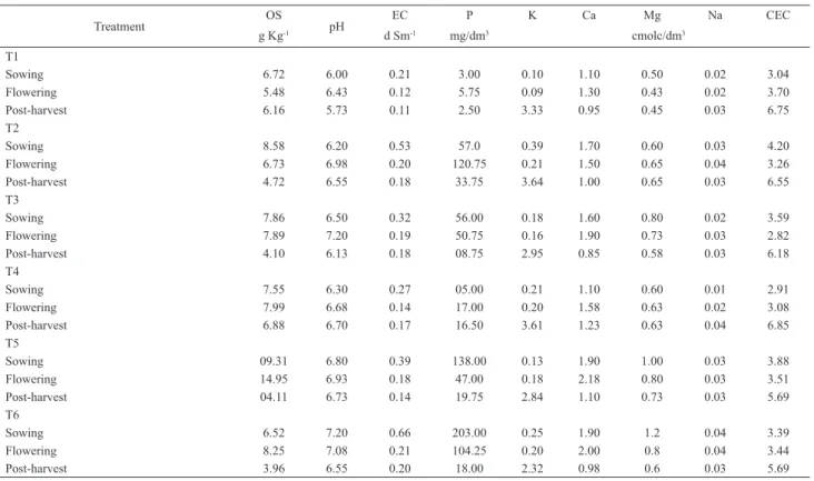
Consi-dering all treatments, a total of 54 soil samples were investigated for the presence of fungi. The soil (Gray Argisol) samples were chemically analyzed at the Soil Laboratory of the Embrapa Semi-Árido (Tab. 1) (Embrapa 1997).
Isolation and identifi cation of the fi lamentous fungi – The fungi were isolated using the serial dilution technique (Mehan et al. 1991): 25 g of each soil sample were suspended in 225 ml of sterilized distilled water (SDW) (1:10); 10 ml of this suspension were added to 990 ml of SDW (1:1000). Then, 1 ml of this suspension was cultured, in triplicate, in Petri dishes containing Sabouraud Agar added of chloranphenicol (500 mg l-1). The plates were kept at room temperature (28ºC) and the development of colonies was observed until 120 h, when the CFU were estimated. After the fi rst 72 h the colonies were transferred to Petri dishes with agar medium (potato dextrose agar, czapeck agar and/or malt extract agar) and kept for 20 days, allowing the formation of reproductive structures. The identifi ca-tion of the species was carried out through macroscopic and microscopic observation of the isolates, consulting the specialized literature (Raper et al.
1949; Ames 1961; Corlett 1966; Rifai 1969; Booth 1971; Ellis 1971; Nicoli & Russo 1974; Samson 1974; Arx 1975; Ellis 1976; Sigler & Carmichael 1976; Carmichael et al. 1980; Domsch et al. 1980; Sutton 1980; Schipper
1984; Arx et al. 1986; Pitt 1988; Udagawa et al. 1989; Klich & Pitt 1994; Hanlin & Menezes 1996).
Statistical analyses – The experimental design was of random blocks in a factorial arrangement 3x6, with three periods of evaluation (sowing, fl owe-ring and post-harvest) and six fertilization treatments (T1, T2, T3, T4, T5 and T6), with three replicates. The values of the CFU were square root (x + 1) transformed before the analysis of variance (ANOVA). The averages were compared by the LSD (least square deviation), 5% of probability, using the program Statistica 5.0 (Statsoft 1997).
The Sorensen index of similarity (Müller-Dombois & Ellemberg 1974) was applied to verify the similarity between the fungal populations isolated in the different sampling periods. Frequency and distribution of each fungal species were calculated for each sampling period according to Brower et. al (1990) and Schnitter & Stephenson (2000).
Treatment OS pH EC P K Ca Mg Na CEC
g Kg-1 d Sm-1 mg/dm3 cmolc/dm3
T1
Sowing 6.72 6.00 0.21 3.00 0.10 1.10 0.50 0.02 3.04 Flowering 5.48 6.43 0.12 5.75 0.09 1.30 0.43 0.02 3.70 Post-harvest 6.16 5.73 0.11 2.50 3.33 0.95 0.45 0.03 6.75 T2
Sowing 8.58 6.20 0.53 57.0 0.39 1.70 0.60 0.03 4.20 Flowering 6.73 6.98 0.20 120.75 0.21 1.50 0.65 0.04 3.26 Post-harvest 4.72 6.55 0.18 33.75 3.64 1.00 0.65 0.03 6.55 T3
Sowing 7.86 6.50 0.32 56.00 0.18 1.60 0.80 0.02 3.59 Flowering 7.89 7.20 0.19 50.75 0.16 1.90 0.73 0.03 2.82 Post-harvest 4.10 6.13 0.18 08.75 2.95 0.85 0.58 0.03 6.18 T4
Sowing 7.55 6.30 0.27 05.00 0.21 1.10 0.60 0.01 2.91 Flowering 7.99 6.68 0.14 17.00 0.20 1.58 0.63 0.02 3.08 Post-harvest 6.88 6.70 0.17 16.50 3.61 1.23 0.63 0.04 6.85 T5
Sowing 09.31 6.80 0.39 138.00 0.13 1.90 1.00 0.03 3.88 Flowering 14.95 6.93 0.18 47.00 0.18 2.18 0.80 0.03 3.51 Post-harvest 04.11 6.73 0.14 19.75 2.84 1.10 0.73 0.03 5.69 T6
Sowing 6.52 7.20 0.66 203.00 0.25 1.90 1.2 0.04 3.39 Flowering 8.25 7.08 0.21 104.25 0.20 2.00 0.8 0.04 3.44 Post-harvest 3.96 6.55 0.20 18.00 2.32 0.98 0.6 0.03 5.69
pH (water) (1:2.5); OS = organic substance; EC = electrical conductivity; CEC = cation exchange capacity. T1 = control, without organic composts; T2 = 77% coconut fi ber, 20% goat manure and 3% K2SO4; T3 = 10% Ricinus communis leaves and stems, 50% Pennisetum purpureum leaves and 40% goat manure; T4 = 77% coconut fi ber, 20% goat manure and 3% termophosphate; T5 = 47% Pennisetum
purpureum leaves, 50% goat manure and 3% K2SO4;T6 = 57% Pennisetum purpureum leaves, 40% goat manure and 3% termophosphate.
Filamentous fungi isolated from the rhizosphere of melon plants (Cucumis melo L. cv. Gold Mine) cultivated...
294
Results
Twenty-fi ve
genera were isolated, including 78 taxa
of fi lamentous fungi, with predominance of
Aspergillus
(15 species) and
Penicillium
(13 species), besides some
Basidiomycota (04) and
Mycelia sterilia
(02), totaling
6,641 x 10
4CFU g
-1(Tab. 2). Most of the species
were anamorphic fungi (68.75%), and the remaining
were Ascomycota (27.5%), Zygomycota (2.5%) and
Basidiomycota (1.25%).
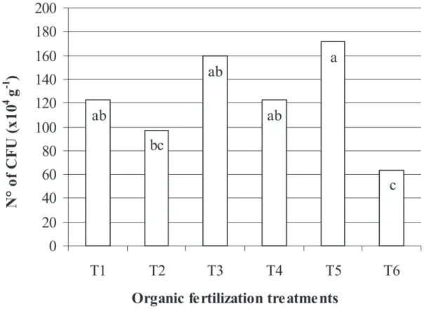
The variance analysis showed differences in the number
of CFU between the sampling periods and between the
organic fertilization treatments. A greater number of CFU was
evidenced in the sowing period (183.5 x 10
4CFU g
-1), differing
signifi cantly from the other periods: fl owering (85.05 x 10
4CFU g
-1) and post-harvest (100.38 x 10
4CFU g
-1). In relation
to the organic fertilization, treatment 5 yielded a greater
number of CFU, followed by treatments 3, 4 and 1 which did
not differ signifi cantly. Signifi cant differences were registered
only between treatments 5, 2 and 6 (Fig. 1).
There was no difference on species diversity between the
sampling periods. A greater number of species was found
in the sowing period (49), without signifi cant difference in
comparison with the other periods: fl owering (39 species)
and post-harvest (41 species).
The soil from treatment 6 presented the highest species
diversity (43), followed by treatments 2 and 5 (39 species),
1 (38 species) and 3 (35 species), which did not differ
signifi cantly. Signifi cant difference was registered only
between treatments 6 and 4 (24 species).
The fungal populations were more similar between
the sowing and fl owering periods (56.8% of similarity),
followed by the flowering and post-harvest periods
(50%), while the similarity between the sowing and
post-harvest periods reached 42.2%. In relation to the organic
fertilization, treatments 2 and 3 showed higher similarity
in all periods: 59.46% (sowing), 68.96% (fl owering) and
68.96% (post-harvest). (“Em relação aos tratamentos de
adubação organica, maior similaridade foi observada entre
os tratamentos 2 e 3, em todos os períodos fenológicos,
59,46 % (fundação), 68,96 % (fl oração) e 68,96 %
(pós-colheita)” veja se fi cou claro)
Sixteen taxa were found in the three sampling periods
(Tab. 2). Most of the taxa identifi ed in the melon rhizosphere
are known as saprobes (96.25%) and only a few (3.75%)
Figure 1. Number of fi lamentous fungi CFU in the rizhosphere of melon plants fertilized with six types of organic compost: T1 = control, without organic composts; T2 = 77% coconut fi ber, 20% goat manure and 3% K2SO4; T3 = 10% Ricinus communis leaves and stems, 50% Pennisetum purpureum leaves and 40% goat
manure; T4 = 77% coconut fi ber, 20% goat manure and 3% termophosphate; T5 = 47% Pennisetum purpureum leaves, 50% goat manure and 3% K2SO4;T6 = 57%
Species Sowing Flowering Post-harvest Total
T1 T2 T3 T4 T5 T6 T1 T2 T3 T4 T5 T6 T1 T2 T3 T4 T5 T6
Alternaria tenuissima (Kunze)
Wiltshire 05 05
Aspergillus fl avipes (Bainier & R.
Sartory) Thom & Church 03 03 06
A. fl avus Link 03 02 05
A. fumigatus Fresen. 13 66 51 09 13 27 16 21 30 10 13 19 16 28 22 21 10 15 400
A. japonicus var. aculeatus (Iizuka)
Al-Musallam 02 02 10 14
A. japonicus var. japonicus Saito 02 04 18 03 03 45 02 04 81
A. nidulans (Eidam) G. Winter 141 90 07 238
A. niger Tiegh. 03 09 61 18 24 02 09 07 01 05 09 09 05 09 06 02 179
A. niger var. niger Tiegh. 89 03 92
A. sydowii (Bainier & Sartory)
Thom & Church 02 02
A. tamarii Kita 02 02 02 06
A. terreus Thom 13 175 18 02 22 18 02 02 03 19 04 09 22 13 09 03 06 339
A. terreus var. aureus Thom & Raper 03 05 15 03 02 34 03 97 06 168
A. ungüis (Emile-Weill &
L. Gaudin) Thom & Raper 08 02 03 07 02 22
A. ustus (Bainier) Thom & Church 02 03 05
A. viridinutans Ducker & Thrower 02 22 13 10 13 60
Chaetomium convolutum Chivers 04 05 03 12
C. cupreum L.M. Ames 02 02
C. leucophorum L.M. Ames 02 02
C. nigricolor L.M. Ames 09 03 12
C. ochraceum Tschudy 02 10 10 22
C. trigonosporum (Marchal & É.J.
Marchal) Chivers 06 02 06 06 03 23
Cladosporium sphaerospermum Penz. 03 02 05
C. tenuissimum Cooke 03 05 08
Curvularia eragrostidis (Henn.)
J.A. Mey. 04 04
Emericella nidulans (Eidam) Vuill. 13 37 561 421 404 03 37 02 06 72 286 121 13 07 45 2028
E. nidulans var. acristata Subram. 300 60 157 31 09 85 22 105 163 145 37 03 21 169 120 34 66 1527
E. nidulans var. echinulata Godeas 07 21 09 06 04 06 02 30 06 91
E. rugulosa (Thom & Raper) C.R. Benj. 02 02
E. variecolor Berk. & Broome 02 02
Eupenicillium brefeldianum (B.O.
Dodge) Stolk & D.B. Scott 06 06
E. crustaceum F. Ludw. 02 02
Eurotium chevalieri L. Mangin 03 03 02 02 10
E. rubrum W. Bremer 02 02
Fusarium equiseti (Corda) Sacc. 02 02
F. merismoides Corda 18 02 20
F. oxysporum Schltdl. 02 02 03 07
F. redolens Wollenw. 02 02
F. solani (Mart.) Sacc. 04 02 04 07 02 02 04 02 02 29
F. stilboides Wollenw. 02 02 04
Gliocladium virens J.H. Mill.,
Giddens & A.A. Foster 03 03
Humicola fuscoatra Traaen 02 04 03 02 02 02 02 17
Monodictys castaneae (Wallr.) S. Hughes 02 02 04
Myrothecium. roridum Tode 07 03 03 13
M. verrucaria (Alb. & Schwein.) Ditmar 02 02 02 06
Neocosmospora vasinfecta var. africana (Arx) P.F. Cannon & D.
Hawksw.
05 03 02 27 43 03 06 02 03 02 02 98
Neoscytalidium dimidiatum (Penz.)
Crous & Slippers 02 02
Paecilomyces carneus (Duché & R.
Heim) A.H.S. Br. & G. Sm. 07 07
P. lilacinus (Thom) Samson 07 21 28
P. variotii Bainier 03 03
Table 2. Filamentous fungi colony forming units (CFU x 104 g-1) isolated from the rizhosphere of melon plants (C. melo cv. Gold Mine), fertilized with organic
composts, during the sowing, fl owering and post-harvest periods.
Filamentous fungi isolated from the rhizosphere of melon plants (Cucumis melo L. cv. Gold Mine) cultivated...
296
are referred to as potential plant pathogens:
Fusarium
oxysporum
,
F. solani
and
Myrothecium roridum
.
Most of the fungi was classified as rare; however
Emericella nidulans
and
E. nidulans
var.
acristata
were
abundant. All the species presented low frequencies of
occurrence.
Discussion
Most of the fungi found in the melon rhizosphere was
registered in rhizosphere and non-rhizosphere soils in
Brazil and other countries (Silva & Cavalcanti 1990, 1991;
Cavalcanti & Maia 1994; Maia & Gibertoni 2002; Mandeel
2002; Souza-Motta
et al.
2003; Ananda & Sridhar 2004;
Cavalcanti
et al.
2006; Costa
et al.
2006; Grishkan
et al.
2006).
Studying the microfungi of the rhizosphere of
Zygophyllum qatarense
Hadidi, in a semiarid environment
of Bahrain, Mandeel (2002) reported the predominance of
species of
Aspergillus
and
Penicillium
. Similarly, Grishkan
et al.
(2006) found species of
Penicillium
and
Aspergillus
to be the most abundant in the rhizosphere of native plants
of Negev, Israel. These results are similar to the fi ndings of
this work, considering that
Aspergillus
and
Penicillium
are
well represented in the studied area, with 15 (18.75%) and
13 (16.25%) species, respectively.
In the Brazilian semiarid region, Silva & Cavalcanti
(1990; 1991) registered
Fusarium
and
Penicillium
as
predominant in the rhizosphere of tomato (
Lycopersicon
esculentum
Mill). Maia & Gibertoni (2002) reported a
Species T1 T2 T3SowingT4 T5 T6 T1 T2 FloweringT3 T4 T5 T6 T1 T2 Post-harvestT3 T4 T5 T6 TotalP. citrinum Thom 02 34 49 02 87
P. corylophilum Dierckx 04 07 11
P. decumbens Thom 03 03 04 02 12
P. dierckxii Biourge 02 03 03 08
P. griseofulvum Dierckx 02 02 02 06
P. janthinellum Biourge 02 03 03 02 10
P. pinophilum Thom 03 02 05
P. restrictum J.C. Gilman &
E.V.Abbott 10 13 24 12 07 66
P. solitum var. crustosum (Thom) Bridge, D. Hawksw., Kozak.,
Onions, R.R.M. Paterson & Sackin 03 03
P. spinulosum Thom 27 27
P. vinaceum J.C. Gilman & E.V.
Abbott 04 02 06 04 51 264 04 335
P. waksmanii K.M. Zalessky 16 16
Rhizopus microsporus var. chinensis
(Saito) Schipper & Stalpers 06 02 03 02 02 03 18
R. microsporus var. microsporus
Tiegh. 03 06 04 02 02 03 02 22
Scopulariopsis brumptii Salv.-Duval 03 03
S. croci J.F.H. Beyma 03 03
S. sphaerospora Zach 04 21 04 03 32
Sordaria fi micola (Roberge ex
Desm.) Ces. & De Not. 02 03 05
Talaromyces trachyspermus (Shear)
Stolk & Samson 02 02 04
Thielavia fragilis (Natarajan) Arx 02 06 02 02 12
T. microspora Mouch. 02 06 07 15
T. terrestris (Apinis) Malloch &
Cain 02 02
T. terricola (J.C. Gilman & E.V.
Abbott) C.W. Emmons 15 42 03 02 07 29 09 12 19 21 06 07 09 21 02 07 07 218
Torula caligans (Bat. & H.P.
Upa-dhyay) M.B. Ellis 04 04 02 12 02 02 02 28
Trichoderma pseudokoningii Rifai 02 02
T. viride Pers. 03 03
Xepicula leucotricha (Peck) Nag Raj 02 18 03 23
Basidiomycota 02 02 04
Black Mycelia sterilia 02 02
CFU TOTAL 655 431 880 503 572 262 213 161 214 296 540 107 237 278 346 308 433 205 6641
T1 = control, without organic composts; T2 = 77% coconut fi ber, 20% goat manure and 3% K2SO4; T3 = 10% Ricinus communis leaves and stems, 50% Pennisetum purpureum leaves and 40% goat manure; T4 = 77% coconut fi ber, 20% goat manure and 3% termophosphate; T5 = 47% Pennisetum purpureum leaves, 50% goat manure and 3% K2SO4;T6 = 57% Pennisetum purpureum leaves, 40% goat manure
and 3% termophosphate.
great diversity of soil fungi in the Brazilian semiarid, with
predominance of:
Aspergillus, Cladosporium, Fusarium,
Humicola, Myrothecium, Paecilomyces, Penicillium,
Rhizopus
and
Trichoderma
. Species of
Aspergillus
and
Penicillium
predominated in soil from a copper mining
impacted area (Costa
et al.
2006) in Bahia State. Likewise,
Cavalcanti
et al.
(2006) registered several fi lamentous fungi
in soils from the Xingó region (Sergipe and Alagoas States),
with predominance of
Penicillium
and
Aspergillus
species.
Several of the fungal species found here were reported in
areas of the Brazilian semiarid region (Maia & Gibertoni
2002; Cavalcanti
et al,
2006; Costa
et al.
2006).
The prevalence of
Aspergillus
and
Penicillium
occurs
probably because these genera have a high number of species
and are capable of surviving in dry environments (Dix &
Webster 1995).
Three of the species found in this study were previously
regarded as pathogen to melon plants:
Fusarium oxysporum,
F. solani
and
Myrothecium roridum
(Marinho
et al.
2002;
Muniz
et al.
2004; Viana
et al.
2001). However, it is not
possible to say if they were in the rhizosphere as saprobe or
pathogen. These species were also registered, respectively,
in the rhizosphere of sugar-cane, tomato and sunfl ower
(Santos & Cavalcanti 1989; Silva & Cavalcanti 1990, 1991;
Souza-Motta
et al.
2003). In the present work these species
showed an occasional distribution.
The organic fertilization in the melon plants favored the
presence of fi lamentous fungi and increased the number
of CFU. It also allowed a wide distribution of saprophytic
fungi, considering that 96.25% of the species isolated in this
study have been described as organic matter decomposers.
Therefore, the introduction of organic composts can improve
the soil quality in the culture of melon by increasing the
diversity and number of fungi.
The phenology of the melon plants did not affect the
diversity of fi lamentous fungi; however, it signifi cantly
decreased the amount of CFU. The highest number of CFU
was observed in the sowing period, probably due to the
processes of incorporation and mineralization of the organic
residues in the soil, resulting in an increase of the cation
exchange capacity (CEC) in the subsequent periods (Tab.
1). Except for treatments 5 and 6, the quantity of organic
matter in the soil was higher in the sowing period, increasing
the offer of substrate for decomposition that was refl ected
in the highest number of CFU in this period.
The analysis of similarity showed a lower index between
the fungi in the sowing and the post-harvest periods.
According to Melnitchouk
et al.
(2005) and Wellbaum
et
al.
(1999), this may be associated with the changes in the
composition of root exudates over the phenological cycle of
the plant, causing variation in the rhizodeposition, and with
the hydrosoluble substances released by decomposition of
organic matter, which represent an energy source promptly
available for the microorganisms. In addition, Parkinson
et
al.
(1963) apud Souza-Motta
et al.
(2003) mentioned that the
young roots are initially colonized by a diversity of soil fungi
which after some days are substituted by a more restricted
mycobiota that remains until the senescence of the roots.
The results attained in this study suggest that anamorphic
fungi dominate the soil mycobiota of cultivated melon fi elds
in the Brazilian semiarid, and that species of
Aspergillus
and
Penicillum
are the most commonly found. Furthermore, the
data indicate that the composition of the rhizosphere interfere
in the fungal succession.
Acknowledgements
The authors would like to acknowledge: Coordenação de Aperfeiço-amento de Pessoal de Nível Superior (CAPES), Conselho Nacional de Desenvolvimento Científi co e Tecnológico (CNPq), Empresa Brasileira de Pesquisa Agropecuária (Embrapa Semi-Árido), Programa de Pós-Graduação em Biologia de Fungos/Universidade Federal de Pernambuco (UFPE) for the support to the project, and Dra. Elaine Malosso and Leonardo Costa for the English review.
References
Ananda, K. & Sridhar, K.R. 2004. Diversity of fi lamentous fungi on de-composing leaf and woody litter of mangrove forests in the southwest coast of India. Current Science of India 87(10): 1431-1437. Ames, L.M. 1961. A monograph of the Chaetomiaceae. Washington,
U.S. Army Research and Development.
Araújo, J.L.P. & Vilela, N.J. 2003. Aspectos Socioeconômicos. Pp. 15-18. In: H.R. Silva & N.D. Costa (eds.). Frutas do Brasil 33: melão, produção e aspectos técnicos. Brasília, Embrapa.
Arx, J.A. von. 1975. On Thielavia and some similar genera of Ascomycetes.
Studies in Mycology8: 1-31.
Arx, J.A. von; Guarro, J. & Figueras, M.J. 1986. The ascomycete genus
Chaetomium. Nova Hedwigia84: 1-162.
Booth, C. 1971. The genus Fusarium. Kew, Commonwealth Mycological Institute.
Brower, J.E.; Zar, J.H. & Von Ende, C.N. 1990. Field and laboratory methods for general ecology. Dubuque, McGraw-Hill.
Carmichael, J.W.; Kendrick, B.; Conners, I.L. & Sigler, L. 1980. Genera of Hyphomycetes. Edmonton, University of Alberta Press. Cavalcanti, M.A.Q. & Maia, L.C. 1994. Cellulolytic fungi isolated from
an alluvial soil in a semi-arid area of the northeast of Brazil. Revista de Microbiologia 25: 251-254.
Cavalcanti, M.A.Q.; Oliveira, L.G.; Fernandes, M.J. & Lima, D.M. 2006. Fungos fi lamentosos isolados do solo em municípios na região Xingó, Brasil. Acta Botanica Brasilica 20(4): 831-837.
Corlett, M. 1966. Perithecium devolopment in Chaetomium trigonosporum.
Canadian Journal of Botany 44: 155-162.
Costa, I.P.M.W.; Cavalcanti, M.A.Q.; Fernandes, M.J.S. & Lima, D.M.M. 2006. Hyphomycetes from soil of an area affected by copper mining activities in the state of Bahia, Brazil. Brazilian Journal of Micro-biology 37: 267-275.
Dias, R.C.S.; Costa, N.D.; Silva, P.C.G.; Queiroz, M.A.; Zuza, F.; Leite, L.A.S.; Pessoa, P.F.A.P. & Terão, D. 1998. A cadeia produtiva do melão no Nordeste. Pp. 441-494. In: A.M.G. Castro; S.M.V. Lima; W.J. Goe-dart; A. Freitas Filho & J.R.P. Vasconcelos (eds.). Cadeias produtivas e sistemas naturais: prospecção tecnológica. Brasília, Embrapa. Dix, N.J. & Webster, J. 1995. Fungal ecology. London, Chapman & Hall. Domsch, K.H.; Gams, W. & Anderson, T.H. 1980. Compendium of Soil
Fungi. London, Academic Press.
Ellis, M.B. 1971. Dematiaceous Hyphomycetes. Kew, Commonwealth Mycological Institute.
Ellis, M.B. 1976. More Dematiaceous Hyphomycetes. Kew, Commonwe-alth Mycological Institute.
Embrapa. 1997. Manual de Métodos de Análise de Solo. Rio de Janeiro, Centro Nacional de Pesquisa de Solos.
Filamentous fungi isolated from the rhizosphere of melon plants (Cucumis melo L. cv. Gold Mine) cultivated...
298
Versão eletrônica do artigo em www.scielo.br/abb e http://www.botanica.org.br/acta/ojs
southward rainfall gradient in desert ecosystems. Journal of Arid Environments 53(3): 409-417.
Hanlin, R.T. & Menezes, M. 1996. Gêneros ilustrados de Ascomicetos. Recife, Imprensa da Universidade Federal Rural de Pernambuco. IBGE. 2006. Produção agrícola. http://www.sidra.ibge.gov.br/bda/tabela/
listabl.asp?c=1612&z=p&o=20 (accessed 20/02/2008).
Klich, M.A. & Pitt, J.I. 1994. A laboratory guide to common Aspergillus species and their teleomorphs. North Ryde, CSIRO Division of Food Processing.
Maia, L.C. & Gibertoni, T.B. 2002. Fungos registrados no semi-árido nor-destino. Pp. 163-176. In: E.V.S.B. Sampaio; A.M. Giulietti; J. Virginio & C.F.L. Guamarra-Rojas (eds.). Vegetação & Flora da Caatinga. Recife, Centro Nordestino de Informações sobre Plantas.
Mandeel, Q.A. 2002. Microfungal community associated with rhizosphere soil of Zygophyllum qatarense in arid habitats of Bahrain. Journal of Arid Environments 50(4): 665-681.
Marinho, R.E.M.; Sales Jr, R.; Maracajá, P.B.; Silva, G.F.; Costa, F.M. & Silva, E.C. 2002. Identifi cação da micofl ora associada a raízes de meloeiro nos estados do Rio Grande do Norte e Ceará. Caatinga 15(1/2): 25-28.
Mehan, V.K.; Mayee, C.D.; Jayanthi, S. & McDonald, M. 1991. Prehar-vest seed infection by Aspergillus fl avus group fungi and subsequent afl atoxin contamination in groundnuts in relation to soil types. Plant and Soil136(2): 239-248.
Melnitchouk, A.; Leinweber, P.; Eckhardt, K.U. & Beese, R. 2005. Qua-litative differences between day-and night-time rhizodeposition in maize (Zea mays L.) as investigated by pyrolysis-fi eld ionization mass spectrometry. Soil Biology & Biochemistry 37: 155-162.
Müller-Dombois, D. & Ellemberg, H. 1974. Aims and methods of vege-tation ecology. New York, John Wiley & Sons.
Muniz, M.F.B.; Gonçalvez, N. & Garcia, D.C. 2004. Qualidade fi siológica e sanitária de sementes de melão (Cucumis melo).Ciência Rural 34(3): 951-953.
Nicoli, R.M. & Russo, A. 1974. Le genre Humicola Traaen et les genres
voisins (Hyphomycetes). Nova Hedwigia 25: 737-781.
Oliveira, A.C. 1991. Impactos econômicos da irrigação sobre o pólo Petrolina-Juazeiro. Recife, UFPE/PIMES.
Pitt, J.I. 1988. A laboratory guide to common Penicillium species. North Ryde, CSIRO Division of Food Processing.
Raper, K.B.; Thom, C. & Fennel, D.I. 1949. A manual of the Penicillia. Baltimore, Williams & Wilkins Company.
Rifai, M.A. 1969. A revision of the genus Trichoderma.Mycological Papers 116: 1-56.
Samson, R.A. 1974. Paecilomyces and some allied Hyphomycetes. Studies in Mycology 6: 1-68.
Santos, A.C. & Cavalcanti, M.A.Q. 1989. Fungos isolados da rizosfera da cana-de-açúcar da zona da mata de Pernambuco. Revista Brasileira de Botânica 12(1): 23-29.
Schipper, M.A.A. 1984. A revision of the genus Rhizopus: I. The Rhizopus stolonifer-group and Rhizopus oryzae. Studies in Mycology 25: 1-34. Schnittler, M. & Stephenson, S.L. 2000. Myxomycete biodiversity in four
different forest types in Costa Rica. Mycologia 92: 626-637. Sigler, L. & Carmichael, J.W. 1976. Taxonomy of Malbranchea and some
other hyphomycetes with arthroconidia. Mycotaxon 4: 349-488. Silva, M.I.L. & Cavalcanti, M.A.Q. 1990. Fungos na rizosfera de sementes
de tomate. Fitopatologia Brasileira 15(4): 323-326.
Silva, M.I.L. & Cavalcanti, M.A.Q. 1991. Hongos associados a semillas de tomate (Lycopersicon esculentum). Boletín Micologico 06(1/2): 59-62. Souza-Motta, C.M.; Cavalcanti, M.A.Q.; Fernandes, M.J.S.; Lima, D.M.M.; Nascimento, J.P. & Laranjeira, D. 2003. Identifi cation and characte-rization of fi lamentous fungi isolated from the sunfl ower (Helianthus annus L.) rhizosphere according to their capacity to hydrolyse inulin. Brazilian Journal of Microbiology 34(3): 273-280.
Statsoft Inc. 1997. Statistica for Windows (Computer Program Manual). Tulsa, Statsoft Inc.
Sutton, B.C. 1980. The Coelomycetes: Fungi imperfecti with pycnidia, acervuli and stromata. Kew, Commonwealth Mycological Institute. Udagawa, S.I.; Horie, Y. & Cannon, P.F. 1989. Two new species of Neo-cosmospora from Japan, with a key to the currently accepted species.
Sydowia 41: 349-359.
Viana, F.M.P.; Santos, A.A.; Freire, F.C.O.; Cardoso, J.E. & Vidal, J.C. 2001. Recomendações para o controle das principais doenças que afetam a cultura do melão na Região Nordeste. http://www.cnpat.embrapa. br/home/down/index.php?pub/ct_12.pdf (accessed 02/07/2007). Wellbaum, C.; Schoenlein-Crusius, I.H. & Santos, V.B. 1999. Fungos