Oral acetylsalicylic acid and prevalence of actinic keratosis
JULIANO SCHMITT1, HÉLIO MIOT11Department of Dermatology, Sao Paulo State University, Botucatu Medical School, Unesp, Campus Universitário de Rubião, Botucatu, SP, Brazil
S
UMMARYStudy conducted at Department of Dermatology, Sao Paulo State University, Botucatu Medical School, Unesp, Campus Universitário de Rubião, Botucatu, SP, Brazil
Article received: 04/05/13
Accepted for publication: 08/30/13
Correspondence:
Departamento de Dermatologia, S/N Faculdade de Medicina da UNESP Campus Universitário de Rubião Jr. ZIP Code: 18618-970 Botucatu – SP – Brazil Phone/Fax: +55 14 3880-1267
http://dx.doi.org/10.1590/1806-9282.60.02.010
Conflict of interest: none
Objective: To investigate the inluence of a regular oral use of acetylsalicylic acid in the prevalence of actinic keratosis.
Methods: A case-control study with dermatologic outpatients above 50 years of age assessed between 2009 and 2011. Cases were deined as those who had been under regular use of oral acetylsalicylic acid for more than six consecutive months. The as-sessment focused on: age, sex, skin-type, tobacco smoking, use of medication, oc-currence of individual or family skin cancer, and sunscreen and sun exposure habits. Actinic keratoses were counted in the medial region of the face and upper limbs. Counts were adjusted by co-variables based on a generalized linear model.
Results: A total of 74 cases and 216 controls were assessed. The median time of acetylsalicylic acid use was 36 months. Cases differed from controls as to the highest age, highest prevalence of use of angiotensin-converting enzyme inhibi-tors and fewer keratosis on the face and on the upper limbs (p<0.05). The mul-tivariate model showed that the use of acetylsalicylic acid was associated to lo-wer counts of face actinic keratosis and upper-limb erythematous actinic keratosis (p<0.05), regardless of other risk factors.
Conclusion: The regular use of oral acetylsalicylic acid for more than six months was associated to a lower prevalence of actinic keratosis, especially facial and erythematous ones.
Key words: actinic keratosis, acetylsalicylic acid, prevalence, risk factors.
I
NTRODUCTIONActinic Keratoses (AKs) are atypical proliferations of ke-ratinocytes induced mainly by ultraviolet radiation (UVR). Besides its potential (0.6-20%) for turning into invasive neoplasms, they relect the accumulated photodamage and indicate a larger risk of developing skin cancers in every region affected by the disease.1-5
They are common in sun-exposed areas of elderly fair-skinned people, representing 5.1% of complaints in der-matological consultations in Brazil. In Australia, it is es-timated that 40-50% of the population above 40 years of age presents at least one lesion.6,7
Considering that AKs can be models of in-vivo stud-ies of skin carcinogenesis, the identiication of factors that can prevent AKs may reveal potential preventive or therapeutic measures related to skin cancer, especially squamous cell carcinoma (SCC).8
For decades, the role of non-steroidal anti-inlamma-tory drugs (NSAI) and mainly acetylsalicylic acid (ASA) has
been studied in an attempt to prevent some types of can-cer, especially colon and skin cancers.9-11 The favorable re-sults in experimental studies and clinical assays were at-tributed mainly to the inhibition of the cyclooxygenase type-2 enzyme (COX-2), which plays an antiapoptotic, an-giogenic, and proliferative role in those neoplasms.12,13
ASA is an old and low-cost medication used as antipyret-ic and anti-inlammatory drug, whantipyret-ich acts by the irrevers-ible inhibition of cyclooxygenase 1 and 2. However, it has been recommended in low doses for preventing thrombo-embolic events. Clinical studies carried out in those popu-lations indicate a reduction in the incidence of some neo-plasms, which opened up new therapeutic possibilities. One believes that, besides COX-2 blockage, other pharmaceuti-cal activities of ASA may compose its antineoplastic effect.8
M
ETHODSWe performed a case-control study involving adult sub-jects above 50 years of age, volunteers, well-informed and consenting, from both sexes, selected among patients from the dermatology outpatient clinic of the Pro-Han-sen Foundation – Curitiba, state of Paraná, Brazil – from April, 2009 to September, 2011.
Cases were deined as those who had used a daily min-imum 80mg of oral ASA, more than four days a week, for more than six consecutive months. The controls were those who reported no previous regular use of ASA.
We excluded: patients with signs of immunosup-pression, genetic syndromes predisposing to cancer, with a communication deficit, bedridden, with diffuse dermatoses in the tegument, skin phototype V or VI, those who had used other AINEs regularly, those who had interrupted the use of ASA more than 2 months before the interview, those who used systemic or top-ical retinoids or immunosuppressant medications, those who treated AKs in some occasion or who had been submitted to phototherapic and radiotherapic treat-ments previously.
A speciic systematization of sample collection or pro-portion between cases and controls was not adopted, and all available patients who itted the inclusion criteria were consecutively included.
Based on a standardized questionnaire, consenting patients were interviewed and examined by an experienced dermatologist.
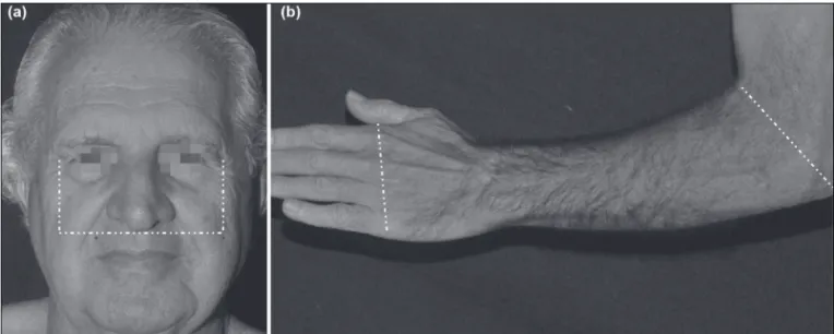
Visible AK lesions on predeined areas on the face - centrally - and on the forearms, from the elbows to the back of the hands (igure 1) were counted. A skin mark-er pen was used to help in the counting process.14
The main variables analyzed were the number and type of AKs (erythematous or hyperkeratotic) present in the standardized areas of the face and upper limbs (ULs), besides oral use of ASA (time and accumulated load). The other independent variables were: age, sex, skin type, smok-ing, use of angiotensin-converting enzyme inhibitors (ACEI), occurrence of personal or family skin cancer, sun-screen and sun exposure habits (occupational or leisure). Quantitative variables were represented by the averag-es and standard deviation, or medians and quartilaverag-es (1st and 3rd) when normality could not be evidenced by the Sha-piro-Wilk’s test.15 When indicated, the comparison between groups was performed by the Student’s t-test or Mann-Whitney test. Quantitative variables of the count type were compared by a negative binomial regression model.16 Cat-egorical variables were represented by their percentages and compared using square tests or linear trend chi-square tests, in case there was an ordinal characteristic. The correlation between quantitative variables was estimated by Spearman’s linear correlation coeficient (rS).17
First, cases and controls were compared bivariately to show homogeneity of the groups. The effect size of the categorical variables was estimated by odds ratio (OR) and the 95% conidence interval (CI 95%).18
Later, an exploratory multivariate model of AKs risk based on the generalized linear counting model (negative binomial) was developed consisting of co-variables that reach p<0.3. The effect size was represented by β estima-tor of the regression.19
Missing data were estimated by the multiple impu-tation technique.20 Two-tailed values of p<0.05 were con-sidered signiicant. Data were tabulated in MS Excel 2003® and analyzed using SPSS 17.0® software.21
The sample design was based on a pre-test consisting of 50 cases and 50 controls, and calculated from a mul-tiple regression model, power estimated at 0.8 and bilat-eral alpha level at 0.05. According to the estimate of vari-ables in the final model, the smallest sample was considered to be 70 cases and 70 controls.22,23
The Project was approved by the institutional review board of the UFPR – Curitiba-Brazil (Nº 299. EXT.005/2009-03).
R
ESULTSTwo hundred and ninety patients were assessed, 74 of them were ASA regular users and 216 were controls. The main clinical and demographic data are presented in ta-ble 1. Cases and controls were not homogeneous regard-ing age, prevalence of the use of ACE inhibitors and num-ber of erythematous AKs.
Among the cases, the median time (quartiles 1 and 3) of ASA use was 36 (17-74) months, 81% used a daily dose of 100mg, and the median accumulated dose was 27.7 (13-59) g.
The sample median (quartiles 1 and 3) of the total number of AKs was 2 (1-5) for face, and 1 (0-3) for ULs; and 80% of the patients presented at least one AK lesion on the face. A slight correlation was observed between the number of lesions and age both for face lesions and UL lesions (rS = 0.3 and 0.4; p<0.01). Similarly, the num-ber of facial lesions correlated to the numnum-ber of UL le-sions (rS = 0.5; p<0.01).
Eighteen percent of the interviewed people had al-ready received a diagnosis of cancer at some time, and they were those who presented a higher median count (quartiles 1 and 3) of AKs both on the face and on ULs (4 (2-7) x 2 (1-5); p<0.01 and 2 (0-10) x 0 (0-2); p<0.01).
TABLE 1. Main clinical and demographic data of cases and controls*
Variables Cases Controls OR (95% CI) pa
Female sex – N(%) 47 (64) 149 (69) 0.78 (0.5-1.4) 0.39b
Age – average (standard deviation) 63.5 (58-70) 59 (55-66) - 0.00c
Skin type – N(%) 0.41d
I-II 20 (27) 44 (20) 1.40 (0.7-2.8)
III 30 (41) 98 (45) 0.94 (0.5-1.8)
IV 24 (32) 74 (34) 1.0
Sunburn history – N(%) 44 (59) 141 (65) 0.78 (0.5-1.3) 0.37b
Personal history of skin cancer – N(%) 11 (15) 41 (19) 0.75 (0.4-1.5) 0.43b
Family history of skin cancer – N(%) 11 (15) 28 (13) 1.17 (0.6-2.5) 0.68b
Sun-exposed occupation (last 10 years) – N(%) 13 (18) 35 (16) 1.10 (0.6-2.2) 0.79b
Sun-exposed leisure – N(%) 30 (41) 63 (29) 1.66 (1.0-2.9) 0.07b
Daily use of sunscreen for more than 2 years – N(%) 10 (14) 46 (21) 0.58 (0.3-1.2) 0.14b
Regular use of ACEI – N(%) 35 (47) 39 (18) 4.07 (2.3-7.2) 0.00b
Smoking now – N(%) 10 (14) 41 (19) 0.67 (0.3-1.4) 0.29b
Face AKs – Median (Q1-Q3) 1 (0-4) 3 (1-6) - 0.02e
Erythematous face AKs 1 (0-4) 3 (1-5) - 0.01e
Hypertrophic face AKs 0 (0-0) 0 (0-0) - 0.59e
UL AKs (Q1-Q3) 0 (0-3) 1 (0-3) 0.03e
Erythematous UL AKs 0 (0-3) 1 (0-3) - 0.02e
Hypertrophic UL AKs 0 (0-0) 0 (0-0) - 0.72e
a Unadjusted values; b Chi-square test; c t-student test; d Tendency chi-square; e Negative binomial regression
OR = Odds Ratio; 95% CI = 95% confidence interval; Q1-Q3 = first and third quartiles; ACEI = Angiotensin converting enzyme inhibitor; AK= Actinic Keratosis; ULs = Upper limbs.
of the ULs among ASA users, in addition to smoking in-luencing basically the ULs count.
The period of ASA use and the ASA load also corre-lated to lower counts of face and UL AKs (erythematous and hypertrophic) (p<0.05).
D
ISCUSSIONThe regular use of ASA was independently associated to lowest AK counts, especially the erythematous forms (face and UL) and the hypertrophic form of the face. The COX-2 inhibition is a possible explanation for such phenome-non.
There is an increase of epidermal COX-2 expression in murine skin submitted to UVB radiation, as well as in AK lesions, squamous cell carcinoma (SCC), and basal cell carcinoma (BCC). Similarly, in humans, there is a signiicantly raised COX-2 expression in SCC lesions (40%), Bowen disease (22%), and AKs (31%) when compared to normal skin.24,26-29
Regular ASA users were older and had a propensity to perform more sun-exposed leisure activities. There were more retired people among the ASA users (23% x 10%; OR = 2.6 (1.3-5.3); p<0.01). Similarly, those who practiced more sun-exposed leisure activities have been more often retired people (24% x 9%; OR = 3.3 (1.7-6.5); p<0.01). When adjust-ed by retirement, cases and controls did not differ regard-ing the practice of sun-exposed leisure (p=0.20).
The co-morbidities more signiicantly associated to ASA regular users were: ischemic heart disease (18% x 2%; OR = 11.3 (3.6-35.9); p<0.01), systemic hypertension (84% x 39%; OR = 8.12 (4.1-16.0); p<0.01), and type 2 diabetes
mellitus (30% x 6%; OR = 6.6 (3.1-14.0); p<0.01).
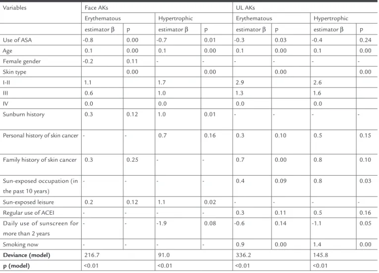
Table 2 presents the regression coeficients of face and UL AK scores. There were different patterns of risk variables for each subtype of neoplasm, the inluence of the age group and of fair skin-types in the incidence of AKs in all groups, a reduction in the number of face AKs (erythematous and hypertrophic) and erythematous AKs
TABLE 2 Generalized linear model (negative binomial) to count AKs adjusted by risk co-variables*
Variables Face AKs UL AKs
Erythematous Hypertrophic Erythematous Hypertrophic estimator β p estimator β p estimator β p estimator β p
Use of ASA -0.8 0.00 -0.7 0.01 -0.3 0.03 -0.4 0.24
Age 0.1 0.00 0.1 0.00 0.1 0.00 0.1 0.00
Female gender -0.2 0.11 - - -
-Skin type 0.00 0.00 0.00 0.00
I-II 1.1 1.7 2.9 2.6
III 0.6 1.0 1.3 1.6
IV 0.0 0.0 0.0 0.0
Sunburn history 0.3 0.12 1.0 0.01 - - -
-Personal history of skin cancer - - 0.7 0.16 0.3 0.10 0.5 0.15
Family history of skin cancer 0.3 0.25 - - 0.7 0.00 0.8 0.10
Sun-exposed occupation (in the past 10 years)
- - - - 0.4 0.09 0.8 0.03
Sun-exposed leisure 0.2 0.12 1.1 0.02 - - -
-Regular use of ACEI - - - - 0.3 0.11 0.5 0.16
Daily use of sunscreen for more than 2 years
- - -1.9 0.08 -0.6 0.14 -1.1 0.05
Smoking now - - - - 0.9 0.00 1.4 0.00
Deviance (model) 216.7 91.0 336.2 145.8
p (model) <0.01 <0.01 <0.01 <0.01
a Composition of each model with variables that result in p<0.3; Model effect: type I analysis.
Transgenic mice, which promote an elevated COX-2 expression, are more susceptible to UV-induced skin tu-mors, while COX-2 deicient mice have a tumor rate 75% lower than normal mice.30,31 Besides, it was observed that the presence of inlamed AKs in humans is associated to the progression to invasive diseases in those lesions.32
COX-2 and prostaglandin E2 are positive regulators of tumor proliferation, mainly through Ras/Raf/MAPK, and Pt13K/Akt.33 The MAPK pathway, on the other hand, seems to induce COX-2 production generating a positive feedback cycle.34 The same pathway may even activate the production of epithelial growth factor (EGF) in colorec-tal tumors.35
Another property of COX-2 related to carcinogene-sis is the promotion of tumor angiogenecarcinogene-sis. Its expres-sion is correlated to tumor vascularity, capillary density, and synthesis of vascular endothelial growth factor (VEGF).29,36 Similarly, in studies with AINE in colon can-cer, a production of angiogenic factors dependent on the activity of COX-2 was observed.13
In addition to stimulating cell proliferation and an-giogenesis, the elevation of COX-2 is related to the resis-tance to apoptosis in various tumors, such as the colon, breast, lung, and prostate tumors. The activity of those enzymes proved to be correlated to the expression of an-tiapoptotic proteins of the Bcl2 family.12,13
A decrease in the formation of UV-induced skin tu-mors in mice that received celecoxib (89% reduction) or indomethacin (78% reduction) was observed.37,38 A reduc-tion in more than 50% of UVR-induced skin tumors in naked mice receiving nimesulide - a selective inhibitor of COX-2 - was also observed.39 Additionally, celecoxib in-hibited the growth of melanoma cells, even in strains not expressing COX-2.40
There was a lower risk of SCC (OR=0.2) and a lower AK count (OR=0.5) among regular users of AINEs for more than one year, even in low doses.41 Similar results concerning SCC were found in a population study with 1.484 participants, observing a risk reduction with vari-ous AINEs, mainly ASA, and more strongly for tumors positive for p53 or with a loss of heterozygosity of the PTCH gene.10
In an observational prospective study involving 2.297 participants, Clouser et cols also identiied an inverse as-sociation between the use of NSAI and SCC and BCC risks. But, in that study, short term use was more protec-tive than long term use.42 Furthermore, Asgari et cols did not observe any association between the use of AINEs and SCC risks in an investigation that included 830 North Americans.43
The oral use of celecoxib, a selective inhibitor of COX-2, was prospectively evaluated in a study with 240 individ-uals for preventing SCC and BCC in carriers of 10-40 AKs. After nine months, there were signiicantly less new SCC (RR=0.4) and BCC (RR=0.4) lesions in celecoxib users, but there was no reduction in the number of AKs.44 Coinci-dentally, a controlled double-blind prospective study ob-served 20% less new BCC lesions in carriers of basal cell nevus syndrome after having used celecoxib for three years.45
Although the inhibition of COX-2 by ASA occurs in a dose-dependent ratio, the oral use of just 81mg daily re-duces signiicantly the prostaglandin E2 levels in the dis-tal colonic mucosa justifying that clinic effect under low dosages, as detected in our sample.46 Besides, COX-2 inde-pendent antineoplastic mechanisms have already been pro-posed for ASA, such as the activation of the kappa beta nu-clear factor (NF-κβ) and stimulus to the expression of DNA repair proteins such as: hMLH1 and hPMS2.47-49
In the present study, the groups were quite similar regarding the variables under study, except for age, prev-alence of co-morbidities, frequency of use of ACEI, and a higher report of outdoor leisure activities among cases. Such characteristics were predictable by the fact the ASA indications presuppose cardiovascular diseases, which are more prevalent with aging. Similarly, cases presented more free time for leisure because most of them are re-tired and can be encouraged to practice physical activi-ties outdoors – such as hiking – for therapeutic and pre-ventive reasons.
In contrast, a more frequent sun-exposed leisure ac-tivity and older age should be factors that would increase the incidence of AKs among cases. A lower count of le-sions in those patients reinforces the evidence that ASA exerts a protective role in the development of AKs. More-over, the multivariate analysis did the adjustment for age, sun exposure and skin type and reinforced the low count-ing of AKs among cases.
The prevalence of AK was 80.3% in this sample (70.3% for ASA users and 83.8% for controls; p=0.01), and AK counting was higher in proportion to age, to low skin types and associated to the occurrence of skin cancer. Such ind-ings corroborate previous epidemiological observations and refer to a probable increase in the incidence of AKs with the increase in longevity of the population and an in-crease in leisure activities exposed to direct sun.2,7
Variations in the prevalence of AKs observed in vari-ous epidemiological studies may be due to the ethnic composition of the population, their sunscreen use and skin-exposure habits, to levels of UV radiation to which they are exposed as well as the counting methodology used by each team. In the region studied - located on par-allel 25-South -, the population is constituted mainly by European descents. In addition, the methodology used promoted an active search for lesions, even though they might not to be signiicant in a speciic population con-sisting of general dermatological patients, contributing to a high prevalence of AKs in the sample.
Tobacco smoking is a well known risk factor for many solid tumors, besides acting as a co-factor in other neo-plasms, for example, in case of tumors related to papillo-mavirus.52,53 There are some studies that relate skin can-cer to tobacco smoking, especially SCC (RR=2.0).54-56 In addition, there is a strong association between tobacco smoking and solitary keratoacanthoma (OR=9.1), which some authors consider as a well differentiated SCC form.57,58
Our data show an association between current smok-ing and the number of AK lesions on the ULs, but not on the face. The explanation for this would be that in the re-gion of origin of those patients - located approximately 1000 meters above sea level, with a cold climate most of the year - there is better sun protection for the ULs than for the face because of the clothes they wear, which per-mits a predominant local action of other carcinogens oth-er than UVR. Such inding must be clariied in studies with speciic designs.
Besides the different AK patterns on the face and on ULs, that can be justiied by the dificulty in quanti-fying skin screening and skin-exposure habits, we iden-tiied differences in the risk proiles for hypertrophic and erythematous AKs. This may be a result of the dis-tribution of hypertrophic lesions, commonly located on the ULs, as well as the fact they occur in smaller ampli-tude of count than the erythematous ones, reducing the exploratory performance of statistical models. Howev-er, it is also possible that hypertrophic AKs present a lower response to ASA, or even that they represent a
lighter risk of evolving or even that they already present an invasive character and may be in more advanced stag-es of carcinogenstag-esis.59 Such data suggest that risk fac-tor studies and even clinical assays in AKs must consid-er the location of lesions and the clinical types when analyzing the obtained results.
There was a negative association between the regular use of sunscreen for more than two years and AK lesions - mainly the hypertrophic ones - which reinforces the rele-vance of sun-exposure in the genesis of AK. The preventive and therapeutic effect of the use of sunscreen on AKs had already been reported (OR=1.5 for remission and RR = 0.6 for incidence), mainly in places and times of year with high levels of UVR.60-62
Our study presents some limitations as a result of a possible inaccuracy in the clinical diagnosis of AKs and also because the manual counting of lesions produces variable reproducibility.14,63
Nevertheless, our results prove to be consistent with the data found in the literature and with the pathophys-iology of the lesions. The differential quantiication be-tween erythematous and hypertrophic AKs also revealed elements that favor an approach by subgroups rather than the total counts of lesions used by other authors, main-ly as long as we have no knowledge of the natural histo-ry and evolution of the genesis and transformation among those subtypes.
As our study was carried out among dermatology patients in a public institution in the south of Brazil, we cannot extrapolate its results to other situations. However, the association between the use of NSAIs and AKs has already been observed in different populations; the consistency between the results of the analyses and the magnitude of the effects found favor the recogni-tion of an associarecogni-tion between the use of ASA and low-er counts of AKs.41 Besides, the strict exclusion criteria and the use of control variables reduced the effect of se-lection biases.
Subsequent clinical trials with representative sample must substantiate our indings and deine the role of the regular use of ASA and other NSAIs in preventing AKs.
C
ONCLUSIONSThe regular use of oral acetylsalicylic acid for more than six months was independently associated to a lower pre-valence of actinic keratosis, especially facial and erythe-matous ones.
R
ESUMOÁcido acetilsalicílico oral e prevalência de queratoses ac-tínicas
Objetivo: Queratoses actínicas são proliferações atípicas de queratinócitos com potencial para se transformarem em carcinoma invasivo. Estudos clínicos e laboratoriais têm observado um efeito preventivo de anti-inlamató-rios não hormonais no desenvolvimento de diversas neo-plasias. Neste estudo, investigamos a inluência do uso regular do ácido acetilsalicílico na prevalência de quera-toses actínicas.
Métodos: Estudo caso-controle com pacientes dermato-lógicos acima de 50 anos de idade, avaliados entre 2009 e 2011. Os casos foram deinidos como aqueles que esta-vam sob uso regular de ácido acetilsalicílico por via oral por mais de seis meses consecutivos. Avaliou-se idade, sexo, fototipo, tabagismo, uso de medicamentos, ocor-rência de câncer de pele no indivíduo ou na família, há-bitos de proteção e exposição solar. As queratoses actíni-cas foram contadas nos membros superiores e região medial da face. As contagens foram ajustadas pelas cova-riáveis em um modelo linear generalizado.
Resultados: Um total de 74 casos e 216 controles foi ava-liado. O tempo médio de uso de ácido acetilsalicílico foi de 36 meses. Casos diferiam dos controles quanto a ida-de mais elevada, maior prevalência ida-de uso ida-de inibidores da enzima conversora da angiotensina e menos querato-ses na face e membros superiores (p <0,05). O modelo multivariado mostrou que o uso do ácido acetilsalicílico esteve associado a contagens mais baixas de queratoses actínicas na face de queratoses não hipertróicas nos mem-bros superiores (p <0,05).
Conclusão: O uso regular do ácido acetilsalicílico por via oral por mais de seis meses esteve associado a uma me-nor prevalência de queratose actínica, especialmente fa-ciais e eritematosas.
Unitermos: queratoses actínicas, ácido acetilsalicílico, prevalência, fatores de risco.
R
EFERENCES1. Schmitt JV, Miot HA. Actinic keratosis: a clinical and epidemiological revision. An Bras Dermatol. 2012;87:425-34.
2. Salasche SJ. Epidemiology of actinic keratoses and squamous cell carcinoma. J Am Acad Dermatol. 2000;42:4-7.
3. Lebwohl M. Actinic keratosis: epidemiology and progression to squamous cell carcinoma. Br J Dermatol. 2003;149(Suppl 66):31-3.
4. Rossi R, Mori M, Lotti T. Actinic keratosis. Int J Dermatol. 2007;46:895-904. 5. Criscione VD, Weinstock MA, Naylor MF, Luque C, Eide MJ, Bingham SF; Department of Veteran Affairs Topical Tretinoin Chemoprevention Trial Group. Actinic keratoses: Natural history and risk of malignant
transformation in the Veterans Affairs Topical Tretinoin Chemoprevention Trial. Cancer. 2009;115:2523-30.
6. Frost CA, Green AC. Epidemiology of solar keratoses. Br J Dermatol. 1994;131:455-64.
7. Sociedade Brasileira de Dermatologia SBD. Peril nosológico das consultas dermatológicas no Brasil. An Bras Dermatol. 2006;81:549-58.
8. Langley RE, Burdett S, Tierney JF, Cafferty F, Parmar MK, Venning G. Aspirin and cancer: has aspirin been overlooked as an adjuvant therapy? Br J Cancer. 2011;105:1107-13.
9. Ruder EH, Laiyemo AO, Graubard BI, Hollenbeck AR, Schatzkin A, Cross AJ. Non-steroidal anti-inlammatory drugs and colorectal cancer risk in a large, prospective cohort. Am J Gastroenterol. 2011;106:1340-50. 10. Torti DC, Christensen BC, Storm CA, Fortuny J, Perry AE, Zens MS, et al.
Analgesic and nonsteroidal anti-inflammatory use in relation to nonmelanoma skin cancer: a population-based case-control study. J Am Acad Dermatol. 2011;65:304-12.
11. Curiel-Lewandrowski C, Nijsten T, Gomez ML, Hollestein LM, Atkins MB, Stern RS. Long-term use of nonsteroidal anti-inlammatory drugs decreases the risk of cutaneous melanoma: results of a United States case-control study. J Invest Dermatol. 2011;131:1460-8.
12. Tjiu JW, Liao YH, Lin SJ, Huang YL, Tsai WL, Chu CY, et al. Cyclooxygenase-2 overexpression in human basal cell carcinoma cell line increases antiapoptosis, angiogenesis, and tumorigenesis. J Invest Dermatol. 2006;126:1143-51. 13. Fecker LF, Stockleth E, Nindl I, Ulrich C, Forschner T, Eberle J. The role of
apoptosis in therapy and prophylaxis of epithelial tumours by nonsteroidal anti-inlammatory drugs (NSAIDs). Br J Dermatol. 2007;156(Suppl 3):25-33. 14. Epstein E. Quantifying actinic keratosis: assessing the evidence. Am J Clin
Dermatol. 2004;5:141-4.
15. Henderson AR. Testing experimental data for univariate normality. Clin Chim Acta. 2006;366:112-29.
16. Coxe S, West SG, Aiken LS. The analysis of count data: a gentle introduction to poisson regression and its alternatives. J Pers Assess. 2009;91:121-36. 17. Norman GR, Streiner DL. Biostatistics. The bare essentials. 3rd ed. Shelton:
People’s Medical Publishing House; 2008.
18. Katz MH. Multivariable analysis. A practical guide for clinicians. 2nd ed.
Cambridge: Cambridge University Press; 2006.
19. Hughes MC, Williams GM, Fourtanier A, Green AC. Food intake, dietary patterns, and actinic keratoses of the skin: a longitudinal study. Am J Clin Nutr. 2009;89:1246-55.
20. Mackinnon A. The use and reporting of multiple imputation in medical research - a review. J Intern Med. 2010;268:586-93.
21. SPSS 17.0 for Windows. In. 17 ed. Chicago (IL): SPSS Incorporation; 2008:Statistical Package for Social Science (SPSS).
22. Ortega Calvo M, Cayuela Dominguez A. [Unconditioned logistic regression and sample size: a bibliographic review]. Rev Esp Salud Publica. 2002;76:85-93. 23. Demidenko E. Sample size determination for logistic regression revisited.
Stat Med. 2007;26:3385-97.
24. Wang CH, Ouyang Q, Tang CW, Huang MH, Li X. [Effects of selective and non-selective cyclooxygenase-2 inhibitor on the growth of colon cancer cells]. Sichuan Da Xue Xue Bao Yi Xue Ban. 2006;37:547-50.
25. Leong J, Hughes-Fulford M, Rakhlin N, Habib A, Maclouf J, Goldyne ME. Cyclooxygenases in human and mouse skin and cultured human keratinocytes: association of COX-2 expression with human keratinocyte differentiation. Exp Cell Res. 1996;224:79-87.
26. Buckman SY, Gresham A, Hale P, Hruza G, Anast J, Masferrer J, et al. COX-2 expression is induced by UVB exposure in human skin: implications for the development of skin cancer. Carcinogenesis. 1998;19:723-9. 27. An KP, Athar M, Tang X, Katiyar SK, Russo J, Beech J, et al. Cyclooxygenase-2
expression in murine and human nonmelanoma skin cancers: implications for therapeutic approaches. Photochem Photobiol. 2002;76:73-80. 28. Wu Y, Liu H, Li J. Expression of p63 and cyclooxygenase-2 and their correlation
in skin tumors. J Huazhong Univ Sci Technolog Med Sci. 2007;27:206-8. 29. Nijsten T, Colpaert CG, Vermeulen PB, Harris AL, Van Marck E, Lambert J.
Cyclooxygenase-2 expression and angiogenesis in squamous cell carcinoma of the skin and its precursors: a paired immunohistochemical study of 35 cases. Br J Dermatol. 2004;151:837-45.
30. Tiano HF, Loftin CD, Akunda J, Lee CA, Spalding J, Sessoms A, et al. Deiciency of either cyclooxygenase (COX)-1 or COX-2 alters epidermal differentiation and reduces mouse skin tumorigenesis. Cancer Res. 2002;62:3395-401.
32. Berhane T, Halliday GM, Cooke B, Barnetson RS. Inlammation is associated with progression of actinic keratoses to squamous cell carcinomas in humans. Br J Dermatol. 2002;146:810-5.
33. Sheng H, Shao J, Morrow JD, Beauchamp RD, DuBois RN. Modulation of apoptosis and Bcl-2 expression by prostaglandin E2 in human colon cancer cells. Cancer Res. 1998;58:362-6.
34. Yano T, Zissel G, Muller-Qernheim J, Jae Shin S, Satoh H, Ichikawa T. Prostaglandin E2 reinforces the activation of Ras signal pathway in lung adenocarcinoma cells via EP3. FEBS Lett. 2002;518:154-8.
35. Yoshimoto T, Takahashi Y, Kinoshita T, Sakashita T, Inoue H, Tanabe T. Growth stimulation and epidermal growth factor receptor induction in cyclooxygenase-overexpressing human colon carcinoma cells. Adv Exp Med Biol. 2002;507:403-7.
36. O’Grady A, O’Kelly P, Murphy GM, Leader M, Kay E. COX-2 expression correlates with microvessel density in non-melanoma skin cancer from renal transplant recipients and immunocompetent individuals. Hum Pathol. 2004;35:1549-55.
37. Fischer SM, Lo HH, Gordon GB, Seibert K, Kelloff G, Lubet RA, et al. Chemopreventive activity of celecoxib, a speciic cyclooxygenase-2 inhibitor, and indomethacin against ultraviolet light-induced skin carcinogenesis. Mol Carcinog. 1999;25:231-40.
38. Orengo IF, Gerguis J, Phillips R, Guevara A, Lewis AT, Black HS. Celecoxib, a cyclooxygenase 2 inhibitor as a potential chemopreventive to UV-induced skin cancer: a study in the hairless mouse model. Arch Dermatol. 2002;138:751-5.
39. Tang X, Kim AL, Kopelovich L, Bickers DR, Athar M. Cyclooxygenase-2 inhibitor nimesulide blocks ultraviolet B-induced photocarcinogenesis in SKH-1 hairless mice. Photochem Photobiol. 2008;84:522-7.
40. Bundscherer A, Hafner C, Maisch T, Becker B, Landthaler M, Vogt T. Antiproliferative and proapoptotic effects of rapamycin and celecoxib in malignant melanoma cell lines. Oncol Rep. 2008;19:547-53.
41. Butler GJ, Neale R, Green AC, Pandeya N, Whiteman DC. Nonsteroidal anti-inlammatory drugs and the risk of actinic keratoses and squamous cell cancers of the skin. J Am Acad Dermatol. 2005;53:966-72.
42. Clouser MC, Roe DJ, Foote JA, Harris RB. Effect of non-steroidal anti-inlammatory drugs on non-melanoma skin cancer incidence in the SKICAP-AK trial. Pharmacoepidemiol Drug Saf. 2009;18:276-83.
43. Asgari MM, Chren MM, Warton EM, Friedman GD, White E. Association between nonsteroidal anti-inlammatory drug use and cutaneous squamous cell carcinoma. Arch Dermatol. 2010;146:388-95.
44. Elmets CA, Viner JL, Pentland AP, Cantrell W, Lin HY, Bailey H, et al. Chemoprevention of nonmelanoma skin cancer with celecoxib: a randomized, double-blind, placebo-controlled trial. J Natl Cancer Inst. 2010;102:1835-44.
45. Tang JY, Aszterbaum M, Athar M, Barsanti F, Cappola C, Estevez N, et al. Basal cell carcinoma chemoprevention with nonsteroidal anti-inlammatory drugs in genetically predisposed PTCH1+/- humans and mice. Cancer Prev Res. 2010;3:25-34.
46. Krishnan K, Rufin MT, Normolle D, Shureiqi I, Burney K, Bailey J, et al. Colonic mucosal prostaglandin E2 and cyclooxygenase expression before and after low aspirin doses in subjects at high risk or at normal risk for colorectal cancer. Cancer Epidemiol Biomarkers Prev. 2001;10:447-53.
47. Stark LA, Reid K, Sansom OJ, Din FV, Guichard S, Mayer I, et al. Aspirin activates the NF-kappaB signalling pathway and induces apoptosis in intestinal neoplasia in two in vivo models of human colorectal cancer. Carcinogenesis. 2007;28:968-76.
48. Goel A, Chang DK, Ricciardiello L, Gasche C, Boland CR. A novel mechanism for aspirin-mediated growth inhibition of human colon cancer cells. Clin Cancer Res. 2003;9:383-90.
49. Din FV, Stark LA, Dunlop MG. Aspirin-induced nuclear translocation of NFkappaB and apoptosis in colorectal cancer is independent of p53 status and DNA mismatch repair proiciency. Br J Cancer. 2005;92:1137-43. 50. Christian JB, Lapane KL, Hume AL, Eaton CB, Weinstock MA. Association
of ACE inhibitors and angiotensin receptor blockers with keratinocyte cancer prevention in the randomized VATTC trial. J Natl Cancer Inst. 2008;100:1223-32.
51. Moscarelli L, Zanazzi M, Mancini G, Rossi E, Caroti L, Rosso G, et al. Keratinocyte cancer prevention with ACE inhibitors, angiotensin receptor blockers or their combination in renal transplant recipients. Clin Nephrol. 2010;73:439-45.
52. Sinha P, Logan HL, Mendenhall WM. Human papillomavirus, smoking, and head and neck cancer. Am J Otol. 2012;33:130-6.
53. Guarisi R, Sarian LO, Hammes LS, Longatto-Filho A, Derchain SF, Roteli-Martins C, et al. Smoking worsens the prognosis of mild abnormalities in cervical cytology. Acta Obstet Gynecol Scand. 2009;88:514-20.
54. De Hertog SA, Wensveen CA, Bastiaens MT, Kielich CJ, Berkhout MJ, Westendorp RG, et al. Relation between smoking and skin cancer. J Clin Oncol. 2001;19:231-8.
55. Sitas F, Yu XQ, O’Connell D, Blizzard L, Otahal P, Newman L, et al. The relationship between basal and squamous cell skin cancer and smoking related cancers. BMC Res Notes. 2011;4:556.
56. Rollison DE, Iannacone MR, Messina JL, Glass LF, Giuliano AR, Roetzheim RG, et al. Case-control study of smoking and non-melanoma skin cancer. Cancer Causes Control. 2012;23(2)245-54.
57. Miot HA, Miot LD, da Costa AL, Matsuo CY, Stolf HO, Marques ME. Association between solitary keratoacanthoma and cigarette smoking: a case-control study. Dermatol Online J. 2006;12:2.
58. Beham A, Regauer S, Soyer HP, Beham-Schmid C. Keratoacanthoma: a clinically distinct variant of well differentiated squamous cell carcinoma. Adv Anat Pathol. 1998;5:269-80.
59. Quaedvlieg PJ, Tirsi E, Thissen MR, Krekels GA. Actinic keratosis: how to differentiate the good from the bad ones? Eur J Dermatol. 2006;16:335-9. 60. Thompson SC, Jolley D, Marks R. Reduction of solar keratoses by regular
sunscreen use. N Engl J Med. 1993;329:1147-51.
61. Hensen P, Muller ML, Haschemi R, Ständer H, Luger TA, Sunderkötter C, et al. Predisposing factors of actinic keratosis in a North-West German population. Eur J Dermatol. 2009;19:345-54.
62. Ulrich C, Jurgensen JS, Degen A, Hackethal M, Ulrich M, Patel MJ, et al. Prevention of non-melanoma skin cancer in organ transplant patients by regular use of a sunscreen: a 24 months, prospective, case-control study. Br J Dermatol. 2009;161(Suppl 3):78-84.