Universidade de Aveiro 2015
Departamento de Biologia
Sara Priscila Lopes de
Abreu
Collagenase for biotechnology applications
Colagenase para aplicações biotecnológicas
DECLARAÇÃO
Declaro que este relatório é integralmente da minha autoria, estando
devidamente referenciadas as fontes e obras consultadas, bem como
identificadas de modo claro as citações dessas obras. Não contém, por isso,
qualquer tipo de plágio quer de textos publicados, qualquer que seja o meio
dessa publicação, incluindo meios eletrónicos, quer de trabalhos académicos.
Universidade de Aveiro 2015
Departamento de Biologia
Sara Priscila Lopes de
Abreu
Collagenase for biotechnology applications
Colagenase para aplicações biotecnológicas
Tese apresentada à Universidade de Aveiro para cumprimento dos requisitos necessários à obtenção do grau de Mestre em Biologia Molecular e Celular, realizada sob a orientação científica da Doutora Ana Sofia Duarte Professora Auxiliar Convidada do Departamento de Biologia da Universidade de Aveiro, e sob a coorientação científica da Doutora Ana Cristina Esteves, Professora Auxiliar Convidada do Departamento de Biologia da Universidade de Aveiro.
o júri
presidente Prof. Doutora Maria Paula Polónia Gonçalves
professora associado do Departamento de Biologia da Universidade de Aveiro
Prof. Doutora Nádia Isabel Almeida Osório
professora equiparada a assistente, em regime de tempo integral, no Departamento das Ciências Biomédicas Laboratoriais na Escola Superior de Tecnologia e Saúde de Coimbra-IPC
Prof. Doutora Ana Sofia Direito dos Santos Duarte
agradecimentos Ao professor António Correia, pela oportunidade de ter realizado o trabalho no MicroLab.
Às minhas orientadoras, Ana Cristina Esteves e Ana Sofia Duarte, por toda a ajuda e orientação, todos os conhecimentos transmitidos, constante
disponibilidade e atenção, foi fundamental na realização deste trabalho. À Eliana Cavaleiro, por toda a paciência e simpatia. Obrigada por todos os conhecimentos e pela maneira como os transmitiste, sem dúvida não o teria conseguido sem esta ajuda.
A toda a equipa do MicroLab, por toda a ajuda ao longo deste ano, pela constante disponibilidade e por todos os bons momentos que tornaram este ano cheio de sorrisos.
Às meninas das proteínas, em especial Carina Félix e Forough Nazapour, contribuíram imenso a todos os níveis ao longo deste ano. Obrigado por todos os conhecimentos, todas as explicações, pela partilha e principalmente por me terem transmitido alegria todos os dias e mostrado como é trabalhar em equipa.
Ao laboratório do RNA, à professora Anabela Rolo do MitoLab (Universidade de Coimbra), ao laboratório do Professor João Rocha (Departamento de Química UA) e da Professora Paula Gonçalves pela disponibilidade e pela cedência de aparelhos e espaço para a realização de alguns dos trabalhos. Em especial à Celeste Azevedo pela constante disponibilidade e simpatia. À pessoa mais importante, a minha mãe. Por todo o esforço para que eu pudesse alcançar os meus objetivos. Pela dedicação, paciência e pela liberdade de me deixar fazer as minhas escolhas, fazendo de mim aquilo que sou hoje. A toda a minha família, por todo o apoio, ajuda e constante presença. Ao Taborda pelo companheirismo, e pelos bons momentos.
Aos meus Aveirenses, Ana, Alina, Zé, Ana V, Rita, Carla, Maria, Maria Helena e Sara, obrigada por estes dois anos, por me terem tão bem recebido e terem feito desta cidade um sítio melhor.
Aos meus amigos de sempre, Inês, Eduarda, Sy, Letícia e Raul, por serem sempre o meu grande apoio a todas as horas e por mostrarem que mesmo longe, nada muda. Vocês serão sempre o melhor! A todos os meus Refoianos, Bracarenses, e todos aqueles que me tenho cruzado ao longo do tempo e que sempre permaneceram.
Um obrigada muito especial, à Duda, Fidalgo, Rita e Valente, por todo o apoio, preocupação e grande ajuda, sem vocês não teria conseguido.
Aos meus meninos de quatro patas, por me transmitirem tanta alegria e causaram os mais sinceros sorrisos, são sem dúvidas o melhor do mundo. Fizeram de mim uma pessoa mais rica e sem dúvida mais feliz. Obrigada.
palavras-chave Colagénio, Colagenases, FALGPA, Purificação
resumo O colagénio é uma proteína abundante em todos os animais, exercendo funções estruturais e reguladoras. Está presente em diversos locais e executa funções específicas, nomeadamente na manutenção dos tecidos e na
conservação da sua estrutura celular, da sua força e integridade estrutural. Possui uma elevada importância na formação da matriz extracelular do tecido conjuntivo ao contribuir para as suas propriedades físicas.
Colagenases são enzimas, mais especificamente metaloproteinases, com um característico domínio de ligação a zinco característico
Atualmente, as colagenases bacterianas possuem diversas aplicações industriais. Por exemplo, na clínica são utilizadas no tratamento de doenças associadas a acumulação excessiva de colagénio, como a Doença de
Dupuytren, no desbridamento de queimaduras e feridas e também em terapia génica oncológica.
O objetivo deste trabalho é a comparação e a otimização de dois métodos de purificação, para avaliar qual dos processos será mais rentável. Para tal, foi utilizado um método de purificação de proteínas recombinantes, que foram marcadas com histidinas, utilizando um método de cromatografia de afinidade com ião metálico imobilizado (IMAC). Para comparação, a purificação foi estabelecida utilizando um método em coluna, HisTrap FF e um método em batch.
Os resultados obtidos através da quantificação de proteína, usando técnica de quantificação por densitometria demonstraram que a purificação quando realizada por coluna, apresenta maior rendimento. Através de ensaios realizados posteriormente foi possível demonstrar a atividade da ColAh por zimografia de gelatina, e quantificar a atividade colagenolitica contra o péptido sintético N-(3-[2-Furyl]acryloyl)-Leu-Gly-Pro-Ala (FALGPA). Os resultados também demonstram que a proteína recombinante apresentou estabilidade, quando armazenada ao longo do tempo, a diferentes temperaturas
keywords Collagen, Collagenases, FALGPA, Purification
abstract Collagen is an abundant protein in all animals and is the main component of numerous tissues, having also structural and regulatory roles. Collagen can be found in various numerous locations presenting specific functions, such as the establishment of the cellular shape, its strength and structural integrity, and is also involved in tissue maintenance. Moreover, collagen has an important role in the formation of the extracellular matrix of the connective tissue, by
contributing to its physical properties.
Collagenases are enzymes, more specifically metalloproteinases, with a zinc-binding domain. These enzymes are able to digest the triple helical region of native collagen, cleaving its peptide bonds. In recent years, bacterial
collagenases have been gaining interest for industry proposes, by offering a variety of applications. In the clinical area, collagenases are used as therapy for diseases associated with excessive deposition of collagen, as for example, in patients suffering from Dupuytren’s disease. The enzymes can also be used to help the debridement of wounds and burns, and also for cancer gene therapy applications.
The objective of this work is the comparison and optimization of two methods of recombinant collagenase purification, evaluating which method provides the most cost-effective purification. For that purpose, a simple and high resolution method for purification of histidine-tagged recombinant protein was used. Immobilized metal ion affinity chromatography (IMAC) with a Ni Sepharose 6 Fast Flow, through a column HisTrap FF 1ml or in batch were used.
Results obtained through protein quantification by densitometry technique, showed that the purification using a column has higher yield. By zimography was possible determine the gelagenolytic activity of ColAh, and was also possible quantify the collagenolytic activity of ColAh, using a synthetic peptide N-(3-[2-Furyl]acryloyl)-Leu-Gly-Pro-Ala (FALGA). Obtained results also showed that the obtained recombinant protein is stable to storage over time and when submitted to different temperatures (room temperature; 4°C; -20°C).
III
I.
ÍNDEX
I. ÍNDEX ... III II. List of Figures ... V III. List of Tables ... VII IV. List of abreviations ... IX
Chapter I 1 1. INTRODUCTION ... 3 1.1. COLLAGEN ... 3 1.2. COLLAGEN DEGRADATION ... 5 1.2.1. Bacterial collagenases ... 5 1.3. APPLICATIONS OF COLLAGENASES ... 7
1.3.1.Life science research ... 7
A. Cell culture ... 7
1.3.2. Food industry ... 8
1.3.3. Health industry ... 9
A. Wound debridement ... 9
1.3.4. Others applications ... 9
1.4. RELEVANCE OF NOVEL COLLAGENASES ... 10
1.5. PURIFICATION OF RECOMBINANT PROTEINS... 11
1.6. AIM OF STUDY ... 15
Chapter II 17 2. MATERIAL AND METHODS ... 19
2.1. EXPRESSION OF RECOMBINANT COLAH ... 19
2.2. PREPARATION OF THE CRUDE EXTRACT ... 19
2.2.1. Addition of salts ... 19
2.2.2. Dialysis ... 20
2.3. PURIFICATION METHODS OF THE HISTIDINE-TAGGED RECOMBINANT COLAH ... 20
2.3.1. Immobilized-metal affinity chromatography (IMAC) ... 20
A. Procedure of stripping and recharging the column of 1 ml. ... 22
IV
A. Resin equilibrations and sample binding ... 22
B. Stripping and recharging with nickel sulfate ... 23
2.4. DESALTING (HIPREP 26/10 DESALTING COLUMN) ... 23
2.5. LYOPHILIZATION ... 23
2.6. PROTEIN QUANTIFICATION ... 23
2.6.1.Colorimetric methods ... 23
A. Pierce 660nm Protein Assay ... 23
B. BCA Protein Assay ... 24
2.6.2. Spectrophotometry assay ... 25
2.6.3. Densitometry analysis ... 25
2.7. EVALUATION OF PURITY AND ENZYMATIC ACTIVITY OF COLAH .... 26
2.7.1. SDS-PAGE – Polyacrylamide gel electrophoresis ... 26
2.7.2. Zymography ... 26
2.8. ENZYMATIC DETERMINATION OF COLLAGENASE ACTIVITY ... 27
2.8.1. FALGPA assay ... 27
2.8.2. Collagen hydrolysis ... 27
2.9. STORAGE STABILITY ASSAY ... 28
2.10. COLAH APPLICATION ON CELL CULTURE PROCEDURE ... 28
Chapter III 29 3. RESULTS AND DISCUSSION ... 31
3.1. ACTIVITY AND STABILITY OF COLAH ... 31
3.3. ACTIVITY ASSAYS OF CRUDE EXTRACT ... 35
3.4. COMPARISON BETWEEN DIFFERENT PURIFICATION METHODS ... 37
3.4.1. Determination of collagenolytic activity ... 40
3.5. COMPARISON BETWEEN DIALYSIS AND SALTS ADDITION IN PREPARATION OF CRUDE EXTRACT ... 41
3.6. COLLAGEN HYDROLYSIS ... 46
3.7. STORAGE STABILITY ... 48
4. CONCLUSION AND FUTURE PERSPECTIVES ... 51
V
II.
List of Figures
CHAPTER I
Figure 1.1. Overview of triple helix of collagen………3
Figure 1.2. Fibrillar procollagens and fibril assembly. ……….5
Figure 1.3. General workflow of affinity purification of tagged recombinant proteins………..…...11
Figure 1.4. Scheme of Affinity chromatography……….…13
CHAPTER III Figure 3.1. Scheme of the process to obtain ColAh until purification..……….31
Figure 3.2. Collagenolytic activity determination using the synthetic peptide FALGPA………...32
Figure 3.3. C2C12 cells detachment by ColAh and trypsin……… 33
Figure 3.4. SDS-PAGE gel with two samples of purified ColAh from two different expressions………...34
Figure 3.5. Zymography 1- Centrifuged crude extract………..36
Figure 3.6. Collagenolytic activity determination using the synthetic peptide: FALGPA………36
Figure 3.7. Different purification schemes used for ColAh………..37
Figure 3.8. Example of an SDS-PAGE for protein quantification through densitometry, with known concentration of BSA as standard………...39
Figure 3.9. Zymogram (10%) of pure ColAh………..40
Figure 3.10. Collagenolytic activity determination of pure ColAh using the synthetic peptide: FALGPA……….40
Figure 3.11. Scheme of two different purification processes tested………41
Figure 3.12. Scheme of crude extract preparation……….42
Figure 3.13. SDS-PAGE for protein quantification by densitometry, with known concentration of BSA as standard………...42
Figure 3.14. Chromatogram of ColAh purification. Recombinant protein sample after salts addition…………..43
Figure 3.15. Collagenolytic activity of ColAh determined by FALGPA hydrolysis………44
Figure 3.16. Collagenolytic activity of ColAh determined by FALGPA hydrolysis………44
Figure 3.17. SDS-PAGE 6% Collagen type I hydrolysis………..46
Figure 3.18. Samples of ColAh and commercial collagenase SERVA during differents storage periods………..……….. 48
VII
III.
List of Tables
CHAPTER II
Table 2.1. Sodium Phosphate Buffer (100mM)……….…19
Table 2.2. Example of contents for salts addition……….20
Table 2.3. Stripping Buffer………20
Table 2.4. Elution Buffer I………...21
Table 2.5. Elution Buffer II………...21
Table 2.6. Binding Buffer………..22
Table 2.7. Diluted Albumin (BSA) Standards ………...25
Table 2.8. Contents for activity assay using FALGPA………...27
CHAPTER III Table 3.1. Values of the used volumes in the purification process and the obtained amount of protein……...38
Table 3.2. Affinity purification by batch and column procedures………..……...38
Table 3.3. Values of the obtained quantity of total protein……….39
Table 3.4. Values of the used volumes in the purification process and the obtained amount of protein……...41
IV.
List of abreviations
ABS - Absorbance
ATCC - American type culture collection BCA - Bicinchoninic acid
BGP – Batch/gravity-flow purification BSA – Bovine serum albumin
ColAh – Purified recombinant collagenase CV – column volume
DMEM - Dulbecco’s modified eagle medium ECM – Extracellular matrix
EDTA - Ethylenediaminetetraacetic acid
FALGPA - N-(3-[2-Furyl]acryloyl)-Leu-Gly-Pro-Ala FBS – Fetal bovine serum
FPLC – Fast protein liquid chromatography
IMAC - Immobilized-metal affinity chromatography IPTG - Isopropyl β-D-1-thiogalactopyranoside LA - Luria agar
LB - Luria broth
MMP – Matrix metalloproteinases SPB - Sodium phosphate buffer
3
1. INTRODUCTION 1.1. COLLAGEN
Collagen is a structural protein, abundant in all animals [1, 2], that exists in all shapes and sizes [3]. Approximately 30% of total proteins in mammalian bodies are various types of collagens, being essential to the structural components of connective tissues [4]. This protein is highly important in the formation of the extracellular matrix (ECM) of connective tissue and has diverse locations and specific functions [1, 5]. It is the major constituent of skin, tendons, blood vessels, and cartilage - being part of the organic component of bones, dentin, cornea and others structures of body, contributing for much of their physical properties [6-10]. As the major structural element of connective tissues, collagens are also found in the interstitial tissue contributing to stability of the tissues and organs and maintain their structural integrity [8]. Fibril-forming collagens, such as collagen type I and III, provide a major biomechanical platform for cell attachment and anchorage of macromolecules [4, 6, 9]. It is a long and rod-like molecule and is composed by three polypeptide chains (figure 1.1). Each chain contains 1050 amino acids wound around one another [7], and contains one or more regions characterized by the repeating amino acid motif Gly-Xaa-Yaa triplets, where Xaa and Yaa can be any amino acid, allowing to form a right-handed triple helical structure [3, 6, 7, 9-11].
Figure 1.1. Overview of triple helix of collagen. First high-resolution crystal structure of collagen.
4
There are several types of collagens with the same function: help tissues withstand stretching. In the body, 80-90 % of collagen consists of collagen types I, II and III [7, 9]. These three types of collagen molecules pack together and form a long thin fibril with a similar structure [7].
The different types of collagens are distinguished by the ability of their helical and non-helical regions to associate into fibrils, to form sheets, or to cross-link different collagen types [7]. They are in general associated to particular tissues, and this heterogeneity may reflect the differential and specific physiological functions of each type [10].
The triplet Gly-Xaa-Yaa can occur in all types of collagen. This types can be categorized in fibrilar and non-fibrilar, however in case of nonfibrillar collagen, triple helical region it is disrupted at some locations within the triple-helical domain, resulting in a nonfibrillar architecture of the connective tissue core [2, 6, 9]. The most non-fibrillar collagen type is type IV and VI [2, 3]. Type IV collagen molecules are longer than the fibrillary collagen and it is the primary component of basement membranes, which represents another type of supramolecular organization which forms three dimensional networks [4, 6]. This membranes are specialized structures, present on the borders of tissues, underlying epithelial, endothelial, fat, muscle and nerve cells [3]. Classical fibrillary collagens account for 80-90% of all collagens in mammals and includes types I, II and III [7, 9]. Collagen type I is the most widely occurring collagen found in skin, tendon, bone, cornea, lung and the vasculature. Type II collagen, has a specific tissue distribution being limited essentially to cartilage, and type III is found in relatively elastic tissues such as embryonic skin, lung and blood vessels [3, 8].
Fibrillar collagens are synthesized and secreted into the extracelular matrix as procollagen, with N- and C- terminal propeptide extentions (figure 1.2) [2]. The triple helix are the basic unit of fibrillary collagen. Have approximately 3000 Å in length and 15 Å in diameter [9]. As in all collagens, each fibrillar collagen molecule consists of three polypeptide chains, called α chains [3].
5
Figure 1.2. Fibrillar procollagens and fibril assembly. N- and C-terminal processing leads to
spontaneous assembly of collagen fibrils, which are subsequently stabilized by the formation of covalent cross-link. (Adapted from Hulmes, 2008).
Fibrillar collagens are synthesized in form of procollagens, with N- and C- terminal propeptide domains. Usually, C-propetides are removed through specific metalloproteinases. N-terminal extent processing depends the type of collagen [3].
Interstitial collagen is assembled by three polypeptide strands [12]. If the residues in the position X and Y are Proline and Hydroxyproline the stability of triple helix increases. Thus, the amino acid sequence is related to the protein stability and their molecular interactions [9].
1.2. COLLAGEN DEGRADATION
Collagen is resistant to most common proteases due to its special structure and only can be degraded by a few types of proteases from mammals or bacteria [4]. During the past decades, the number of microorganisms capable to produce enzymes that degrade collagen have been increasing [7]. Degradation of collagen, occurs during various physiological and pathological conditions. However, only a limited number of proteases with unique characteristics can trigger collagen degradation [10, 13].
1.2.1. Bacterial collagenases
Over the years, substantiating evidence has been assembled to the hypothesis that growth and proliferation of pathogenic bacteria is dependent on the proteolytic enzymes action [5, 10]. Collagenases are endopeptidases that degrade native collagen in the triple helix region [11, 14]. They have a great ability to disaggregate connective tissues [15, 16] and have a characteristic multidomain MMP structure which is composed by a signal peptide, a propeptide, a catalytic domain, a hinge region, and a hemopexin domain [17].
6
Clostridial collagenases are large multi-modular zinc metalloproteases consisting of four to six domains and they belong to the MEROPS peptidase family M9, which comprises microbial metalloproteases with presumable collagenolytic activity [12, 18] . The first ones to be identified and characterized are the reference enzymes for comparison of newly discovered collagenolytic enzymes [9].
The pioneer studies about clostridial collagenases’ characteristics were carried by Mandl, Seifter, Harper and their associates [19] and later by Van Wart and Bond [20] the classification work, and the first commercially available collagenase isolated from
Clostridium histolyticum appeared in 1959 [14]. When the study of collagenases began,
researchers have recognized the involvement of proteolytic enzymes from Clostridium in tissue putrefaction. Consequently, a Clostridium histolyticum extracellular enzyme, which was capable to digest tendons was isolated [10]. Thus, collagenases expressed by
Clostridium histolyticum are preferred to others, since they are active practically on all
mammalian collagen isoforms, even in their native conformation [15, 16]. These enzymes also completely degrade the obtained fragments into a mixture of small oligopeptides [12].
Based in kinetic and structural characteristics, clostridial collagenases can be classified into two classes: class I and II. This was established according its activities towards collagen, gelatin and synthetic substrates, freeze-thawing stability and the primary, secondary and tertiary structures [20, 21]. Collagenases from class I are highly active against collagen and moderately active against the synthetic peptide FALGPA used to kinetic studies of collagenases due to its characteristics of other proteinases are not capable of hydrolyzing it [22]. Class II enzymes show moderate activity towards collagen but high activity against synthetic peptides [9].
Collagenases can be further classified into two groups. The first are the ‘classic’ collagenases as the enzymes that cleave interstitial collagens, and the other group in ‘type-specific’ collagenases, which is constituted by enzymes with capacity to degrade one or more specific collagens [23]. Collagenases are capable to degrade native triple helical collagen [12]. The enzymes demonstrate preference towards different fibrillar collagen subtracts cleaving only at well-defined site [12]. Under physiological conditions of pH and temperature, true collagenases cleave helical regions of collagen molecules in fibrillar form [4, 10, 16].
7
The degradation of native triple helical collagen, for animal collagenases is crucially dependent to type of collagen and the species of origin. True collagenases, gelatinases or other bacterial proteases, are difficult to differentiate between them [9]. However, vertebrate gelatinases and collagenases are very easy to distinguish. First, vertebrate gelatinases do not hydrolase collagen fibers, only denatured collagen (gelatin). Vertebrate collagenases cut the collagen molecule at a single peptide bond, across the three α chains, forming specific 3/4 and 1/4 fragments. The fragments tend to uncoil into random polypeptides, being more susceptible to other proteases [9]. Therefore, vertebrate collagenases differ from bacterial collagenases in that they exhibit broader substrate specificity [14].Bacterial collagenases can digest all collagen types, being capable to make multiple cleavages within triple helical regions, and degrade water-insoluble native collagens and water-soluble denatured ones [12, 14].
1.3. APPLICATIONS OF COLLAGENASES
Nowadays, important progresses have been made in the identification of new bacterial collagenases, and their mechanisms of collagen degradation [4].
Microbial collagenases have several applications. These enzymes are used in diverse areas such as food industry [24], cosmetics, pharmaceutical and clinical areas [4] or in preparation of cells [9].
1.3.1.Life science research A. Cell culture
Cell culture has been widely used as an essential tool in diverse areas, as in molecular biology, toxicology and biotechnology, to address important scientific, technical and economic issues in biology [9].
Enzymes are frequently used for tissue disaggregation in the first step of cell isolation and for cell dissociation during subculturing procedures to maintain in vitro cell cultures. Trypsinization is a procedure commonly used for dissociation and disaggregation of adherent cells from solid substrates during subculturing or in order to harvest cells to being extensively used in laboratory experiments to re-suspend cells adherent to the Petri dish wall during the process of harvesting cells [9, 25]. Trypsin, originally was isolated from pancreas, and has functions on the digestion of food and in the degradation of proteases inhibitors [26]. Furthemore, trypsin cleaves peptide bonds at the carboxyl side of lysine
8
and arginine residues, and its proteolytic activity may damage cells by cleaving the cell surface growth factor receptors or membrane proteins [9].
The choice of the most suitable enzymes in the appropriate concentration, in order to obtain a maximum performance of functionally viable single cells, is crucial. Furthermore, fibrous tissues, with high collagen contents, are especially resistant to the action of trypsin digestion. In this case where trypsin would not be effective or may be damaging, collagenases are especially valuable [9].
1.3.2. Food industry
In food industry, the meat tenderness is one of the most concerning characteristics. Tenderness it is an important sensory quality in the meat. This can be influenced by various factors, as the length of the sarcomeres, the integrity of myofibrils that affect the actomyosin toughness and the connective tissues integrity which contribute to the background toughness [24].
Over the years, different ways to improve physical and chemical methods for meat tenderization have been investigated. Proteolytic enzymes constitute one of the most progressive methods to increase meat tenderness. Nowadays, the enzymes as mesophilic and thermophilic proteases are used in meat tenderness [24].
Collagen constitutes 80% of the connective tissue and it is partly responsible by the toughness of red meats, being a significant factor influencing the texture of meat. Therefore, collagen digestion is an inducer of meat tenderness [9, 24]. Due to their characteristics, bacterial collagenases can be profitable in the process of meat tenderization [9].
However, pathogenicity concernings have limited the industrial use of bacterial collagenases in the process of meat tenderization. To be an ideal meat tenderizer, the enzyme should be specific, with high activity at room temperature and easily inactivated during cooking [9, 24].
In food industry, productions of recombinant collagenases in non-pathogenic strains would simplify their purification and avoid the presence of undesired virulence factors in collagenases.
9
1.3.3. Health industry
The health industry is probably the most relevant area for bacterial collagenases’ application, due to their ability matrix remodelling ability useful to treat several human diseases. These enzymes are successfully applied in the debridement treatment of wounds and burns, cancer genetic therapy or electro-genetic therapy, treatment of lumbar disc herniation, to the treatment of chronic total occlusions, and Dupuytren's disease [4, 9]
Collagenases from Clostridum hystoliticum are widely used in the clinical area, due to their ability to disaggregate connective tissues, to facilitate the isolation of cells of interest from multiple tissues, and used as a therapeutic drug for the removal of necrotic wound tissues for several decades [4]. These enzymes are preferred to others, because of their proteolytically active practically on all mammalian collagen isoforms, even in native conformation [15, 18]. Currently, clostridial collagenases were approved to be used for Peyronie’s disease as a non-surgical treatment [4].
A. Wound debridement
For more than half century, enzymatic debridement was suggested as a therapy and has been continuously applied till the current days [9, 27].
Wound healing can be delayed if necrotic tissues are left in the wound site. Therefore, wound debridement is necessary to remove this necrotic tissue, cellular waste, and harmful exudate. Among various debridement methods, enzymatic wound debridement uses proteolytic enzymes to hydrolyze the denatured proteins (mostly collagens) in wound scar tissue [28].
According to the study of Waycaster and Milne, which purpose was to determine the cost-effectiveness of enzymatic debridement using a collagenase ointment derived from C.
histolyticum, but the cost of collagenase ointment is high [27].
Nonetheless, the use of a collagenase ointment, in a long-term care setting treatment of necrotic pressure can result in substantial cost savings compared to autolysis with a hydrogel dressing [27].
1.3.4. Others applications
Collagenases have a variety of applications as described above. In addition, collagenases have also applications in the leather industry [9].
10
In the leather industry there are two processes where the action of enzymes is useful. One is in the dyeing process, which is an important process in the leather industry that uses a large number of colorants. Other process is the unhairing process [9].
Dyeing process uses synthetic colorants which are an important pollution source [9]. This process is an inevitable step of imprinting color to the leather, carried out in post-tanning operations. Leather is a difficult substrate to dye due to the unique nature of the raw material that has variations within the matrix [29]. Employing collagenases in this process can be an advantage due to their application, aiming pollution reduction. After leather tanning, the use of collagenases results in opening-up of the fibrous leather network enhancing the diffusion of dyes into the leather matrix. This approach is more eco-friendly, uptaking up to 99% of the dye in leathers with improved bulk properties like softness, fullness, grain smoothness, feel and general appearance [9, 29].
Unhairing is the first step in leather making, which consists in the removal of hair and
epidermis from the pelt and the keratinous material that needs to fill the hair follicles. Since this process is highly pollutant, the use of enzymes has been studied, showing that they are capable to reduce the loaded pollution. Application of enzymes in the skin-unhairing step accomplishes many weighty advantages, mainly on the final leather product quality and over comes all the environmental pollution by chemicals [30]. However, the costs of enzymes production, scale up and bioprocessing, suitability and efficacy of enzyme preparation to process different raw materials are concerning factors to implement enzymes in dehairing process [31].
1.4. RELEVANCE OF NOVEL COLLAGENASES
Currently, bacterial collagenases are still largely unknown and understudied. However, the identification of novel collagenases is extremely important to a variety of applications in many fields, as the wide application in research, medicine, food processing and their enormous biotechnological potential [4, 5, 9].
A large number of potential “true collagenases” is still unknown. With that, the characterization of novel bacterial collagenases will certainly expand the range of applications of these enzymes [9].
11
1.5. PURIFICATION OF RECOMBINANT PROTEINS
Proteins have many potential applications in therapeutic, diagnosis and industry. A high throughput screening approaches are being performed in order to identify proteins with these potentials and applications [32, 33].
In several areas as genomics, or proteomics, the number of experiments produced using recombinant techniques have been grown [32, 34]. Recombinant proteins’ production and purification are intimately related [34]. Nowadays, there are a large number of alternative methods for the same purpose. For example, the competition method is widely used to identify specific binding proteins among the proteins retained on an affinity resin [35].
His-tag is the most commonly purification tag used worldwide and a variety of different techniques using this purification method have been developed [33]. Purification of recombinant proteins by affinity chromatography is one of the most efficient strategies due to the high recovery yields and purity achieved [32, 34, 36].
Recombinant proteins can be manual purified or by using a chromatographic system, in order to save time and efforts [37].
Chromatography techniques are used to purify proteins, having the purpose to separate the proteins according to the differences in specific properties. Affinity chromatography is designed to specifically recognize proteins, namely recombinant tagged proteins [37].
Affinity tags are useful for purifying individual proteins and especially protein complexes, allowing diverse proteins to be purified in a single step [36, 37]. Usually, affinity purifications involve less chromatography steps resulting in a decrease of time to perform the methodology (figure 1.3). Affinity chromatography can have high yields, making this alternative economically favorable [32].
Figure 1.3. General workflow of affinity purification of tagged recombinant proteins. (Adapted from
12
Developments in affinity chromatographic methods, such as Immobilized Metal Ion Affinity Chromatography (IMAC), are thus extremely important if the desired proteins are to be isolated in high yield and purity from complex biological feed stocks generated by fermentation or cell culture techniques [38]. IMAC is a simple and fast technique which has been successfully used for the purification of many recombinant proteins, produced by different microorganisms [39].
Affinity tags can be defined as exogenous amino acid sequences with a high affinity for specific biological or chemical ligand [32]. Purification methods using affinity tags is an advantageous method due to its stability of the immobilized metal ions, high capacity of protein loading and reduced costs [40].
These tags (namely the His-tag) usually have a high selective affinity towards a metal ion such as Ni2+ [37]. In general, Ni2+ is the preferred metal ion for purification of histidine-tagged proteins [30]. His-tagged proteins are enriched by interaction with the immobilized metal ions [40]. The tagged protein will be selectivelly bound to a metal ion charged media such as Ni Sepharose High Performance (HP) or Ni Sepharose 6 Fast Flow (FF) while others proteins will bind weakly. Generally, histidine-tagged proteins are the strongest binder among all the proteins in a crude sample extract.
Currently, Escherichia coli is still the preferred organism for large-scale production of recombinant proteins. Fusion proteins have helped considerably in enhancing the solubility of heterologous proteins and their purification with affinity chromatography [41]. Histidine-tagged proteins expressed in E. coli can accumulate in two main forms: i) as inclusion bodies that are aggregates of denatured or partly denatured protein that lack biological activity but often allow high expression levels of the recombinant protein, or ii) as biologically functional soluble proteins[37].
Figure 1.4 shows an overview of a common purification workflow for histidine-tagged protein, under native conditions.
13
As refered above, purification can be performed in many scales, having a number of columns with different sizes, and the columns can be prepacked with a chromatography medium or can be manually packed [37]. Ni Sepharose 6 Fast Flow is a bio process IMAC medium which contains 90 μm beads of highly cross-linked agarose, to which a chelating group has been coupled and after charged with Ni2+. Its properties of high flow makes it an excellent choice for scale-up and also its low Ni2+ leakage, high protein binding capacity, and its compatible with a wide range of additives used in protein purification [30].
In summary, engineered purification tags can facilitate very efficient purification of recombinant proteins, resulting in high yields and purities in a few standard steps [33].
1 2 3
Figure 1.4. Scheme of Affinity chromatography. 1) Equilibration: IMAC medium are equilibrated in
binding buffer containing a low concentration of imidazole; 2) Samples application: sample applied under conditions favoring specific binding of tagged protein to the ligand, and unbound material are washed away 3) Elution: concentration of imidazole increase to promote elution of the tagged protein. (Adapted from recombinant protein purification principles and methods – GE Healthcare)
15
1.6. AIM OF STUDY
In the present work, the main purpose was to
i) Optimize a purification protocol for the recombinant collagenase ColAh a. Comparedifferent purification methods
ii) Characterize the recombinant protein – ColAh.
In order to accomplish these objectives was also evaluated: - Gelatinolytic and collagenolytic activity;
- ColAh application in the dissociation of cells in culture; - Perfomance ofColAh in collagen hydrolysis;
19
2. MATERIAL AND METHODS
2.1. EXPRESSION OF RECOMBINANT COLAH
For the expression of the recombinant collagenase (ColAh), 10 µl of a positive transformant (clone A stored in 15% glycerol, -80ºC) was used to inoculate a LA plate containing kanamycin (50 µg/ml) and grown overnight at 37ºC. Clone A was previously obtained by cloning in E. coli and consists of an open reading frame that codes for a putative Aeromonas collagenase [42].
Once clone A was grown, 15 ml of LB+50 µg/ml kanamycin were inoculated with a single bacterial colony from a fresh LB-agar plate and incubated at 37 °C with shaking (170-180 rpm) overnight. 50 µl of the overnight cultures were diluted 1:1.000 in fresh LB medium, containing the appropriate antibiotics, and incubated at 37 °C with shaking in 250-ml baffled flasks until the bacterial cultures reached the planned optical density.
One milliliter of the overnight culture was diluted in 50 ml LB+50 µg/ml kanamycin in a 250-ml blaffed flasks and incubated at 37ºC (170–180 rpm) until the bacterial cultures reached the planned optical density (OD600nm = 0.6).
After induction with 0.5 mM IPTG, cultures were transferred to 26ºC (130rpm) and allowed to grow for additional 18 h.
Cells were separated from the supernatant by centrifugation at 4ºC, for 10 minutes, at 10.000 rpm. After centrifugation, the supernatant was collected and immediately processed for buffer exchange.
2.2. PREPARATION OF THE CRUDE EXTRACT 2.2.1. Addition of salts
Buffers
100mM Sodium Phosphate Buffer (SPB)1
Table 2.1. Sodium Phosphate Buffer (100mM)
Final Volume 500 ml
NaH2PO4*H2O 1.55 g
Na2HPO4 5.45 g
The pH was adjusted to 7.4 and the final volume was brought to 500 mL with ultrapure water.
1 Sodium Phosphate Buffer: NaH
20
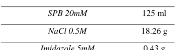
Crude extracts were supplemented with the appropriated volume of SPB stock solution (Table 2.1) containing salts (Table 2.2.), to the final concentrations: 20 mM SPB, 0.5 M NaCl and 5 mM imidazole.
The mixture was homogenized by stirring for 30 minutes and the pH was adjusted to 7.4. Afterwards, the samples were filtered using a 0.22 µm of cellulose acetate membrane syringe filter (sterile).
Table 2.2. Example of contents for salts addition. Final volume of 625ml (500ml of supernatant – 125ml stock salt solution (100mM SPB)
2.2.2. Dialysis
Alternatively to salt addition, crude extracts (prepared, as described in section 2.2) were dialyzed in cellulose dialysis tubing with a molecular weight cut-off =14.000Da (Sigma-Aldrich) against binding buffer 10 mM2. For efficient buffer exchange, dialysis was performed in a large stirred beaker overnight at 4°C with at least 2 buffer changes.
2.3. PURIFICATION METHODS OF THE HISTIDINE-TAGGED
RECOMBINANT COLAH
2.3.1. Immobilized-metal affinity chromatography (IMAC)
Buffers
Stripping Buffer3
Table 2.3. Stripping Buffer
2 Binding Buffer 10mM: 20mM sodium phosphate; 0.5M NaCl; 10mM imidazole; pH 7.4. 3 Stripping Buffer: 20 mM Sodium Phosphate, 0.5 M NaCl, 50 mM EDTA, pH 7.4
Final Volume (Crude extract) 625 ml
SPB 20mM 125 ml
NaCl 0.5M 18.26 g
Imidazole 5mM 0.43 g
Final volume 500 ml
21
The pH was adjusted to 7.4 and the final volume was brought to 500 mL with ultrapure water.
Elution Buffer I4
Table 2.4. Elution Buffer I
Final volume 500 ml
Sodium phosphate (100mM) 100 ml
NaCl 14.61 g
Imidazole 17.02 g
The pH was adjusted to 7.4 and the final volume was brought to 500 mL with ultrapure water.
Elution Buffer II5
Table 2.5. Elution Buffer II
The pH was adjusted to 7.4 using and the final volume was brought to 500 mL with ultrapure water.
4 Elution Buffer I: 20mM sodium phosphate; 0.5M NaCl; 500mM imidazole; pH 7.4 5 Elution Buffer II: 20mM sodium phosphate; 0.5M NaCl; 75mM imidazole; pH 7.4
EDTA (500mM) 50 ml NaCl 14.61 g Final volume 500 ml Sodium phosphate (100mM) 100 ml NaCl 14.61g Imidazole 2.553g
22
Binding Buffer1 Table 2.6. Binding Buffer
The pH was adjusted to 7.4 and the final volume was brought to 500 mL with ultrapure water.
Purification was carried out in a pre-equilibrated (Binding buffer2, 1ml/min) HisTrap FF 1ml column (GE Healthcare), in an AKTA FPLC system using the Unicorn 5.11 software. Crude extracts (20ml), pre-prepared in the appropriated salt conditions (prepared by dialysis or salt addition) were injected onto the resin. The target protein was eluted with 75mM imidazole using a step gradient of imidazole (75, 100 and 500 mM imidazole) at a flow rate of 1 ml/min. A blank run was established before every purification procedure. All the water and buffer solutions were degassed [with Helium(g)] and filtrated (0.22 µm pore) before applied to the system. Collected fractions were stored at -20°C, until further characterization.
A. Procedure of stripping and recharging the column of 1 ml.
To improve protein purification, the column was stripped and recharged between every purification procedure, according to the manufacturer’s instructions.
The prepacked column was washed with at least five column volumes with ultrapure water, buffer containing imidazole (10 mM imidazole), stripping buffer and at last with ultrapure water. In the second part of this step, the resin was recharged with NiSO4 0.1M,
washed with ultrapure water and the column was equilibrated with binding buffer1.
2.3.2. Batch/gravity-flow purification (BGP) A. Resin equilibrations and sample binding
Batch purification was carried out with pre-equilibrated (binding buffer1) Ni Sepharose 6 Fast Flow resin (GE Healthcare). Crude extract was loaded onto the resin with binding
Final Volume 500 ml
Sodium phosphate (100mM) 100 ml
NaCl 14.61g
23
buffer. The resin was washed with buffer containing imidazole (Binding Buffer 10mM imidazole). The target protein was eluted with elution buffer II5 with 75 mM imidazole.
B. Stripping and recharging with nickel sulfate
After each purification, the resins were submitted to stripping and recharging with NiSO4. First the resins were washed at least twice 5-10 column volumes with stripping
buffer2, then 5-10 column volumes of binding buffer1 and finally 5-10 column volume with ultrapure water (three - four times).
The resins were recharged with NiSO4 0.1M and washed at least three times with
ultrapure water.
2.4. DESALTING (HIPREP 26/10 DESALTING COLUMN)
Desalting procedure was performed according to the manufacturer’s instructions. Desalting was performed using a Sephadex G-25 Fine column (HiPrep 26/10 – GE Healthcare) coupled to an ÄKTA FPLC system. Before loading the sample, the system and column were washed with ultrapure water. The filtrated protein sample was loaded onto the column and the target protein was collected when the ABS (mAU) values started to increase. Sample collection was stopped when the conductivity reached 0.040 mS/cm. All the solutions (water and buffers) used in the HiPrep 26/10 desalting column were degassed [with Helium(g)] and filtrated (0.22 µm) before used onto the column.
2.5. LYOPHILIZATION
As final step, the samples were concentrated by lyophilization. This is a process where the water is removed from a product after it is frozen (-80°C) and placed under a vacuum, allowing the ice to change directly from solid to water vapor without passing through a liquid phase. The samples had approximately 15/20 ml each one. In this step a Snijders scientific type 2040 lyophilizer was used.
2.6. PROTEIN QUANTIFICATION 2.6.1.Colorimetric methods
A. Pierce 660nm Protein Assay
Pierce 660nm Protein Assay from Thermo Scientific was used to quantify the total protein concentration. This methodology is compatible with high concentrations of many
24
detergents, reducing agents and other commonly used reagents. Is a methodology based on the binding of a proprietary dye-metal complex to protein in acidic conditions. This causes a shift in the maximum absorption of the dye at 660nm, reproducing a colorometric response, dye-metal complex is reddish-brown and changes to green upon protein binding [43]. In this method bovine serum albumin (BSA) was used as standard.
A standard curve was prepared with BSA final concentration from 25 μg/ml to 2000 μg/ml and was measured with an absorbance at 660nm. The 25 µg/ml standard was prepared by mixing 10 μl of the 1000 μg/ml standard with 390 μg of 0.9% saline. 0.1ml of each replicate of standard, unknown sample and blank sample was added into an appropriately labeled test tube. Protein assay reagent (1.5ml) was added to each tube and vortexed to mix. The labeled test tubes were cover and incubated for 5 minutes at room temperature. Afterwards, total protein concentration was determined by measuring the absorbance at 660nm.
B. BCA Protein Assay
To determine the protein concentration of ColAh samples, the BCA Protein Assay Reagent Kit, from Thermo Scientific, was used. Bovine serum albumin (BSA) was used as a standard with a final concentration from 5 μg/ml to 250 μg/ml and was measured with an absorbance at 562nm, following the manufacturer’s instructions.
This is a high-precision and detergent-compatible assay reagent, based on bicinchoninic acid (BCA) for the colorimetric detection of the total protein concentration compared to a protein standard. It consists in the reduction of Cu2+ into Cu1+ by proteins in an alkaline medium creating a biuret reaction. The amount of Cu2+ reduced is proportional to the protein concentration present in the samples. This method has a high working range (5-250 µg/ml) [44].
Total protein concentration of each sample was determined using a standard curve. The standards were prepared as described in the following table 2.7.The protocol was based on the manufacturer instructions.
25
Table 2.7. Diluted Albumin (BSA) Standards: (2mg/ml) used in the BCA protein assay.
Volume (μl) BSA volume (μl) Final concentration of BSA (μg/ml) A 700 100 μl of stock 250 B 400 400 μl of A dilution 125 C 450 300 μl of B dilution 50 D 400 400 μl of C dilution 25 E 400 100 μl of D dilution 5 F 400 0 blank 2.6.2. Spectrophotometry assay
The NanoDrop spectrophotometer software (Thermo Scientific) uses the absorbance at 280nm in combination with the molar extinction coefficient to calculate the protein concentration. The system was calibrated using 2 µl of ultrapure water, and loading 2 µl of sample gently homogenized, avoiding bubbles when mixing and pipetting. The concentration of the ColAh was calculated using the Beer-Lambert Law:
The ColAh molar extinction coefficient () was estimated using ProtParam – ExPASy [45].
2.6.3. Densitometry analysis
Densitometry analysis on SDS-PAGE (10% of acrylamide gel) was also used to quantify protein. The gel analysis was performed after staining the gel with Comassie Brilliant Blue R-250 and scanning on a GS-800 Calibrated Densitometer (Bio-Rad). To estimate the quantity of ColAh, the software Quantity One 5.11 (Bio-Rad) was used, using
26
known concentrations of BSA (1µg, 2 µg, 3 µg, 4 µg, 5 µg, and 10 µg) as standard which were loaded to the same SDS-PAGE gel. For each gel were performed replicates.
2.7. EVALUATION OF PURITY AND ENZYMATIC ACTIVITY OF COLAH 2.7.1. SDS-PAGE – Polyacrylamide gel electrophoresis
This assay is adapted from the method described by Laemmli (1970). It is an electrophoretic separation of proteins in a polyacrylamide gel on the presence of SDS.
In this methodology, the presence of ColAh was characterized on a 10% polyacrylamide gel. The samples were denaturated at 100°C for 5 minutes. Mini-Protean 3 Cell (Bio-Rad), linked to an electrical potential difference generator [PowerPac 300 (Bio-Rad)] were used. The gels were electrophorized for 100-120 minutes at 120Volts. After the run, the gel was emerged into a stain solution, Coomassie Brilliant Blue R-250 for 1 hour. The staining solution contains 0.25% (w/v) of staining, 10% of acetic acid (v/v) and 50% of ethanol (v/v). After, the gel was incubated into a destaining solution with 25% of ethanol (v/v) and 5% of acetic acid (v/v), until the stains become visible. Afterwards, the gel was scanned on a Gel Image Analyzer GS-800 Calibrated Densitometer (Bio-Rad) using the software Quantity One (Bio-Rad). For each assay, were tested three replicates for each sample.
2.7.2. Zymography
Zymography is an electrophoretic assay, which implies a co-polymerization of a substrate (gelatin) to the 10% polyacrylamide gel, allowing detection of enzymes and of their activity. The samples were partially and reversibly desnaturated by SDS, but not reduced, and applied into a gel for separation. The run was performed at 4°C, 120 volts, for 120 minutes. When the electrophoresis ends, the gel was washed with a non-ionic detergent, Triton X-100 (0.25%, v/v). Afterwards, the gel was incubated with a reaction buffer6 for 2 hours, at 37ºC, allowing the digestion of substrate by ColAh. The result was visualized after emerged the gel into a staining solution, Coomassie Brilliant Blue R-250. The gel was incubated in a staining solution with 0.25% (w/v) of staining, 10% of acetic acid (v/v) and 50% of ethanol (v/v), for 1 hour. Destaining step was performed by soaking the gel in 25% of ethanol (v/v) and 5% of acetic acid (v/v). The gel was incubated until digested areas become visible. For each assay, were tested three replicates for each sample.
6 Reaction Buffer: Tris 1.5M; NaCl 1M; CaCL
27
2.8. ENZYMATIC DETERMINATION OF COLLAGENASE ACTIVITY 2.8.1. FALGPA assay
Collagenase activity was determined according to Van Wart and Steinbrink (1981). This assay is used to determine the activity of collagenases and is based on the hydrolysis of synthetic peptide: 2-furanacryloyl-leu-gly-pro-ala (falgpa).
For each assay, FALGPA reaction buffer7 and FALGPA were preincubated on a water bath at 37ºC, for approximately 20 minutes. Before each assay, a recommended test to the peptide was performed (negative control) [12, 22].
FALGPA reaction buffer was used as a blank to calibrate the spectrophotometer. The assays mixtures were prepared by adding the FALGPA peptide to the FALGPA reaction buffer. The reaction was started by adding the adjusted volume of the enzyme sample (table 2.8). The continuous spectrophotometric rate determination (A345, Light path = 1 cm) for each enzymatic assay was recorded for 10 minutes. Were performed three replicates for each sample.
Table 2.8. Contents for activity assay using FALGPA.
2.8.2. Collagen hydrolysis
Collagen type I was prepared according to Duarte et al., 2005, and used as complex substrate for evaluating ColAh collagenolytic activity. ColAh was purified through a prepacked HisTrap FF 1 ml column, as previously described (section 2.3). NanoDrop
7 FALGPA buffer: 50 mM Tricine buffer; 10 mM CaCl
28
spectrophotometer software (Thermo Scientific) was used to quantify the ColAh. Hydrolysis of collagen was carried out by incubating type I collagen (148 µg) with ColAh (2.5 µg) for 24h at temperature between 25-30ºC. After digestion, the samples were analyzed by SDS-PAGE polyacrylamide gel 6% of acrylamide, allowing to the detection of the hydrolysis of collagen by ColAh. The gel was running for 80 minutes at 100volts. Clostridial collagenase SERVA was used as positive control [46].
2.9. STORAGE STABILITY ASSAY
To evaluate the storage stability of ColAh, it was incubated at diferent temperatures: room temperature, 4°C and -20°C, over time. At day 1 and 18 of storage, SDS-PAGE gels (7.5% of acrylamide) was performed, loaded with the samples (three replicates each) stored at differents temperatures. The gels were running for 45minutes at 80Volts.
The assay was performed accordingly the conditions stablished by the Patent NO.: US
8,725,985 B2 – Clostridium histolyticum recombinant collagenase and method for the manufacture thereof [47].
2.10. COLAH APPLICATION ON CELL CULTURE PROCEDURE
C2C12 cell line derives from Mouse C3H muscle myoblasts. This cell line was used in
order to evaluate the capacity of the recombinant collagenase (ColAh) for detachment of adherent cells. The maintenance of the cell line was carried out according to the protocol recommended by American Type Culture Collection (ATCC). Cells were grown in culture flasks at 37°C in a humidified atmosphere with 5% CO2 and maintained in Dulbecco’s
Modified Eagle Medium (DMEM) supplemented with 3.7g/l NaHCO3, 1% of antibiotic
and 10% of Fetal Bovine Serum (FBS). Cell detachment was performed by using trypsin (control) or the ColAh in 0.1mg, 0.25mg, 0.5 mg and 1mg were added to each well. The culture plate was placed one minute at 37°C. To inhibit the action of trypsin and the ColAh, DMEM with 10% of FBS was added. Finally, cell culture morphology was evaluated by TC10 Automated Cell Counter, Bio-Rad Laboratories, Hercules,
31
Figure 3.1. Scheme of the process to obtain ColAh until purified
3. RESULTS AND DISCUSSION
In the present study, the main purpose was to evaluate the possible biotechnological potential of a recombinant protein – ColAh. For that, several assays were performed to evaluate different parameters, such as collagenolytic activity and purification yield.
3.1. ACTIVITY AND STABILITY OF COLAH
After ColAh expression and salt addition, different methodologies were implemented until reaching its pure (and still active) form. Immobilized metal affinity chromatography purification was carried out through batch procedure with an equilibrated Ni Sepharose 6 Fast Flow resin (nominated batch gravidity protocol - BGP) (figure 3.1).
One of the steps to evaluate and characterize the purified recombinant collagenase – ColAh, is to verify its activity. In this case, after purification by batch, ColAh was tested to assess its collagenolytic activity against a synthetic peptide.
The assay requires the use of a spectrophotometer, able to automatically make readings over time, and with thermostated cell. To measure collagenolytic activity, the synthetic peptide 2-furanacryloyl-Leu-Gly-Pro-Ala (FALGPA) [22], that mimics the primary structure of the collagen molecule was selected and its hydrolysis was monitored over time., The Serva Collagenase NB 1 from Clostridium histolyticum was used as control [48]. The concentration of FALGPA degradation was determined spectrophotometrically and calculated using the Beer- Lambert Law.
32 T i m e A B S ( 3 4 5 n m ) 0 2 0 0 4 0 0 6 0 0 0 .1 2 0 .1 4 0 .1 6 0 .1 8 a ) b ) T i m e A B S ( 3 4 5 n m ) 0 2 0 0 4 0 0 6 0 0 0 .1 2 0 .1 4 0 .1 6 0 .1 8
Figure 3.2. Collagenolytic activity determination using the synthetic peptide FALGPA. a) Pure
recombinant protein – ColAh - with salts addition and purification through batch. b) Commercial collagenase from Serva as control.
Synthetic peptide FALGPA mimics the primary structure of the collagen molecule. It is considered a good substrate for collagenases [22], which hydrolyzes FALGPA results in a decrease in spectrophotometric absorvance at 324 nm [49].
Figure 3.2 shows that ColAh is able to degrade the synthetic peptide, demonstrating that this recombinant protein retains collagenolytic activity. However, the collagenolityc activity of ColAh (1.16×10-04 mM/sec) is lower than the control (3.39×10-04 mM/sec), indicating that peptide degradation is lower than expected.
33
3.2. CELL CULTURE
In this assay, the detachment abilities of ColAh were assayed using C2C12 cell line from Mouse C3H muscle myoblast. Trypsin was used as control.
Figure 3.3. C2C12 cells detachment by ColAh and trypsin.
Trypsinization is a frequently used for the dissociation of adherent cells, in order to allow their harvest and use for a wide range of analysis [25]. Trypsin can be ineffective in when tissues are too fibrous or may cause damage in sensitive cells. The use of commercial collagenases for the cell detachment influences yield, viability and cells function. Viability and function of isolated cells are essential for several applications, as in diabetes therapy, pharmacological tests, scientific research and others [48].
The purpose was to evaluate the cell release performance of ColAh from the culture plate and to compare its effects with trypsin’s performance.
Figure 3.3 shows that, contrasting to trypsin, the recombinant protein was unable to completely dissociate C2C12 cells, even using higher concentrations of pure ColAh. Nevertheless, the results revealed slightly cell detachment by using 1mg of ColAh. This observation suggests that the ColAh added to the cells might not be at the required concentration to obtain a full detachment of the cells.
One possible explanation is that the concentration used may not be sufficient to obtain the expected result. However, if higher doses of ColAh were used (higher than the commercial collagenases concentration required) the process would not be rentable. This may be due to the fact that the protein quantification (made using the BCA Protein Assay Reagent Kit) [44] was not the apropriate, estimating a larger protein concentration than the real purified amount of ColAh.
Therefore, other methods for protein quantification were tested, such as the Pierce 660 nm Protein Assay (Thermo Scientific) [43] and absorbance measurement at 280nm (using
34
a NanoDrop) [50]. However, again the protein quantification obtained from these methods was not in accordance with the corresponding analysis by SDS-PAGE (figure 3.4). The SDS-PAGE gel allows separating proteins according to their molecular weight [51], as well confirming the quantity of protein electrophoresed on the gel.
The protein quantification methods used differ in several parameters. BCA Protein Assay is a method based in the reduction of Cu2+ to Cu1+ by proteins in an alkaline medium, the called biuret reaction. It is a highly sensitive and selective colorimetric detection of Cu1+ by bicinchoninic acid (BCA). The amount of Cu2+ reduced is proportional to the sample protein concentration. This method is highly sensitive to the presence of reducing agents [52].
The Pierce 660 nm Protein Assay is also based in a colorimetric protein quantification. Bovine serum albumin was used as standard. This method is compatible with most detergents even in high concentration, reducing agents and other commonly reagents. However, it is known that certain substances interfere with the method [43]. This is the case of imidazole: the maximum compatible concentration is 200mM and EDTA (maximum compatibility concentration is 20mM) [43].
The quantification by absorbance measurement (in a NanoDrop) allows to quantify purified proteins that contains Trp, Tyr residues or Cys-Cys disulphide bonds [50, 53]. All the methods performed did not corroborate with the quantities added to the SDS-PAGE gels. This is well illustrated in figure 3.4, where the same protein quantity, determined by absorvance measurement (NanoDrop) is not confirmed by SDS-PAGE analysis.
Figure 3.4. SDS-PAGE gel with two samples of purified ColAh from two different expressions. 1) Colah
10µg - expression 19-05-15; 2) Colah 10µg - expression 22-04-15
In figure 3.4, the SDS-PAGE gel shows the presence of ColAh, to relate the SDS-PAGE with protein quantification by NanoDrop. After protein quantification, 10 µg of ColAh
2 1 MW
35
were load. As it is possible to observe, the amount loaded onto the gel did not match the quantification result.
The results obtained until this stage of the work suggest that the ColAh maybe co-purified with an interferent. From this point of view, the interferent might have also been quantified, giving erroneous values of ColAh concentration. This explains the divergent result obtained by SDS-PAGE analysis.
Until here, the protein purification was carried by batch gravity purification (BGP). So, it is possible that the purification of ColAh by this method does not yield a pure enzyme at the end of the process. With that arises another hypothesis for the unexpected results, demonstrated in the first tasks. The method used for ColAh purification is not effective. In order to clarify this hypothesis collagenolytic activity of crude extract was assayed.
3.3. ACTIVITY ASSAYS OF CRUDE EXTRACT
In the previous tasks, it was demonstrated that the recombinant protein, in its pure form, did not exhibit the expected activity (figure 3.2), suggesting that during some phase of the protocol, expression or purification, the protein is losing activity. For that reason, a hypothesis that the purification process may not be carried as predictable was emerged. In this task, gelatinolytic and collagenolytic activity of crude extract was tested.
Firstly, aiming to assess the deviation from the initial planning, a zymography assay was performed. This technique allows determining the apparent molecular weight and the gelatinolytic activity of a protein. Two samples of centrifuged crude extracts from two independent expression assays were loaded into a gelatin zymogram gel. In this way, it is possible to verify if the recombinant protein expression is being successful, and if not, this test allows the identification of the problem that was observed on the first presented results.
Zymography is an electrophoretic assay with an appropriate substrate, in this case gelatin [13].
36
Figure 3.5. Zymography assay. 1- Centrifuged crude extract – Expression of day 17.04.15
2- Centrifuge crude extract – Expression of day 20.04.15
Figure 3.5 shows the two samples of crude extract from different expression and purification procedures. This result confirms that crude extract has gelatinolytic activity, shown in the zymogram as clear bands (substrate degradation) against dark background [13]. Thus, the expression step was being successful. This result, may suggest that the protein may be losing its activity in some part of the purification process. Collagenolytic activity of the crude extract was further assessed. As described above, the activity was measured using the synthetic peptide FALGPA; as control the commercial collagenase from Serva was used. This assay was performed using a spectrophotometer with readings over time, with a thermostated cell.
T i m e ( s e c ) A B S ( 3 4 5 n m ) 0 2 0 0 4 0 0 6 0 0 0 .4 4 1 0 .4 5 1 0 .4 6 1 0 .4 7 1 a ) T i m e ( s e c ) A B S ( 3 4 5 n m ) 0 2 0 0 4 0 0 6 0 0 0 .1 5 7 0 .1 6 7 0 .1 7 7 0 .1 8 7 b )
Figure 3.6. Collagenolytic activity determination using the synthetic peptide: FALGPA. a) Centrifuged
crude extract of ColAh; b) Commercial collagenase Serva as control. 100 75 50 250 150 37 25 20 10 15 MW 1 2 kDa