Universidade de Aveiro 2006
Secção Autónoma de Ciências da Saúde
António José
Monteiro Amaro
Factores Biomecânicos e estrutura do gluteus
medius
na artrose da anca: Indicadores clínicos e
radiográficos
Biomechanical factors and gluteus medius structure
in hip osteoarthritis: clinical and radiographic
Universidade de Aveiro 2006
Secção Autónoma de Ciências da Saúde
António José
Monteiro Amaro
Factores Biomecânicos e estrutura do gluteus
medius
na artrose da anca: Indicadores clínicos e
radiográficos
Dissertação apresentada à Universidade de Aveiro para cumprimento dos requisitos necessários à obtenção do grau de Doutor em Ciências Biomédicas, realizada sob a orientação científica do Professor José Alberto Duarte,
Professor Catedrático da Faculdade de Desporto da Universidade do Porto e do Professor Francisco Amado, Professor Auxiliar do Departamento de Química da Universidade de Aveiro.
This study was supported by a grant for health research (“Projectos I&DT em Ciências e Tecnologias da Saúde – 2002”) conceded by the University of Aveiro.
Agradecimentos Este trabalho só foi possível com o apoio e colaboração de numerosas pessoas a quem quero agradecer e das quais destaco: O Professor José Alberto Duarte, que aceitou orientar esta tese e cujo suporte e entusiasmo em todas as etapas da mesma foram fundamentais para a sua conclusão. O Professor Francisco Amado, pela sua colaboração, incentivo e total
disponibilidade ao longo do desenvolvimento do estudo. A Professora Helena Nazaré, Reitora da Universidade de Aveiro e o Professor Nelson Rocha, Pró-Reitor para a Saúde, os grandes “responsáveis” por todo o processo que levou à minha decisão de elaborar este trabalho; o seu apoio e estímulo constantes foram essenciais; desde o nosso primeiro encontro há cerca de 6 anos, que fiquei convicto de que a Universidade de Aveiro não só tinha um projecto ambicioso para as Ciências da Saúde, mas acima de tudo tinha as pessoas certas para o liderar. O Prof. Hans Joachim Appell pelo seu incentivo e colaboração nas várias etapas deste trabalho. Os Médicos dos Serviços de Ortopedia e de Medicina Física e de Reabilitação do Hospital Infante D. Pedro, com particular destaque para os seus directores Dr. António Meireles e Drª Acilda Mendes, pela sua colaboração e disponibilidade para garantir todos os meios necessários à execução dos estudos. A Mestre Sandra Calado e todos os Técnicos bem como o Director do Serviço de Anatomia Patológica do Hospital Infante D. Pedro, pela sua colaboração no tratamento das amostras de biópsia muscular. A Drª Alexandra Ferreira, o Mestre Luís Sancho, a Professora Joana Silva, a Terapeuta Gabriela Oliveira, o Dr. José Oliveira e o Dr. Carlos Neri pela sua generosa colaboração. De forma particular a Família, que é o meu reduto, a minha verdadeira “circunstância” sem a qual o “Eu” e todo o seu trabalho não teria qualquer sentido.
o júri
presidente Prof. Dr. Maria Helena Vaz de Carvalho Nazaré
Reitora da Universidade de Aveiro
Prof. Dr. José Alberto Ramos Duarte
Professor Catedrático da Faculdade de Desporto, Universidade do Porto (orientador)
Prof. Dr. António Martins da Silva
Professor Catedrático Convidado do Instituto de Ciências Biomédicas Abel Salazar, Universidade do Porto
Prof. Dr. Hans-Joachim Appell
Professor Catedrático do Departamento de Fisiologia e Anatomia da Universidade Alemã de Desporto, Colónia
Prof. Dr. João Espregueira Mendes
Prof. Associado Convidado da Escola de Saúde da Universidade do Minho
Prof. Dr. José António Pereira da Silva
Professor Auxiliar da Faculdade de Medicina da Universidade de Coimbra
Prof. Dr. Francisco Manuel Lemos Amado
Professor Auxiliar, Departamento de Química da Universidade de Aveiro (co-orientador)
palavras-chave Osteoartrose, glúteo médio, atrofia muscular, amplitude de movimento,
biomecânica da anca, função abdutora, Índice de Lequesne, Índice de Kellgren, osteófitos, entrelinha articular, prótese total da anca
resumo O presente trabalho teve como objectivo o estudo da relação entre a função,
os factores biomecânicos e o grau das alterações radiográficas em doentes com diferentes graus de osteoartrose (OA) da anca. Foi efectuado protocolo de avaliação clínica e radiográfica a ambas as ancas de 65 doentes a aguardar cirurgia para colocação de prótese total da anca (PTA). Durante a cirurgia foram efectuadas, em todos os doentes, biópsias do músculo glúteo médio (GM) ipsilateral. A reavaliação 6 meses após cirurgia foi efectuada em 18 destes doentes. Utilizando a soma das medidas da entrelinha articular (EA) efectuadas em ambas as ancas e em 3 pontos da região de suporte de carga (lateral, superior e axial), obteve-se uma correlação mais forte com a função das ancas medida pelo índice de Lequesne (r=0.67, p<0.05), relativamente a qualquer valor individual incluindo a EA mínima; esta correlação foi também mais marcada com todas as amplitudes articulares com destaque para a abdução (r=0.60, p<0.05) e a rotação externa (r=0.57, p<0.05). O comprimento dos osteófitos acetabulares superiores (OAS) teve uma correlação positiva significativa com o score de dor do índice de Lequesne (r=0.38, p<0.05) e negativa com a abdução máxima (r=-0.50, p<0.05); a abdução teve a correlação mais forte com o ângulo de abdução livre radiográfico (r=0.60, p<0.05), situado entre o colo do fémur e a extremidade dos OAS e com vértice no centro da cabeça do fémur. Após mais de 6 meses da cirurgia, a anca operada apresentou correlação significativa dos parâmetros radiográficos (braço de alavanca dos músculos abdutores e índice disfuncional) com o tempo de permanência na abdução activa máxima (respectivamente r=0.61 e r=0.63; p<0.05). A soma das 3 medidas da EA teve correlação significativa com a área média das fibras do GM, sendo esta mais marcada com a EA da anca oposta (r=0.49, p<0.05) do que da anca a aguardar cirurgia (r=0.32, p<0.05). Este trabalho sugere que, nos doentes submetidos a PTA, o braço de alavanca abdutor é determinante nos resultados funcionais pós-cirurgia. A soma dos valores da EA em 3 pontos estandardizados da área de suporte de carga tem a maior correlação com o grau de evolução da OA da anca. Os OAS limitam a amplitude da abdução da anca. A atrofia do GM correlaciona-se com a medida da EA de ambas as ancas. O défice de força dos abdutores pode condicionar o desgaste articular da anca contralateral. Estes resultados apontam para a importância do reforço dos músculos abdutores e da adopção de estratégias que diminuam a sobrecarga mecânica articular de forma a prevenir e limitar a evolução da OA da anca.
keywords Osteoarthritis, gluteus medius, muscle atrophy, range of motion, hip
biomechanics, abductor function, Lequesne Index, Kellgren Index, osteophytes, joint space width, total hip replacement
abstract This study investigated the possible relationship between function,
biomechanical factors and radiographic degenerative changes in patients with several degrees of hip osteoarthritis (OA). A clinical and radiographic
evaluation protocol was performed on both hips of 65 patients awaiting surgery for total hip replacement (THR). During surgery a biopsy of the ipsilateral gluteus medius (GM) muscle was done in all patients. Post-surgery follow up was performed in 18 patients. The sum of radiographic joint space width (JSW) made in three points of the load bearing region (lateral, superior and axial) in both hips, evidenced a stronger correlation with hip function measured by the Lequesne Index (r=0.67, p<0.05), when compared to any individual value, including minimum JSW. This correlation was also stronger with all joint ranges, particularly abduction (r=0.60, p<0.05) and external rotation (r=0.57, p<0.05). Cranial acetabular osteophytes (CAO) length correlated positively and significantly with pain score of Lequesne Index (r=0.38, p<0.05) and negatively with maximal abduction (r=-0.50, p<0.05); abduction had the strongest
correlation with the radiographic abduction free angle (r=0.60, p<0.05), located between the femoral neck and CAO lateral extremity, and with the apex at the centre of the femoral head. Follow up conducted over 6 months showed a significant correlation between post-surgery radiographic parameters (abductor muscle lever arm and hip dysfunction index) and time of maximal active abduction (r=0.61, r=0.63 and p<0.05, respectively). The sum of the three measures of hip JSW was correlated significantly with the mean GM fibre area. This correlation was stronger with the JSW of contralateral hip (r=0.49, p<0.05) than in hip awaiting surgery (r=0.32, p<0.05). This study suggests that in THR the abductor lever arm length is critical to post-surgery functional results. The sum of three standard measurements of JSW in hip weight-bearing area has the strongest correlation with the clinical development of hip OA; the hip abduction range of motion is limited by CAO; the GM atrophy correlates with JSW of both hips. The abductor weakness leads to the increase of mechanical impact during load shift, which may determine the wearing-off of the
contralateral hip. Therefore, the present results point to the importance of reinforcing the abductor muscles, together with strategies to decrease the joint mechanical overload, in order to prevent and limit the evolution of hip OA.
T
ABLE OF
C
ONTENTS
GENERAL INTRODUCTION 1
Objectives 7
EXPERIMENTAL WORK 9
I.
Characteristics of the gluteus medius muscle in an asymptomatic patient with radiographic signs of coxarthrosis
II.
Joint space width and functional impairments in osteoarthritic hips
III.
Cranial acetabular osteophytes limit the maximal amplitude of hip abduction
IV.
Radiographic geometric measures of the hip joint and abductor muscle function in patients after total hip replacement
V.
Does gluteus medius muscle atrophy constitute a primary risk factor to the development of contralateral hip joint
osteoarthritis? 11 23 35 49 67 OVERALL DISCUSSION 85 CONCLUSIONS 95 REFERENCES 97 APPENDICES
Clinical evaluation protocols Radiographic evaluation protocol
113 115 127
L
IST OF
A
BBREVIATONS
ACR American College of Rheumatology
AP Anteroposterior
BMI Body mass index
CA Coxarthrosis
CAO Cranial acetabular osteophytes
DI Dysfunction Index
FA Free Angle
GM Gluteus Medius
IPH Infante D. Pedro Hospital JSW Joint Space Width
MHC Myosin heavy chains MJS Minimal joint space
OA Osteoarthritis
ROM Range of motion
ROPM Range of passive motion
SPSS Statistical Package for the Social Sciences
G
ENERAL
I
NTRODUCTION
Osteoarthritis (OA) is the most common joint pathology and has a multifactorial, complex and still relatively unknown etiology (Dieppe, 1984, 1991, 1997; Hart and Spector, 1995a; Altman, 1997; Altman et al., 2000; Felson et al., 2000; Doherty and Dougados, 2001; Hunter and Felson, 2006). In particular the hip and knee OA develop with functional limitations and reduced quality of life associated to their clinical signs and symptoms, such as pain, stiffness and periarticular muscle strength deficit (Cooper et al., 1996; Oberg and Oberg, 1996; Hopman-Rock et al., 1997; Felson and Zhang, 1998; Doherty and Dougados, 2001; Arokoski et al., 2004).
Hip OA (also named coxarthrosis) has a great social, economic and functional impact and is considered a major public health problem in developed countries (Klippell and Dieppe, 1997; Murray and Lopez, 1997; The Consensus Document, 1998; Yelin, 1998; Felson and Zhang, 1998; Felson et al., 2000); its prevalence increases with age (Lawrence et al., 1966; Danielsson et al., 1984; Hamerman, 1993) reaching 3% to 6% in adult caucasian population (Heliövaara et al., 1993; Hoaglund et al., 1995; Felson and Zhang, 1998; Hoaglund and Steinbach, 2001). Unlike the knee OA, the hip OA has been relatively forgotten in basic biomechanical investigation and in its potential clinical implications (Sun et al., 1997; Robertson et al., 2003). However, both personal (pain and disability) and socioeconomical (job absences, related surgeries and hospitalisation days) hip OA repercussions (Carr, 1999; Brauer et al., 2005; Gupta et al., 2005) seem to constitute important reasons to improve the knowledge related to this pathology.
The study of the radiographic and functional parameters and their correlations to hip OA clinical research may lead to a better diagnosis and treatment and to a better understanding of the factors underlying this
pathology. Joint space narrowing, subchondral sclerosis, osteophyte and subchondral cyst formation are the main radiological OA features used to assess OA severity in global classification indexes, being Kellgren & Lawrence’s Index (Kellgren and Lawrence, 1957) the better known. According to the American College of Rheumatology (ACR), the clinical criteria to define OA consists of hip pain in most days of the prior month and degenerative joint changes evident in radiographs (Altman et al., 1991); the format of the ACR classification also includes clinical criteria alone, with hip pain and range of motion (ROM) limitations in internal rotation and flexion (Altman et al., 1991). In clinical practice, the diagnosis and grading of severity of hip OA is based mainly on the narrowing of the joint space width (JSW) or global radiographic indexes such as the Kellgren’s. However, in hips without radiographic changes and with OA symptoms, a significant percentage of degenerative arthroscopic alterations can be found (Santori and Villar, 1999) suggesting that the radiographic features have a low sensitivity to detect joint damage before it becomes extensive (Altman, 1995; Dieppe, 1995).
The functional evaluation of the osteoarthritic hip is frequently assessed using global indexes aiming to quantify a set of items related to pain, stiffness or functional impairments, being the Lequesne Index (Lequesne et al., 1987) and WOMAC (Bellamy et al., 1988; McConnel et al., 2001) the most frequently used; in addition, ROM and functional evaluation of periarticular muscles must be included in hip examination (Klippell and Dieppe, 1997). Although the ROM can be considered an indicator of joint stiffness, a commonly standard for major ROM deficits of the OA hip joint is actually absent; this situation may result from the great variability in normal hip ROM among healthy subjects (Allander et al., 1974; Boone and Azen, 1979; Roaas and Andersson, 1982; Svenningsen et al., 1984; Roach et al., 1991; Greene and Heckman, 1994; Escalante et al., 1999) and also from the different techniques used to assess joint ROM (American Academy of Orthopaedic Surgeons, 1966; Stratford et al., 1984; Gajdosik and Bohannon, 1987; Greene and Heckman, 1994). On the other hand, the methodology commonly used to assess functional repercussions of hip OA is static and poorly related to the complex dynamics of human activities
(Arokoski et al., 2004). These multiple variables and methods to measure physical function and disability of hip OA patients are a great problem in clinical research, needing a standardized methodology. Moreover, the hip disability must be considered according to the International Classification of Functioning, Disability and Health (ICF) as impairments of articular function and structure, which limit performance activities and restrict daily life situations (World Health Organisation, 2001).
The hip is a ball-and-socket joint and forms a functional unit consisting of bones, cartilage, synovium and capsule with blood, lymphatic and nerve supply and is involved by burse, ligaments and muscles (Vilensky, 1998; Dewire and Einhorn, 2001); the OA, often known as a degenerative “wear and tear” disease of articular cartilage (Dieppe, 1984, 1999), seems to affect all these tissues (Vilensky, 1998; Felson et al., 2000; Hurley, 2002; Hunter and Felson, 2006). It has been assumed that hip OA would result from an imbalance between the mechanical efforts, to which the hip is subjected, and osteo-cartilagineous tissue resilience and repair capacity. (Tanaka et al., 1998; Sandell and Aigner, 2001). Thus, both systemic and local biomechanical factors interfere with OA’s pathophysiology (Felson et al., 2000; Hunter and Felson, 2006). Among local biomechanical factors, the strength deficits associated to disuse atrophy caused by pain (O’Reilly et al., 1997; O’Reilly et al., 1998; Hurley, 1999; Felson et al., 2000) and other periarticular muscle alterations (Sirca and Susec-Michieli, 1980; Sims, 1999; Howell et al., 2001; Sims et al., 2002) have been referred; even so, longitudinal studies in knee OA suggest that quadriceps strength deficits might not only result from pain, but may actually precede radiographic OA signs, thus representing a risk factor for joint degeneration, presumably due to a reduced stability and decreased mechanical shock absorption ability (Slemenda et al., 1997, 1998; Hurley, 1999).
In the knee joint, quadriceps weakness is a better determinant of pain and disability than any radiographic changes (Lankhorst et al., 1985; Hurley and Newham, 1993; Madsen et al., 1995). In the hip joint, few studies have assessed the relationship between muscular function and OA (Jandric, 1998; Arokoski et al., 2002), although strength deficit of periarticular muscles,
particularly in abductors, has been found in patients with this type of OA (Murray and Sepic, 1968; Jandric, 1998; Arokoski et al., 2002). The functionality of the hip abductor muscles is determinant in the global function of this joint, namely during walking, because of their pelvic stabilizing role (Inman, 1947; Pauwels, 1980; Kapandji, 1996; Kumagai et al., 1997; Fetto et al., 2002). In this way, the importance of the gluteus medius (GM) with their ability to generate abductor momentum proportional to its length and lever arm (Pauwels, 1980; Kapandji, 1996) must be considered. So, as with the quadriceps muscle for the knee OA, one might postulate that the main hip abductor muscle (i.e. gluteus medius) dysfunction, might affect the OA onset and development (Hurley, 1999; Sims, 1999; Sims et al., 2002).
For instance, in OA patients subjected to total hip replacement (THR), a deficit in the abductor muscles might be related to an intrinsic strength loss of the periarticular muscle, or an indirect result of biomechanical parameter change due to surgical intervention (McGrory et al., 1995; Downing et al., 2001; Asayama et al., 2002), particularly the shortening of the lever arm of the abductor muscles leading to higher tension development to support the body weight during unipodal stance (Johnston et al., 1979; Delp et al., 1996). In fact, despite the general consensus of the benefits for patients subjected to arthroplasty, particularly in pain control and improvement of joint mobility (Laupacis et al., 1993; NIH Consensus Conference, 1995; Altman et al., 2000; Zhang et al., 2005; Hunter and Felson, 2006), this intervention does not always result in muscular strength recovery or normal daily activities fulfilment (Gore et al., 1977; Borja et al., 1985; Skinner, 1993; Nilsdotter et al., 2003). The pending question is whether hip abductor function deficits found in THR patients are more closely related to abductor muscle intrinsic changes, or rather more intimately associated with surgery induced biomechanical parameter alteration, which can be measured through radiographic means.
Macroscopic (Howell et al., 2001) and microscopic (Sirca and Susec-Michieli, 1980) changes of the GM were already described in a significant percentage of THR-subjected patients. Despite the obvious association, no cause-effect
relationship has been so far established between hip joint degenerative alterations and periarticular muscle dysfunction; moreover, the relationship between these macro and microscopic findings in the GM of THR submitted patients and post-surgery functional results is far to be comprehended. Nevertheless, based on empirical evidences, post-THR hip muscle strengthening training has been suggested by several authors (Minns et al., 1993; Ueda and Tohkura, 1993; Vaz et al., 1993; Horstmann et al., 1994; Shih et al., 1994; Giurea et al., 1998).
Additionally, the hip biomechanical special characteristics make the functional evaluation difficult, namely the aforementioned role of abductor muscles in pelvic stability during walking; in this case, abductor muscle function of one side is essential for a harmonious load reception and transfer, thus avoiding mechanical shock in both hips; therefore, by studying an abductor muscle like GM, one must hypothesise that its dysfunction may have repercussions both on the ipsilateral and on the contralateral hip. Theoretically, hip OA disability is mainly explained by the degree of joint degeneration (Bagge et al., 1991; Dekker et al., 1992; Dieppe, 1997), despite hip radiological features and disability not always being well correlated (Lawrence et al., 1966, 1989; Spector and Hochberg, 1994). As an hypothesis, it may be suggested that degenerative changes which modify joint structure, shape, and position with adverse joint biomechanical repercussions are the main responsibles for hip disability, specifically the presence and dimensions of osteophytes, and the intensity and pattern of joint space narrowing; this hypothesis will be supported by the existence of a parallel correlation of these radiographic features with ROM and with hip disability (Dekker et al., 1992; Escalante et al., 1999; Steuljens et al., 2000; Arokoski et al., 2004).
JSW was suggested to be the most important individual characteristic in radiological evaluation of hip OA and on monitoring its progression (Dougados et al., 1996, 1997; Goker et al., 2000; Bierma et al., 2002); the JSW was even considered a better indicator of current clinical situation than universal global scales (Croft et al., 1990), while osteophytes do not seem to be related to the radiological or symptomatic progression of hip OA
(Danielsson, 1966; Dougados et al., 1996). Joint space is also the best relating radiographic measurement to hip pain intensity (Croft et al., 1990; Scott et al., 1992); unlike the hip, however, in knee OA the pain shows a better correlation to osteophytes than to JSW (Spector et al., 1993).
The radiographic cartilage loss pattern in hip OA can be classified in three sub-types (superolateral, concentric and medial), the superolateral being the most frequent (Solomon, 1976; Ledingham et al., 1993); these patterns are associated with femoral head migration in different directions, with subsequent alteration of joint geometry, remaining to be characterised the biomechanical and functional effects of this migration.
Although the JSW narrowing may constitute an important sign of hip OA, there is no consensus concerning the minimum value to radiographically define hip OA since the proposed values vary between 1,5 and 4,0mm (Croft et al., 1994; Spector and Hochberg, 1994). Moreover, the minimum JSW measurement is referred only to a single point of the joint, disregarding location or how much cartilage is left; this difficulty may be overcome using the sum of measurements in three standard points (lateral, superior and axial), located in the weight-bearing region of the joint (Solomon, 1976; Hodge et al., 1986; Jacobsen et al., 2004a; Jacobsen et al., 2004c). This method of evaluating remaining cartilage may be useful in finding joint space narrowing in people subjected to high joint impact, namely certain professionals and athletes to whom counselling is necessary, as well as the timely enforcement of preventive measures for joint sparing. Thus, supposing that the sum of the above mentioned measurements exhibits a better correlation with hip function than the existing one with isolated values, like minimal JSW, the utilization of this methodology may be useful not only for the follow up of hip OA patients, but also in the definition of new radiographic criteria for hip OA diagnosis.
On the other hand, OA Kellgren’s Index has been criticised for emphasizing osteophytes, as they are considered part of the normal ageing process (Danielsson, 1966) or because, despite being one of the most characteristic aspects of the OA joint (Altman et al., 1986), their role, meaning and clinical impact are not fully understood (Sandell and Aigner, 2001; Neuman et al.,
2003). Osteophytes were proposed to be an endogenous repair attempt of the OA joint, i.e., a physiological response to the instability or mechanical overload (Gelse et al., 2003; Neuman et al., 2003); nevertheless, both humoral and mechanical factors seem to be involved in their origin, stimulating new cartilage and bone development in the sinovial-periosteon joint (Aigner et al., 1995; Mow et al., 1995; Matyas et al., 1997; Felson and Zhang, 1998; Felson et al., 2000; Uchino et al., 2000; Sandell and Aigner, 2001; Gelse et al., 2003; Blom et al., 2004).
Although the joint ROM deficit is normally used as a diagnosis criterion in hip OA (Cyriax, 1957; Lequesne, 1980; Altman et al., 1991; Bierma et al., 1999), there are few studies correlating this parameter with other radiographic characteristics of hip OA, such as JSW or osteophytes dimension and location. According to the hip joint shape, maximal abduction must be limited by bone impact of femoral neck at the upper acetabular edge, although this normally does not occur because of the adductor muscles and iliofemoral and pubofemoral ligaments (Kapandji, 1996); so, it may be argued that the presence of cranial acetabular osteophytes (CAO) would constitute a particular limitation of abduction, since these osteophytes, by prolonging the acetabular edge, might limit this hip movement before the mentioned muscles and ligaments reach their maximal length. On the other hand, if there is a correlation of CAO with abduction, this is probably dependent on the pelvic shape, namely the free-angle (FA), whose apex is located in the centre of the femoral head, and is limited by a line drawn from the lateral extremity of the CAO to the femoral neck point where it hits the CAO during maximum abduction.
Objectives
Considering the above referred, the main objective of this work was to improve the pathophysiology knowledge of hip OA through the characterization and correlation of radiographic, biomechanical, functional, and structural parameters, in order to reach a better clinical diagnosis and treatment management.
The specific objectives were:
1. To study the relationship between pain, ROM, functional deficit and the radiographic hip OA features (narrowing of JSW and osteophytes).
2. To establish the main biomechanical factors of post-THR surgery abductor function.
3. To assess the relationship of abductor muscle structure (gluteus medius) with hip OA radiographic findings, age and physical activity.
This work was supported by the following original studies:
1. Amaro A, Sousa L, Sancho L, Meireles J, Calado S, Vitorino R, Amado F, Appell HJ, Duarte JA (2004). Characteristics of the gluteus medius muscle in an asymptomatic patient with radiographic signs of coxarthrosis. Eur J Orthop Surg Trauma 14(3): 182-185.
2. Amaro AJ, Amado FM, Appell HJ, Duarte JA (2006). Joint space width and functional impairments in osteoarthritic hips. Clin Orthop Relat Res (submitted).
3. Amaro AJ, Amado FM, Vitorino R, Appell HJ, Duarte JA (2006). Cranial acetabular osteophytes limits the maximal amplitude of hip abduction. Eur J Orthop Surg Trauma (in press).
4. Amaro AJ, Amado FM, Mendes A, Oliveira J, Malheiro A, Meireles A, Appell HJ, Duarte JA (2006). Radiographic geometric measures of the hip joint and abductor muscle function in patients after total hip replacement. Eur J Orthop Surg Trauma (in press).
5. AmaroAJ, AmadoFM, AppellHJ, DuarteJA (2006). Does gluteus medius muscle atrophy constitute a primary risk factor to the development of contralateral hip joint osteoarthritis? Clin Anat (submitted).
Experimental Work Paper I
C
HARACTERISTICS
OF
THE
GLUTEUS
MEDIUS
MUSCLE IN AN ASYMPTOMATIC PATIENT WITH
RADIOGRAPHIC SIGNS OF COXARTHROSIS
A. Amaro1, L. Sousa2, L. Sancho1, J. Meireles2, S. Calado1, R. Vitorino3, F. Amado3, H.J. Appell4,6, J.A. Duarte1,5,6
1Health School, University of Aveiro, Portugal
2Department of Orthopaedics, Hospital Infante D. Pedro, Aveiro, Portugal 3Chemistry Department, University of Aveiro, Portugal
4Department of Physiology and Anatomy, DSHS Cologne, Germany 5CIAFEL, FCDEF, University of Porto, Portugal
6Muscle Atrophy Research Group (MARG)
Abstract
This case study describes the micromorphology and some biochemical features of gluteus medius muscle in a 79-year-old woman with radiographic signs of coxarthrosis but with no clinical symptoms, who was initially admitted in the orthopaedic emergency service with a non-displaced subcapital fracture of the femoral neck due to a domestic accident (fall). The X-ray of the hip showed some characteristic features of coxarthrosis, classified grade 2 of the Kellgren criteria. After informed consent, it was decided to carry out the functional evaluation according to the indexes of Lequesne and WOMAC (Western Ontario and McMaster Universities Osteoarthritis Index) and to take a biopsy of the gluteus medius muscle for microscopical examination and myosin heavy chain isoform identification during hip replacement surgery. For the Lequesne Index (score 0–24), the total score was 0, and for the WOMAC (score 0–96), the total score was also 0, both speaking in favour of full joint and muscle function. All the structural features observed in muscle were considered not to have any pathological relevance. The composition of the myosin heavy chains in the gluteus medius muscle was 48% MHC I, 41% MHC IIa, and 11% MHC IIx. The muscle characteristics do not support earlier concepts about muscle weakness as a predisposing factor for osteoarthritis. It is moreover concluded that the diagnosis should rather consider clinical symptoms than radiographic signs of osteoarthritis.
Keywords: Osteoarthritis, Osteoarthrosis, Muscle structure, Muscle function, Myosin heavy chain isoform
Introduction
Osteoarthritis (OA), synonymously named osteoarthrosis, is the most common joint pathology usually related to advanced age and may have a multifactorial and complex aetiology [2, 9, 19]. It is frequently associated with strength deficits among the periarticular muscles [11, 12, 18], which has been attributed to some disuse caused by pain [9, 12]. Longitudinal studies of OA of the knee suggested that quadriceps strength deficits might not only result from pain of the knee but may precede radiographic signs of OA and may represent a risk factor for the development of joint degeneration [23, 24]. Concerning the hip joint, the coxarthrosis (CA) has especially been associated with weakness of the stabilising muscles [4, 16, 21], among which the gluteus medius is of special importance [10].
If the idea holds true that weak periarticular stabilising muscles predispose a joint for OA, signs of atrophy or any pathological alterations at the ultrastructural level should be present in patients with radiographic signs. In this case report, we describe the structural and some biochemical features of gluteus medius muscle in a patient who showed radiographic signs of coxarthrosis, but was clinically asymptomatic.
Case report
A 79-year-old woman with a body weight of 66 kg and body height of 1.60m was admitted to the orthopaedic emergency service of the local hospital with pain and functional impairment of the right hip due to a domestic accident (fall). A supine X-ray of the pelvis showed a non-displaced subcapital fracture of the femoral neck (Fig. 1). It was decided to submit her to surgery for a cemented total hip replacement. The patient had a prior history of osteoporosis (medicated with calcium and vitamin D3), diabetes (medicated with glibenclamide and metformin chlorinate) and ischaemic myocardiopathy (medicated with nimodipine, captopril, and isosorbide mononitrate). The X-ray (Fig. 1) further showed some characteristic features of CA in the right hip, classified grade 2 (score 0–4) of the Kellgren criteria [13]. Evaluating the alterations by the Altman OA Atlas (score 0–3) [1], it exhibited:
Fig. 1 Supine AP X-ray of the right hip joint with a non-displaced subcapital fracture of the femoral neck showing signs of osteoarthritis, a superior acetabular osteophyte, discrete narrowing of the axial articular joint space and acetabular and femoral subchondral sclerosis
– Grade 2 signs—superior acetabular osteophytes
– Grade 1 signs—narrowing of the axial articular joint space, superior femoral osteophytes, acetabular and femoral subchondral sclerosis
– Grade 0 (normal) signs—narrowing of the superior articular joint space, femoral and acetabular subchondral lucencies.
Considering the X-ray signs and the fact that upon anamnestic interrogation the patient had been asymptomatic before this episode, it was decided, after informed consent, to carry out clinical evaluation and to collect a biopsy of the gluteus medius muscle during surgery for microscopical examination and determination of myosin heavy chain (MHC) composition. Since functional tests were not feasible due to the clinical situation, we decided to estimate the functional situation according to the indexes of Lequesne [14] and WOMAC (Western Ontario and McMaster Universities Osteoarthritis Index) [6].
For the Lequesne Index (score 0–24), which aims at evaluating the degree of symptoms and joint functionality, the total score was 0, since there were neither prior complaints nor functional deficits while walking, climbing stairs, or doing other daily activities. For the WOMAC (score 0–96), which evaluates
the degree of pain, stiffness, and functional deficits caused by OA, the total score was also 0, supporting again the results of the previous anamnestic interrogation.
Muscle biopsies were processed using routine histological methods and electrophoretic method [5] for MHC isoform determination. At the light microscopical level (Fig. 2), the muscle fibres appeared normal in diameter and revealed a normal structural pattern. Some fibres, however, showed sarcoplasmic vacuolisation, and the interstitial space contained some adipocytes. At the electron microscopical level (Figs. 3 and 4), some minor ultrastructural alterations were detected, such as focal alterations of the striation pattern. The mitochondrial density appeared to be relatively low, but droplets were frequently found in muscle fibres. These lipid-droplets should explain the above-mentioned sarcoplasmic vacuolisation, in which case some of them had been washed out due to the histological procedure. A high concentration of glycogen particles was observed in the sarcoplasma. Some inclusions, suggesting lipofuscin-like bodies, were observed, typical for aged muscle. All the structural features observed were considered to be typical for elderly muscle but not to have any pathological relevance.
Fig. 2 Light micrograph of the right gluteus medius muscle showing a normal morphology. Note the sarcoplasmic vacuolisation of some fibres and the presence of adipocytes in interstitial space (original magnification ×800)
Fig. 3 Electron micrograph of the right gluteus medius muscle shows two muscle fibres containing inter-myofibrillar lipid droplets. In general, both fibres display a normal ultrastructure (original magnification ×8,000)
Fig. 4 Electron micrograph of the right gluteus medius muscle showing a muscle fibre with lipid droplets and two sub-sarcolemmal inclusions of lipofuscin-like bodies and small mitochondria between normal myofibrils. The insert shows abundant glycogen within that muscle fibre (original magnification ×12,500 and ×16,000 for the insert)
The composition of the MHC isoforms in the gluteus medius muscle was 48% MHC I, 41% MHC IIa, and 11% MHC IIx, speaking in favour of regular muscle activity, normal muscle strength abilities, and the absence of a typical age-related slowing of this muscle [15].
Discussion
In this case study, we did not find morphological nor biochemical signs in the gluteus medius muscle that should justify any attempt to explain the radiological signs of CA being caused by weak or insufficient muscle. In contrary to what could have been expected [21, 23, 24], the features of muscle structure, including abundant glycogen as energy source, should favour the idea that the gluteus medius muscle of this 79-year-old woman was in good functional condition and in regular use. Although the frequent occurrence of lipid droplets (assumably associated with mitochondrial ageing) and the existence of lipofuscin-like bodies would favour the idea of a
senescent muscle, the percentage of the MHC isoform, with an almost equal distribution of MHC I and MHC IIa, rather resembled the situation of a muscle at its best age. The MHC isoform distribution has been described to show a shift towards the slow MHC I in senescent muscles [15].
The obviously good structural condition of the muscle is supported by the outcome of the functional scores. The index of Lequesne and WOMAC showed that the patient was pain free, had no functional deficits, and was able to do all daily activities like stair climbing and others. The notion of a selected type II muscle fibre atrophy in patients with CA [22] cannot be supported by the present case, and it remains to be some kind of chicken-and-egg problem as to whether OA provokes muscle alterations or whether muscle alterations provoke OA, or finally, whether a type II fibre atrophy is simply an effect of ageing not directly related to OA. If the theory that muscle weakness initiates OA disturbances [23, 24] holds true, our patient should not have developed radiological signs of CA. It has again to be emphasised that the patient had been completely asymptomatic and would not have been subjected to radiography as a diagnostic tool to detect CA if she had not experienced the accident.
Many factors have to be taken into account for the development of OA; for example, systemic and local biomechanical factors and a complex articular pathophysiology [9]. Among those, osteoporosis was also present in our patient. The observed radiographic signs of OA according to the Kellgren criteria [13] and to the Altman OA Atlas [1] might rather be interpreted [17] as the attempt to reinforce the joint in a generally weak bone situation than as typical OA signs in the sense of a degenerative disease [19].
Many joints with radiographic signs of OA do not exhibit pain or other symptoms. In the case of the hip, the degree of narrowing of the joint space seems to be more closely related to pain than other radiographic findings, such as osteophytes or subchondral sclerosis [7, 20]. This aspect gains extra relevance if we establish a hierarchy of radiological findings considering that, in this case, the most significant sign in the X-ray was the superior acetabular osteophytes (grade 2 on the Altman OA Atlas) and not the narrowing of the articular space. Thus, the emphasis on osteophytes given
by the Kellgren classification had been brought into question, suggesting that hip osteophytes, namely when no other alterations are present, may be a part of the normal process of ageing [8].
This case clearly demonstrates, according to the criteria of the American College of Rheumatology [3], that the symptomatic clinical situation should receive more attention than an X-ray when diagnosing osteoarthritis.
Acknowledgment
This work was supported by a grant supplied by Aveiro University.
References
[1] Altman RD, Hochberg M, Murphy WA, Wolfes F (1995) Atlas of individual radiographic features in osteoarthritis. Osteoarthritis Cartilage 3: 3-70
[2] Altman RD, Hochberg MC, Moskowitz, RW, Schnitzer TJ (2000) Recommendations for the medical management of osteoarthrits of the hip and knee. Arthritis Rheum. 43: 1905-1915
[3] Altman R, Alarcon G, Appelrouth D, Bloch D, Borenstein D, Brandt K, Brown C, Cooke TD, Daniel W, Feldman D (1991). The American College of Rheumatology criteria for the classification and reporting of osteoarthritis of the hip. Arthritis Rheum 34: 505-514
[4] Arokoski MH, Arokoski JPA, Haara M, Kankaanpaa M, Vesterinen M, Niemitukia LH, Helminen HJ (2002) Hip muscle strength and muscle cross sectional area in men with and without hip osteoarthritis. J Rheumatol 29: 2185-2195
[5] Bamman MM, Clarke MSF, Talmadge RJ, Feeback DL (1999) Enhanced protein electrophoresis technique for separating human skeletal muscle myosin heavy chain isoforms. Electrophoresis 20: 466-468.
[6] Bellamy N, Buchanan WW, Goldsmith CH, Campbell J, Stitt LW (1988) Validation study of WOMAC: a health status instrument for measuring clinically important patient-relevant outcomes following total hip or knee arthroplasty in osteoarthritis. J Rheumatol 1: 95-108
[7] Bierma-Zeinstra SMA, Oster JD, Bernsen RMD, Verhaar JAN, Ginai AZ, Bohnen AM (2002). Joint space narrowing and relationship with symptoms and signs in adults consulting for hip pain in primary care. J Rheumatol 29: 1713-1718
[8] Danielsson L (1966) Incidence of osteoarthrosis of the hip (coxarthrosis). Clin Orthop 45: 67-72
[9] Felson DT (2000) Osteoarthritis: New insights, part 1: The disease and its risk factors. Ann Intern Med 133: 635-646
[10] Fetto J, Leali A, Moroz A (2002) Evolution of the Koch model of the biomechanics of the hip: clinical perspective. J Orthop Sci 7: 724-730
[11] Howell GED, Biggs RE, Bourne RB. (2001) Prevalence of abductor mechanism tears of the hips in patients with osteoarthristis. J Arthroplasty 16: 121-123
[12] Hurley MV (1999) The role of muscle weakness in the pathogenesis of osteoarthritis. Rheum Dis Clin North Am 25: 283-298
[13] Kellgren JH, Lawrence JS (1957) Radiological assessment of osteoarthrosis. Ann Rheum Dis 16: 494-502
[14] Lequesne MG, Mery C, Samson M, Gerard P (1987) Indexes of severity for osteoarthritis of the hip and knee. Validation-value in comparison with other assessment tests. Scand J Rheumatol Suppl 65: 85-89
[15] Lexell J (1995). Human aging, muscle mass, and fiber type composition. J Gerontol A Biol Sci Med Sci. 50: 11-16
[16] Murray MP, Sepic SB (1968) Maximum isometric torque of hip abductor and adductor muscles. Phys Ther 48: 1327-1335
[17] Neuman P, Hulth A, Lindén B, Johnell O, Dahlberg L (2003). The role of osteophytic growth in hip osteoarthritis. Int Orthop 27: 262-266
[18] O’Reilly SC, Jones A, Muir KR, Doherty M (1998) Quadriceps weakness in knee osteoarthritis: the effect on pain and disability. Ann Rheum Dis 57: 588-594
[19] Rogers J, Shepstone L, Dieppe P (2004) Is osteoarthritis a systemic disorder of bone? Arthritis Rheum 50: 452-457
[20] Scott JC, Nevitt MC, Lane NE, Genant HK, Hochberg MC (1992) Association of individual radiographic features of hip osteoarthritis with pain. Arthritis Rheum 35: S81
[21] Sims KJ, Richardson CA, Brauer SG (2002) Investigation of hip abductor activation in subjects with clinical unilateral hip osteoarthritis. Ann Rheum Dis 61: 687-692
[22] Sirca A, Susec-Michieli M (1980) Selective type II fibre muscular atrophy in patients with osteoarthrits of the hip. J Neurol Sci 44: 149-159
[23] Slemenda C, Brandt KD, Heilman DK, Mazzuca S, Braunstein EM, Katz BP Wolinsky FD (1997) Quadriceps weakness and osteoarthritis of the knee. Ann Intern Med 127: 97-104
[24] Slemenda C, Heilman DK, Brandt KD, Katz BP, Mazzuca S, Braunstein EM, Byrd D (1998) Reduced quadriceps strength related to body weight: a risk factor for knee osteoarthritis in women? Arthritis Rheum 41: 1951-1959
Résumé
Cette étude de cas décrit la micromorphologie et quelques caractéristiques biochimiques du muscle gluteus medius (moyen fessier), chez une patiente âgée de 79 ans avec des signes radiographiques de coxarthrose mais sans symptomatologie, qui avait été admise dans le service d’urgence avec une fracture sous-capitale alignée du col du fémur, à la suite d`un accident domestique (chute). La radiographie de la hanche montrait quelques signes de coxarthrose, (degré 2 de Kellgren). Après information et acceptation de la patiente, on a effectué en utilisant les Index de Lequesne et WOMAC, l’évaluation fonctionnelle ainsi qu`une biopsie du muscle gluteus medius pendant l’intervention chirurgicale pour examen microscopique et identification des isoformes da la chaîne lourde de myosine. Pour l’Index de Lequesne (score 24), le total a été 0 et pour celui de WOMAC (score 0-96), le score total a aussi été zéro, les deux indices étant favorables à une bonne fonctionnalité musculaire et articulaire. Toutes les caractéristiques structurales observées ont été considérées sans relevance pathologique. La composition des chaînes lourdes de myosine dans le muscle gluteus medius a été de: 48% MHC I, 41% MHC II et 11% MHC IIx. Les caractéristiques du muscle n’appuient pas l’hypothèse que la dysfonction musculaire serait un facteur qui prédisposerait à l’arthrose. On a conclut également que le diagnostic de coxarthrose doit plus prendre en compte les symptômes cliniques plutôt que les signes radiographiques d’arthrose.
Mots Clés: Ostéoarthrite, Ostéoarthrose, Muscle structure, Muscle fonction, Myosine heavy chain isoform
Experimental Work Paper II
J
OINT
S
PACE
W
IDTH AND
F
UNCTIONAL
I
MPAIRMENTS IN
O
STEOARTHRITIC
H
IPS
A. Amaro, MD1, F. Amado, PhD2, H.J. Appell, PhD3,4, J.A. Duarte, PhD1,4
1Health School, University of Aveiro, Portugal
2Chemistry Department, University of Aveiro, Portugal
3Department of Physiology and Anatomy, DSHS Cologne, Germany 4CIAFEL, FCDEF, University of Porto, Portugal
Abstract
Radiological signs are frequently used to determine the degree of hip osteoarthritis (OA), especially joint space narrowing. The aim of the study was to correlate clinical measures of hip function with radiological signs, introducing a new proposal to measure joint space width (JSW). Fourty patients were included with 71 hips showing osteoarthrosis. Hip pain and function was assessed using the Lequesne Index and passive range of motion, and lateral, superior, and axial as well as minimum JSW was measured in radiographs. The data were analysed using the Pearson correlation coefficient. A significant correlation was found among almost all variables. The highest correlation existed between all functional variables and the sum of lateral, superior, and axial JSW. It is concluded that this new measure representing the weight-bearing area of cartilage may be useful to predict developing hip disabilities.
Keywords: Kellgren Index, Lequesne Index, Hip Biomechanics, Joint Cartilage, Hip function
Introduction
Joint space narrowing, subchondral sclerosis, subchondral cysts and the formation of osteophytes are the most frequent radiological features of osteoarthritis (OA). They are useful to assess the severity of hip OA in a global radiological classification such as the widely used Kellgren & Lawrence`s grading system [10]. Especially the measurement of joint space width (JSW) has been found to well reflect the clinical status and the progression of hip OA [3, 7, 8, 9], since it is the radiographic measurement most closely associated with reported hip pain [5, 15]. Despite its obvious importance, the critical threshold of JSW to define OA is not unequivocal, ranging from 1.5 to 4.0 mm [6, 18].
The radiographic pattern of cartilage loss in hip OA has been classified into three subtypes according to its location, i.e. superolateral, concentric and medial [13, 16], but it remains uncertain whether they represent distinct pathophysiologic entities. Biomechanical studies have shown that the superior pole of the hip is the main area of contact for weight-bearing, which is in agreement with the greater frequency of superolateral narrowing of the JSW in OA hips [16].
Current knowledge suggests that the extent of joint space narrowing and its location are correlated with hip disability, but most studies sought to identify the location of the narrowest JSW. An isolated location with a small JSW does not necessarily allow for the prediction of hip dysfunction related to OA, if other parts of the hip joint are well covered with cartilage. Therefore, not only the smallest JWS might be of interest, but especially its localisation considering the biomechanical situation and the JSW across larger weight-bearing areas of the hip joint.
The aim of the present study was to evaluate the correlation of clinical measures related to hip function and pain with a more global assessment of weight-bearing JSW in patients with OA of different severity.
Materials and methods
Fourty patients (25 males and 15 females, with a mean age of 67.6 +/- 7.7 years, ranging 50-82 years and a BMI of 28.4 +/- 4.0, ranging 20.6-44.6)
were referred to the Orthopaedic Department with hip osteoarthritis and gave their informed consent to the objectives and protocol of the study. All of them met the radiographic inclusion criterion of uni- or bilateral hip OA (Kellgren`s score ≥ 2). History of hip surgery or fractures, neurological or muscular disturbances that might interfere with hip joint function, childhood hip disorders or rheumatoid arthritis of any joint constituted the exclusion criteria. From all patients, 71 hip joints match the selection criteria. According to the clinical criteria of the American College of Rheumatology [1] based on the presence of pain in the articular region during the last month and on radiological alterations, 47 (66%) of hip joints met these criteria but 24 (34%) did not.
Clinical examination
The hip pain and function were assessed using the Lequesne Index [14]; the function was also evaluated by the measurement of the passive range of motion (PROM) with a standard goniometer using well-established methods [2]. All tests were done by the same examiner without knowing the respective radiographic findings. Height and weight were recorded and body mass index (BMI [kg/m2]) was calculated for each individual.
Radiographic evaluation
From each patient, a pelvic radiograph was taken in the standard anteroposterior (AP) view, with the patients in supine position, legs in neutral position, and the X ray tube centered to the pubic symphysis 100 cm above the table. The severity of OA was graded using the Kellgren and Lawrence grading scale and both hips of a given patient were eligible and included if they met the inclusion criterion.
JSW was measured at three locations (lateral, superior and axial); the lateral JSW was measured at the lateral margin of the acetabular subchondral sclerotic line; the superior JSW was measured at the apical transection by a vertical line through the centre of the femoral head; the
axial JSW was measured just superior from the fovea (Fig. 1). These quantitative measurements were standardized and performed by the same observer using a 0.1 mm graded ruler. When the minimal joint space (MJS) was not encountered in any of these three locations, a fourth measurement was taken at the point of maximum narrowing. The sum of the lateral, superior and axial JSW was calculated as a marker of the available cartilage to resist to weight-bearing activities.
Statistical analysis
All statistical analyses were performed using the Statistical Package for the Social Sciences (SPSS for Windows v.13.0, Chicago, IL). The respective data for the radiographic and functional parameters were correlated for each hip using the Pearson correlation coefficient with a significance level of 0.05.
Results
The JSW varied considerably among the population studied, as did clinical symptoms and range of motion (Tab. 1). The distribution of Kellgren and Lawrence grade showed that 17 hips (23.9%) had score 2, 31 hips (43.7%) score 3, and score 4 was found in 23 hips (32.4%). With regard to the minimal JSW determined on the radiographs, only 8 hips (11.3%) showed the minimum JSW out (medial) of the weight-bearing surface. Among those remaining 63 (88.7%) hips showing the minimal JSW in the area of weight-bearing, this point was located supralateral in 50 hips (70.4%), it was found in the axial region in 13 hips (18.3%).
A significant correlation was found between the radiographic and functional parameters for almost all variables (Tab. 2). The highest correlation with the Lequesne score was found for the sum of the three (lateral, superior and axial) JSW measurements (r=-0.67, p<0.05). This superiority of the sum of the three JSW measurements also holds true for the correlations with the other functional parameters (Table 2, printed in bold).
Discussion
Although some studies were not able to show strong correlations between hip pain and OA radiographic signs [11, 12, 18], such correlations appear to become stronger with increasing severity of OA [3, 4, 17]. Seventy-six percent of the patients examined in the present study were considered to have severe OA with a Kellgren index of 3 or more. Not surprisingly, significant correlations existed between radiographic signs of interarticular space narrowing and functional parameters for almost all of the variables studied.
The diagnosis and grading of OA is mainly based on narrowing of the articular space width or on global scales [5, 10]. When determining the minimal JSW, one might be faced with some difficulties to select the exact point where to measure. This may account for the wide variations in defining the threshold of JSW, at which a hip joint is considered to have OA [6, 18]. It appears obvious that a decision based on a single point measurement might not guarantee for great confidence.
This new measure to systematically evaluate the existing articular cartilage across the weight-bearing region of the hip joint is therefore proposed. The sum of the three measurements turned out to show the strongest correlations with the functional parameters and should be considered a valid diagnostic tool in radiographic assessment of hip OA. Since it is strongly correlated with functional impairment and pain that usually forces a patient to seek for the advice of a physician, this measure might also be helpful for the detection of developing yet asymptomatic OA. In this context it also may be meaningful to do repeated measurements over longer periods of time to follow a potential evolution leading to severe symptomatic hip OA or to predict hip disability, especially in middle-aged populations.
Table 1 General characteristics of the sample (JSW joint space width)
Table 2 Pearson correlations (PC) between radiographic and functional hip joint parameters (JSW joint space width); (*) Correlation is significant at the 0.05 level
Fig. 1: Radiograph of a hip joint with the points measured for JSW in the weight-bearing area indicated: Lateral, superior, and axial.
Lateral
Axial Superior
References
[1] Altman R, Alarcon G, Appelrouth D, Bloch D, Borenstein D, Brandt K, Brown C, Cooke TD, Daniel W, Feldman D: The American College of Rheumatology criteria for the classification and reporting of osteoarthritis of the hip. Arthritis Rheum 34:505-514, 1991.
[2] American Academy of Orthopedic Surgeons: Joint motion: Method of Measuring and Recording. Edinburgh, Churchill Livingstone 1966.
[3] Bierma-Zeinstra SMA, Oster JD, Bernsen RMD, Verhaar JAN, Ginai AZ, Bohnen AM: Joint space narrowing and relationship with symptoms and signs in adults consulting for hip pain in primary care. J Rheumatol 29:1713-1718, 2002.
[4] Birrell F, Croft P, Cooper C, Hosie G, Macfarlane G, Silman A and the PCR Hip Study Group: Predicting radiographic hip osteoarthritis from range of movement. Rheumatology 40:506-512, 2001.
[5] Croft P, Cooper C, Wickham C, Coggon D: Defining osteoarthritis of the hip for epidemiologic studies. Am J Epidemiol 132:514–522, 1990.
[6] Croft P, Cooper C, Coggon D: Case definition of hip osteoarthritis in epidemiologic studies. J Rheumatol 21:591–592, 1994.
[7] Dougados M, Gueguen A, Nguyen M, Berdah L, Lequesne M, Mazieres B, Vignon E: Radiological progression of hip osteoarthritis: definition, risk factors and correlations with clinical status. Ann Rheum Dis 55:356–362, 1996.
[8] Dougados M, Gueguen A, Nguyen M, Berdah L, Lequesne M, Mazieres B, Vignon E: Radiographic features predictive of radiographic progression of hip osteoarthritis. Rev Rheum Engl Ed 64:795–803, 1997.
[9] Goker B, Doughan AM, Schnitzer TJ, Block JA: Quantification of progressive joint space narrowing in osteoarthritis of the hip: longitudinal analysis of the contralateral hip after total hip arthroplasty. Arthritis Rheum 43:988–994, 2000.
[10] Kellgren JH, Lawrence JS: Radiological assessment of osteoarthrosis. Ann Rheum Dis 16: 494-502, 1957.
[11] Lawrence JS, Bremner JM, Bier F: Osteoarthrosis: prevalence in the population and relationship between symptoms and X-ray changes. Ann Rheum Dis 25:1– 24, 1966.
[12] Lawrence RC, Hochberg MC, Kelsey JL, McDuffie FC, Medsger TA, Felts WR, Shulman L: Estimates of the prevalence of selected arthritic and musculoskeletal diseases in the United States. J Rheumatol 16:427–441, 1989.
[13] Ledingham JM, Dawson S, Preston B, Doherty M: Radiographic progression of hospital referred hip osteoarthritis. Ann Rheum Dis 52:263-267, 1993.
[14] Lequesne MG, Mery C, Samson M, Gerard P: Indexes of severity for osteoarthritis of the hip and knee. Validation-value in comparison with other assessment tests. Scand J Rheumatol Suppl 65:85-89, 1987.
[15] Scott JC, Nevitt MC, Lane NE, Genant HK, Hochberg MC: Association of individual radiographic features of hip osteoarthritis with pain. Arthritis Rheum 35:S81, 1992.
[16] Solomon L: Patterns of osteoarthritis of the hip. J Bone Joint Surg 58B:176-183, 1976.
[17] Spector TD, Hart DJ, Byrne J, Harris PA, Dacre JE, Doyle DV: Definition of osteoarthritis of the knee for epidemiological studies. Ann Rheum Dis 52:790-794, 1993.
[18] Spector TD, Hochberg MC: Methodological problems in the epidemiological study of osteoarthritis. Ann Rheum Dis 53:143–146, 1994.
Experimental Work Paper III
C
RANIAL ACETABULAR OSTEOPHYTES LIMIT THE
MAXIMAL AMPLITUDE OF HIP ABDUCTION
A. Amaro1, F. Amado2, R. Vitorino2, H.J. Appell3,5, J.A. Duarte1,4,5
1Health School, University of Aveiro, Portugal
2Chemistry Department, University of Aveiro, Portugal
3Department of Physiology and Anatomy, DSHS Cologne, Germany 4CIAFEL, FCDEF, University of Porto, Portugal
5Muscle Atrophy Research Group (MARG)
Abstract
The aim of the study was to establish a relationship between the existence of cranial acetabular osteophytes (CAO) in hip joints showing varying degrees of degenerative alterations and the range of maximal hip abduction. Seventy-six hip joints from 38 patients (42-82 years old) expecting hip joint replacement because of primary osteoarthritis (OA) were examined; out of those, 50 hip joints showed different levels of OA according to the American College of Rheumatology criteria. The radiological evaluation included the measurement of the length of the CAO and the amplitude of “free-angle” (FA, centered in the femoral head), remaining between the lateral extremity of the CAO and the point where the femoral neck would hit the CAO during abduction. The range of passive motion (ROPM) for abduction was assessed using a goniometer. The correlation between abduction and radiological alterations was calculated with the Pearson coefficient. ROPM in abduction was negatively correlated with the length of CAO (r=-0.50; p<0.01) and positively correlated with the FA (r=0.60; p<0.01). The data suggest that the length of CAO plays a key role to limit the ROPM during abduction and that the length of CAO and FA may be useful measures to predict hip function during abduction.
Introduction
Osteoarthritis (OA) of the hip joint is associated with functional impairments and is frequently encountered especially in elderly people [8]. The function of the hip with regard to its range of motion has been correlated with the degree of OA assessed by radiography [4, 5]. Moreover, limitations in hip movements have speculatively been associated with several factors like an increased tightness of the ligamento-capsular complex, muscular contractures, or a remodellation of osteophytes [12], but this context has not systematically been studied.
In spite of the limited value to assess hip joint function per se as a diagnostic criterion of OA [2, 3, 6], it may be at least an indicator for the severity of AO. However, functional measures have never been correlated with the existence or extent of cranial acetabular osteophytes (CAO). Theoretically, maximal hip abduction is structurally limited by the impact of the femoral neck at the upper acetabular edge, although this in reality does not happen because of muscular and ligamentous protective mechanisms [10]. In the case of CAO as a structural extension of the acetabular edge, it may be argued, that this physical limitation reduces the range of abduction. If this hypothesis will be established, a less radical surgical approach, mainly focused to the ablation of the osteophyte, should be considered as a therapeutic tool in hip osteoarthritis.
It was therefore the aim of the present study to establish potential relationships between the existence and extent of CAO and the range of passive motion (ROPM) during hip abduction in hip joints showing varying degrees of osteoarthritis.
Materials and methods
The studied sample consisted of 76 hip joints from 38 patients (25 males, 13 females) with a mean age of 66.2 ± 9.2 years (range 42-82 years). Their major characteristics are individually depicted in table 1. All patients were expecting surgery for total hip replacement, had given their informed
consent to the objectives and protocol of the study, and met the inclusion criterion: Diagnosis of uni- or bilateral primary hip OA according to the American College of Rheumatology (ACR) criteria [3]. Exclusion criteria were: Any prior hip surgery or fractures, any neurological or muscular disturbances that might interfere with hip joint function, or sharp hip joint pain during the clinical examination that would inhibit the maximal range of passive motion.
According to the clinical ACR criteria based on the presence of pain in the hip region during the last month and on radiological alterations, 50 (65.8%) hip joints met these criteria, but 26 (34.2%) did not.
Clinical examination: The hip pain was assessed using the pain score of the Lequesne Index [13] with a range from 0 to 8. The ROPM of both hip joints was assessed using a standard goniometer with the patient in supine position. Hip abduction was measured with both hips abducted simultaneously, the goniometer centered over the spina iliaca anterior superior, the stationary arm placed perpendicular to a line between both iliac crests, and the moving arm aligned with the longitudinal axis of the femur in the midline of the anterior thigh. The same physician did all tests without knowing the respective radiographic findings.
Radiographic evaluation: The radiographs (a.p.) of the hip joints were taken in a standardized manner, with the patients in supine position, the legs in neutral position, and the X ray tube centered above the pubical symphysis 100 cm above the table. The severity of OA was graded using the Kellgren and Lawrence grading scale [11].
The length of the CAO extending from the cranial acetabular edge was determined in these radiographs following its inferior border (Fig. 1). Moreover, a line was included from the center of the femoral head to the lateral extremity of the CAO, and the length of this line was measured. This distance was taken as the radius, centered in the femoral head, and was
projected downwards until its peripheral end overlaid the lateral femoral neck. This point was regarded to hit the CAO during maximum abduction. The sector between the first line and the second line allowed for the determination of a “free-angle” indicative for the maximum passive abduction until impact of the femoral neck at the CAO (cf. Fig. 2).
Statistical analysis: All statistical analyses were performed using SPSS for Windows v.13.0. The results are expressed as range and median. The respective data for the length of the CAO, free-angle, and ROPM during abduction were correlated for each hip using the Pearson correlation coefficient with a significance level of 0.05.
Results
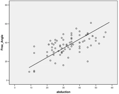
Individual characteristics of the 38 patients studied concerning general, functional and radiographic hip joint parameters are shown in table 1, that was arranged with regard to the individual subjects. However, for statistical analysis hips were considered individually. In terms of general characteristics the used sample shows a distribution of age ranging from 42 to 82 years old with a male:female ratio of 2:1.The length of the CAO ranged from 0 to 30 mm with a median of 8 mm (mean of 8,9±6.3). The range of the free-angle for abduction was 9 to 61 degrees with a median of 38 degrees (mean of 36.9±10.2) while the ROPM for abduction ranged from 9 to 56 degrees with a median of 32 degrees (mean of 32.8±10.7). The presented parameters are normally distributed.
The Pearson coefficient shows a positive correlation between ROPM in abduction and the free-angle (r=0.60, p<0.01) and a negative correlation between ROPM and the length of the CAO (r=-0.50, p<0.01). Taking into account the Kellgreen index as a general tool to radiographically evaluate osteoarthritis, a strong correlation is found with the pain score of Lequesne index (r= 0.65, p<0.01) and with CAO (r= 0.51, p<0.01). Moreover, a