Faculdade de Engenharia da Universidade do Porto
Conjugates of peptide nucleic acid-biotin for the
detection of bacteria using in situ hybridization
Diogo Henrique Fernandes Constante
Dissertation performed under the
Integrated Master in Bioengineering – Biomedical Engineering
Supervisor: Prof. Nuno Azevedo, PhD
Co- supervisor: Prof. Carina Almeida, PhD
Porto, July 2013
iii
Abstract
The microbiological detection and identification is an important tool in any clinical laboratory. Culture-based approaches are still valid and useful for microbial identification, but nowadays the inclusion of rapid technologies, such as immunological and nucleic acid-based methods, caused a dramatic impact on the routine diagnostic procedures.
The fluorescence in situ hybridization (FISH), a nucleic acid-based detection method, has been subjected to several studies and improvements, but the need for an epifluorescence microscope (or a cytometer) is still the biggest barrier to the adoption of this technology in microbiology laboratories.
The continuing need for new methods of detection led to the design of new detection tests. Keeping this in mind an alternative in situ hybridization (ISH) method was designed, where a different type of reporter molecule was used. A biotin molecule attached to the PNA probe was added to allow the inclusion of a conjugate, which contains a molecule with high affinity to biotin, streptavidin, and a enzyme, horseradish peroxidase (HRP), that is able to generate a colorimetric signal. Using this new strategy, the presence or absence of the target species can be determined by using only a spectrophotometer, similarly to an enzyme-linked immunosorbent assay test.
In this work the new ISH protocol was characterized in terms of sensitivity, specificity and detection limit; and improved by increasing the signal to noise ratio of the procedure. A previously described PNA probe, EUB338Bio, that targets all eubacteria, was used.
Firstly, the specificity/sensitivity of the EUB338 probe was verified using a standard PNA-FISH test. A set of microorganism including gram-negative bacteria (Escherichia coli, Pseudomonas fluorescens and the Delftia tsuruhatensis), gram-positive bacteria (Listeria monocytogenes, Staphylococcus aureus and Bacillus cereus), the yeast Saccharomyces cerevisiae and the archaea bacterium Methanobacterium formicicum, was used. Differences between samples and controls were only observed in eubacteria, thus proving the specificity/sensitivity of the probe.
Next, it was also tested if the introduction of a blocking reagent on the protocol could decrease the noise values found in the negative samples. The results showed no significant differences.
The HRP conjugate concentration and the incubation period in the HRP subtract (Tetramethylbenzidine - TMB) was also evaluated in order to optimize the results readings. Three conjugate concentrations (0.2, 0.5 and 1% wt/vol) and three incubation periods in TMB (5, 15 and 30 minutes) were selected. The combination of 0.2% of conjugate and 15 minutes in TMB shown to be the most effective. It was also found that the increasing of HRP concentration does not improve the signal obtained, regardless the incubation period. Also, the signal intensity increases with the TMB incubation period up to 15 minutes.
The protocol specificity/sensitivity test of the ISH procedure showed the method was unable to detect gram-positive bacteria probably due to permeabilization issues.
To find the protocol detection limit several cellular concentrations of E. coli (ranging from 10 to 109 cfu/ml), were tested. It was founded that the protocol has a detection limit of 2.5x10⁷ cfu/ml.
In its current stage, the described method could only be used to analyze the presence of gram-negative bacteria, with a very poor detection limit. Although technical improvements are still needed to increase the performance of ISH procedures, this work have shown that DNA mimics may easily be adapted to this type of protocols.
v
Acknowledgements/Agradecimentos
A realização do presente trabalho não teria sido possível sem o apoio de várias pessoas, de formas distintas ajudaram-me e guiaram-me durante todo o decorrer desta fase.
Primeiramente, gostaria de agradecer à Faculdade de Engenharia da Universidade do Porto, em especial ao Laboratory for Process, Environmental and Energy Engineering, por me terem acolhido nas suas instalações, pelas boas condições que me disponibilizaram e por me terem proporcionado o melhor ensino possível. O meu sincero agradecimento aos meus orientadores, Professores Nuno Azevedo e Carina Almeida, por terem partilhado o seu conhecimentoe por me terem apoiado e orientado durante todo este percurso.
Gostaria de agradecer toda a equipa do laboratório E007, pela camaradagem e apoio que me proporcionaram. Permitindo que a minha inclusão fosse fácil e da forma mais divertida possível.
À Joana e à Elisabete um agradecimento especial, pela amizade que permitiu suportarmo-nos mutuamente suportarmo-nos bons e maus momentos desta fase da suportarmo-nossa vida.
À minha irmã Andreia e à minha namorada Ana, pelo suporte incondicional independentemente da quantidade de trabalho com que se encontravam. A sua ajuda e suporte emocional foram mais importantes que nunca.
Por fim, mas não menos importante aos meus pais, José e Maria Constante, por sempre terem sido, literalmente, a Constante da minha vida. Nada seria possível sem o seu apoio.
vii
Table of contents
Abstract iii
Acknowledgements/Agradecimentos v
Table of contents vii
Figures list ix Abbreviations list xi Chapter one 1 Introduction 1 1.1 - Thesis organization 2 Chapter two 3 Literature review 3
2.1 - Microbiologic detection and identification 3
2.1.1 - Growth on selective and differential medium 3
2.1.2 - Immunodiagnostics 4
2.1.2.1 - Immunofluorescent tests 5
2.1.2.2 - Enzyme Immunoassay and Radioimmunoassay 5
2.1.3 - Nucleic acid-based detection 6
2.1.3.1 - Target amplification 7
2.1.3.2 - In situ hybridization 8
2.2 - An alternative in situ hybridization method using PNA molecules 11
Chapter three 15
Material and methods 15
3.1 - Microorganisms selection 15
3.2 - Culture maintenance 15
3.3 - Probe specificity/sensitivity test 16
3.5 - Effect of the blocking reagent 18 3.6 - Effect of HRP concentration and the incubation period of TMB 18
3.7 - Specificity/sensitivity of the ISH procedure 19
3.8 - Detection limit of the ISH procedure 19
3.9 - Statistical analysis 19
Chapter four 21
Results and discussion 21
4.1 - Probe specificity/sensitivity test 21
4.2 - Effect of the blocking reagent 24
4.3 - Effect of HRP concentration and the incubation period of TMB 25
4.4 - Specificity/sensitivity of the ISH procedure 26
4.5 - Detection limit of the ISH procedure 27
Chapter five 29 Conclusions 29 Chapter six 31 Future work 31 References 33 Appendix 39
ix
Figures list
Figure 1 - Basic steps of FISH: fixation, hybridization and washing. [20]
Figure 2 - Schematic representation of the molecular links and reactions that occur in
the new protocol.
Figure 3 - Oxidation process of TMB. [49]
Figure 4 - Fluorescence microscopy images resultants of PNA-FISH performed on
different species with and without the EUB338 probe. The samples tested without probes worked as controls. Images were obtained with equal exposure times, with an original magnification of x 100.
Figure 5 - Results for the inclusion of a BR in the procedure before probe hybridization
or before HRP conjugate biding. Results for standard protocol (without the BR) and a reduced protocol without cells (and also without fixation, hybridization and washing), are also present. Controls and samples refer to the test with and without probe respectively. Statistically significant differences between values indicated by a and b (p<0.05). n=6
Figure 6 - Results obtained by varying the conjugate concentration and the TMB
incubation period. Controls and samples refer to the test with and without probe, respectively. Statistically significant differences between values indicated by a and no differences between values indicated by b (p <0.05). n=6
Figure 7 - Results obtained in the specificity/sensitivity test of the ISH procedure.
Controls and samples refer to the test with and without probe, respectively. Statistically significant differences between values indicated by a and b (p <0.05). n=6
Figure 8 - Results obtained in detection limit test of the designed protocol. Controls and
samples refer to the test with and without probe respectively. n=6
Figure 9 - Photograph of detection limit test (partial results) of the ISH protocol.
Different concentrations (cfu/ml): A-109; B-5.0x10⁸; C-5.0x10⁷; D-2.5x10⁷. The controls are marked with c and the samples with s.
xi
Abbreviations list
125I Isotope iodine-125
AEEA 8-amino-3,6-dioxaoctanoic acid B. cereus Bacillus cereus
BR Blocking Reagent
D. tsuruhatensis Delftia tsuruhatensis
DNA Deoxyribonucleic acid
E. coli Escherichia coli
EIA Enzyme Immunoassay
ELISA Enzyme-linked immunosorbent assay FISH Fluorescent in situ hybridization FRET Fluorescence resonance energy transfer
HRP Horseradish peroxidase
L. innocua Listeria innocua
LNA Locked nucleic acid
M. formicicum. Methanobacterium formicicum
OD Optical density
P. fluorescens Pseudomonas fluorescens
PBS Phosphate buffered saline
PCR Polymerase chain reaction
PNA Peptide nucleic acids
qPCR Quantitative real-time polymerase chain reaction
RIA Radioimmunoassay
RNA Ribonucleic acid
rRNA Ribosomal ribonucleic acid
S. cerevisiae Saccharomyces cerevisiae
Tm Melting temperature
1
Chapter one
Introduction
The microbiologic diagnostic is a major area of microbiology. In this area, the microbiologist detects, identifies, and characterizes the microorganisms from a variety of samples, either from clinical, food or environmental origin. [1, 2]
There are several ways to make microbiological detection and identification, which can be divided into three groups. The most basic and primary technique involves the use of selective and differential media. A selective medium selectively inhibits the growth of certain microorganisms, and a differential medium differentiate specific biochemical reactions using a dye. These reactions are able to detect the presence or absence of enzymes involved in the catabolism of the culture substrate [1, 2]. A major drawback of these tests is the time associated with the culture methods, which usually require several days to reach to a definitive result. There is increasing awareness on the importance of precise identification of a pathogen for proper treatment and/or prevention of infectious disease, and new methods are being continually developed.
Recently, the inclusion of rapid technologies, such as immunological and nucleic acid-based methods, caused a dramatic impact on the routine diagnostic procedures. Actually, these technologies are nowadays well implemented in clinical/food laboratories. More recently, the introduction of the DNA mimics in fluorescence in situ hybridization has provided a valid and simple alternative for microbiological tests. FISH is a technique that allows the direct visualization of the cells by using fluorescently labelled probes that bind specifically to conserved regions in the ribosomal RNA (rRNA). The needs for epiflurescence microscope or cytometer for results reading, which are expensive and require a specialized technician, are the biggest barrier to the adoption of this technology in microbiology laboratories. As such, an alternative reporter molecule can provide important benefits to these type of procedures.
Chapter one
In this work we tested the introduction of a different report molecule in the hybridization procedure. With this new strategy it may became possible to analyze the presence or absence of the target species using only a spectrophotometer, similarly to an enzyme-linked immunosorbent assay (ELISA) test.
The methodology mentioned is still at an early stage of development, therefore this study will try to improve the protocol and find its technical limits. To achieve this goal, different factors possibly affecting the performance of the protocol, were evaluated. Then, specificity and detection limit tests have been performed.
1.1 - Thesis organization
This section aims to provide an overview of the general content and organization of this thesis.
The first chapter is the introduction. In this section the theoretical context behind this scientific study and the objectives to be achieved with it are exposed.
On chapter two a literary review from the most important topics on microbiological detection is presented. Here techniques applied to microbiological detection like selective and differential medium, immunological techniques or the nucleic acid-based techniques are discussed, together with their technological fundamentals, advantages and disadvantages. The principles of the alternative procedure proposed in this study are also explained.
Chapter three aims to describe on the best way all laboratory protocols performed to achieve the present results. All materials and methods used are described so that an interested researcher can perform them in the best way possible and get similar results.
Following the previous chapter, in chapter four the results are exposed and discussed. The methodology features are compared with the characteristics described for the competing technologies.
3
Chapter two
Literature review
2.1 - Microbiologic detection and identification
The most established technique for microbiological detection involves the use of selective and differential media, but nowadays the most preeminent technologies are immunological and nucleic acid-based methods [1-3]. These techniques are already well implemented in clinical/food laboratories but they also present well-known disadvantages, such as an expensive laboratory protocol and the need of complex equipments and specialized technician. These technologies, their characteristics, advantages and disadvantages will be discussed in detail in the next sections.
The usefulness of any diagnostic test depends on the test’s specificity and sensitivity. Specificity refers to the ability of a test to recognize only a single microorganism, i.e. the test must be specific for a single microorganism, and will not identify any other microorganism. High specificity prevents false-positive results. In this context, sensitivity refers to the capacity of the used method to detect all the strains of specific species. This prevents false-negative reactions. It is also important to highlight the concept of detection limit, which is defined by the lowest numbers of a microorganism, or its product, that can be detected. A good limit of detection also prevents false-negative reactions [1-3].
2.1.1 - Growth on selective and differential medium
One of the earliest forms used for detecting and identifying microorganisms is using selective and differential media for growth. Because, in a first stage, the microorganism identity is unknown, the microorganism is subcultured in several diagnostically relevant culture media to evaluate different biochemical reactions. Many of these tests are available
Chapter two
in miniaturized kits containing a number of different media in separate wells, which can be inoculated all at one time [3].
The media can be selective, differential, or both. A selective medium is able to selectively inhibit the growth of certain microorganisms by incorporating in its composition antimicrobial agents, or others compounds, that inhibit the indigenous flora while allowing the growth of the microorganisms resistant to these compounds. For example, the MacConkey agar allows the growth of most Gram-negative rods but inhibits the Gram-positive growth [4]. The differential medium normally uses a dye to differentiate and measure different biochemical reactions carried out during growth. The procedure takes advantage from the different carbohydrate fermentation processes performed by the microorganisms. The Eosin methylene blue, which is able to differentiate between lactose and sucrose fermentation, is a good example of a differential medium. MacConkey agar medium is also a differential medium for lactose fermentation, but it also works as a selective medium since it includes, for instance, bile salts to inhibit most Gram-positive bacteria [5].
After the isolation in agar medium, a confirmation step is usually performed using biochemical tests. The most well-known of these biochemical tests are summarized in appendix 1. It is possible to measure many different biochemical reactions by measuring the presence or absence of enzymes involved in the catabolism of the substrate or substrates in the differential medium. [1, 2] Currently the recommended standards for microbiological analysis are based on these biochemical tests.
Sometimes, in case of a dubious match, it is necessary to use other tests with more sophisticated identification procedures, normally required for microorganisms with similar growth characteristics and similar biochemical profiles [2]. It is also important to note that in this method the time required to perform culture can be a major drawback, mostly for clinical purposes. Moreover, it also fails to detect non-cultivable organisms and, occasionally, isolates exhibit a biochemical behaviour that does not fit into the typical biochemical pattern of the species, resulting in inaccurate results.
2.1.2 - Immunodiagnostics
Immunological detection is used in clinical/food and research laboratories to detect specific pathogens or pathogen products, like bacterial cells, spores, viruses and toxins. Several immunoassays are performed using a specific antibody to detect individual infectious agents (pathogens or their products). These in vitro methods are also used to obtain evidence if a patient was exposed or infected by a pathogen. Virtually any chemical compound can serve as an antigen if it is able to trigger an immunogenic response. [1-3]
In fact, the binding of an antibody to its target is the key to the immunologic detection techniques. The ability to detect the occurrence of this event enables us to infer about the target presence [6]. It can be done by labelling the antibody with other molecule that
Conjugates of peptide nucleic acid-biotin for the detection of bacteria using in situ hybridization
5
produces a measurable signal either by a colour change, production of light/fluorescence, an electrical or other optical output.
There are several strategies that use that knowledge to detect microorganisms. Among all categories, the immunofluorescent tests, the enzyme Immunoassay (EIA) and radioimmunoassay, should be highlighted.
2.1.2.1 - Immunofluorescent tests
Antibodies are able to be chemically modified with fluorescent dyes, like rhodamine B (red) or fluorescein isothiocyanate (yellow-green). These dyes do not alter the specificity of the antibody but make them very powerful on the detection of antigens on intact cells, because a fluorescence microscope can be used for detection, once the antibody has bound to cell or tissue surface antigens. The cell-bound fluorescent antibodies emit a bright fluorescent color when excited with light of particular wavelengths, making them visible at fluorescence microscope [1, 2].
Fluorescent antibodies are widely used for diagnostic microbiology, because they allow the identification of a microorganism directly in a patient specimen (in situ), and they are also very useful in microbial ecology as a method for the direct visualization and identification of microbial cells. This technique avoids the need for the isolation and culture of the organism. Fluorescent antibodies can also be used to separate mixtures of cells into relatively pure populations or to define the numbers of individual cell types in complex mixtures such as blood, when combined with flow cytometry [1, 2].
Fluorescent antibody-staining methods can be performed in a direct or indirect way. In the direct method the antibody, that interacts with the surface antigen, is itself covalently linked to the fluorescent dye. In the indirect method the presence of a non-fluorescent antibody, on the surface of a cell, is detected by the use of a fluorescent antibody directed against the nonfluorescent antibody [2]. Fluorescent antibodies, under appropriate conditions, can provide rapid and highly specific information about a variety of clinical conditions.
2.1.2.2 - Enzyme Immunoassay and Radioimmunoassay
Enzyme immunoassay (EIA) or enzyme-linked immunosorbent assay (ELISA), and radioimmunoassay (RIA) methods are two very sensitive immunological assays and, for that reason, they are widely used in clinical and research applications. EIA and RIA employ covalently bonded enzymes and radioisotopes, respectively, to antibodies, allowing the detection of very small quantities of antigen–antibody complexes [2].
The EIA is a high specific and sensitive method of immunological detection that uses an enzyme covalently attached to an antibody molecule. The bond does not modify the enzyme’s catalytic properties and the antibody’s specificity. The typical enzymes used to bind to
Chapter two
antibodies are the horseradish peroxidase, alkaline phosphatase, and β-galactosidase. All this enzymes interact with substrates to form reaction products that can be detected in very low amounts [2]. More specifically, these enzymes catalyze the conversion of the substrate into a colored product. The color produced is proportional to the amount of antigen present and can be detected with a spectrophotometer [1, 2]. From these three enzymes, the horseradish peroxidase conjugates are known to have the lowest detection limits and highest analytical sensitivity at shortest periods of substrate reaction when compared to the other two enzymes [7, 8].
In the EIA, the specimen is typically added to the wells of a microtiter plate, previously coated with specific antibodies for the antigen to be detected. If it’s present in the sample it will bound to the antibodies. Next, a washing step is performed and a second antibody (conjugated with an enzyme) is added. This second antibody is also specific for the antigen, but it binds to other exposed specific antigen. After a second wash, the enzyme activity is determined by adding its substrate. This method has been described as simple, economic and with a good limit of detection [2]. Nonetheless, several studies have also pointed some drawbacks, such as: cross-reactivity with similar antigens, implications of the animal antibodies usage and the interference of the matrix used [9-11].
The RIA is implemented just like the EIA but it employs radioisotopes in the antibody or antigen conjugates, instead of the enzymes used in EIA. Antibodies or antigens can be modified covalently, commonly with the isotope iodine-125 (125I), without disrupting their immune specificity. This method is less used than the EIA because it requires expensive equipment. [2]
2.1.3 - Nucleic acid-based detection
By definition, any self-replicating biological entity can be discriminated on the basis of nucleic acid sequences unique to that particular organism (exception to prions, which don’t have associated nucleic acids). A number of different strategies have been explored to identify unique sequences for lower eukaryotes and prokaryotes, to initiate the development of an assay. The challenge is to make these assays totally inclusive, specific and robust [3].
Very sensitive methods, based on nucleic acid analyses, are widely used in clinical microbiology to detect pathogens. These methods do not depend on pathogen isolation or growth. They only depend on detection of species-specific nucleic acid sequences in DNA or RNA genes [2].
The success of DNA-based diagnostic is based on four principles: nucleic acids can be readily isolated from contaminated samples; the nucleic acid sequence of a given microorganism’s genome is unique; nucleic acid sequences can be amplified to increase the amount of material available; nucleic acids, when present in sufficient amounts, can be readily visualized and measured. [2]
Conjugates of peptide nucleic acid-biotin for the detection of bacteria using in situ hybridization
7
Nucleic acid-based assays can be classified into different categories. In the next section we will focus on two very common categories in microbiological identification: target amplification, mostly used in polymerase chain reaction (PCR), and in situ hybridization.
2.1.3.1 - Target amplification
In the field of nucleic acid-based detection there was a great improvement by developing amplification processes which effectively increase the number of targets in vitro. These assays should be able to detect a single target organism by generating enormous copies of target nucleic acid, starting with a single copy of it. Beyond the development of several amplification methods, many post-amplification detection systems have also been developed [3].
Polymerase chain reaction
The polymerase chain reaction is essentially DNA replication in vitro. The PCR amplifies nucleic acids, forming multiple copies of the target sequences. PCR techniques can use primers for a microorganism specific gene to examine DNA derived from suspected contaminated samples, even in the absence of an observable, culturable microorganism [2]. PCR-based tests are widely used for identification of several pathogens and are particularly useful for identifying viral and intracellular infections, where culturing the agents may be difficult or even impossible [3].
This method involves the enzymatic replication of a target region of nucleic acid defined by a set of oligonucleotide primers, which are typically oriented in a convergent manner. Briefly, the PCR process can be divided into three steps. First, double-stranded DNA (dsDNA) is separated at temperatures above 90°C. Second, oligonucleotide primers generally anneal at 50–60°C to the target sequence, and, finally, optimal primer extension occurs at 70–78°C, temperature at which the DNA polymerase works. The newly formed strand will work as a template for a subsequent round of replication [3, 12]. For conventional PCR methods, the amplification products are analyzed by performing an electrophoresis and an ethidium bromide staining.
To simplify the results analysis, researchers developed quantitative real-time PCR (or qPCR), which involves the amplification and detection of amplified products coupled in a single reaction vessel, greatly increasing the speed of detection. This allows for the direct detection of the PCR product during the exponential phase of the amplification reaction, combining amplification and detection in one step. The accumulation of target DNA is monitored during the qPCR process by adding fluorescent probes/dyes to the PCR reaction mixture. The level of fluorescence increases proportionally to the DNA target amplification. Because qPCR amplification can be monitored continuously it does not require other method
Chapter two
to confirm amplification. The monitoring also allows determining accurately the amount of DNA target present in the original sample [2]. The three most common probes used on qPCR are the Taqman, the molecular beacons and the fluorescence resonance energy transfer (FRET) probes [13, 14].
The Taqman probes, typically with 23-30 nucleotides, have a 5’ fluorescent reporter molecule and a 3’ quencher molecule [15]. In this probe the signal is emitted when the reporter and quencher molecules are separated, by the 5’ exonuclease activity of the enzyme, and the reporter is detected by the qPCR instrument. In the molecular beacons probes, the secondary structure of the probe keeps the reporter and the quencher molecules close enough to prevent any fluorescent signal. The signal only occurs when the probe anneals to target DNA, causing a sufficient gap between the molecules and allowing the fluorescent signal [16]. Unlike the previous two methods on FRET-based assays the key is the energy transfer between a 3’ donor and a 5’ reporter fluorophores on separate probes [17, 18].
The PCR method is considered the most sensitive between the rapid methods for microbial detection, particularly when the specimen is not easily cultured or requirer a long cultivation period. However, this method also has some disadvantages due to its susceptibility to inhibitors, cross-contaminations and sensitivity to small experimental variations, which may cause the occurrence of false positives and negatives. [19].
2.1.3.2 - In situ hybridization
When DNA is denatured, the single strands can form hybrid double-stranded molecules with other single-stranded DNA/RNA molecules by complementary base pairing. This is called in situ hybridization (ISH), and is widely used in detecting, characterizing, and identifying segments of DNA. Segments of single-stranded nucleic acids, with known identity, that are used in hybridization, are called nucleic acid probes. Probes can be general (bind by complementary base pairing to conserved sequences in the DNA/rRNA of all organisms) or specific (react only with the DNA/RNA of species in a single domain/species). Nucleic acid probes are usually 100-1000 bases long, when applied to cytogenetic techniques [2, 3]
If a sample contains DNA/RNA sequences complementary to the probe, the probe will hybridize, forming a double-stranded molecule. To detect the binding, the probe is labelled with a reporter molecule (a radioisotope, an enzyme, or a fluorescent compound). [2, 3, 20] The stability of the hybrid complexes is correlated with the temperature at which the hybrids dissociate, i.e. melting temperature (Tm). As a general rule, the Tm is equivalent to the sum
of 2°C for each A-T base pair and 4°C for each G-C base pair. Other factors that can affect the Tm are ionic concentration, pH, and mismatches between the strands. [3]
This kind of technique offers several advantages over immunological assays: the nucleic acids are much more stable than proteins at high temperatures and high pH, and are more resistant to organic solvents and other chemicals. Also, nucleic acid probe technology can
Conjugates of peptide nucleic acid-biotin for the detection of bacteria using in situ hybridization
9
even be used to positively identify organisms that are no longer culturable; the probes may be more specific than antibodies and are able to detect single nucleotide differences between DNA sequences. [2, 20]
In addition to clinical diagnostics applications, the nucleic acid probes are also used in food industries to monitor contamination of foods by pathogens. Nucleic acid probes can be sensitive enough to detect less than 1 CFU per ml/g of sample [2]
For signal generation and therefore to allow its detection, the probe can be labelled with a variety of reporter molecules including radioisotopes, fluorophores or enzymes. The radioisotopes were the first to be utilized, but they have drawbacks due to limited half-life and handling concerns. The most used reporter molecules in situ hybridization techniques are fluorochromes, since they eliminate post-hybridization steps.
Fluorescent in situ hybridization (FISH)
There was an urgent need for the development of new methods to analyse microorganisms and their communities in situ. [21] The exact quantification of microbial populations remains a challenging task, and it is this context that marks the recent progress in fluorescence in situ hybridization, a cultivation-independent method for the in situ analysis of the composition of microbial communities and their dynamics. [20]
FISH has become a widely used method for the identification, quantification and phylogenetic characterization of microbial populations in complex environment. [22] FISH can be used to target DNA or rRNA molecules, but the last one is more widely used. rRNA molecules are well suited for the identification of microorganisms for several reasons. One of the most significant is that, because all cells require ribosomes for translation, each cell contains a high number of rRNA copies [20, 23]. In addition, the evolutionary conservation of rRNA sequences is higher than the one found in most of the protein-encoding genes, enabling the design of oligonucleotide probes for large taxonomic entities [20].
The FISH method usually comprises three steps: fixation/permeabilization, hybridization and washing (Figure 1). [20] Firstly the microbial cells are fixed by using a chemical fixative, such as formalin, paraformaldehyde and ethanol [22, 24, 25]. This step not only stabilizes cell morphology (preserve its integrity and shape, and prevent cell lysis), but also permeabilizes the cell membrane for subsequent hybridization (allowing the oligonucleotides to diffuse to their rRNA targets). The membrane integrity is intrinsically linked to cell viability and, consequently, fixed FISH-stained cells are no longer viable [20]. The challenge in this step is that, because of cell wall diversity composition of bacteria and archaea, there is not a standard permeabilization protocol for all microbial cells. [20] This step may include enzymatic digestion of thick peptidoglycan layers by lysozyme, digestion of proteinaceous cell walls by proteases, the removal of wax by solvents, the use of detergents and even short-term incubations in hydrochloric acid. [26-28]
Chapter two
During the hybridization step, the temperature, pH, ionic strength and formamide concentrations are all well defined to guarantee that the probe accesses and hybridizes with the target sequence [23]. In this step the microbe is incubated with a probe, usually for a few hours, during which the labelled oligonucleotide diffuses to its intracellular targets and forms specific hybrids [20].
Finally, the washing step ensures that all loosely bound or unbound labelled probes are removed from the sample, providing specificity to the detection. After this stage the sample is ready for single-cell identification and quantification by either epifluorescence microscopy or flow cytometry [23].
The typical fluorescent labels include cyanine and fluorescein molecules, but a new group of fluorophore families, which includes de Alexa Fluors and of nanosized crystal particles, named quantum dots, is gaining widespread acceptance. The advantages of these new dyes are based on an increased photostability and brightness. [29, 30]
Typically the hybridization step includes a DNA probe (DNA FISH); however DNA FISH shows several limitations, such as low specificity [31] and low affinity between the probe and the target [32]. Because of these problems, researchers started to search for alternatives to improve the robustness of this method. The solution appears to have arrived in the form of nucleic acid analogues [33, 34], a new class of molecules that mimics DNA and that has made the FISH procedures easier and more efficient [2, 35]. The most common, in microbiology, are peptide nucleic acids (PNA) [33] and locked nucleic acid (LNA) [36]. PNA has been by far the molecule most applied to microbial identification and there are already a large number of PNA FISH protocols described for the detection of pathogenic bacteria [23, 24].
FISH techniques using PNA molecules involve a simple procedure, but an important disadvantage lies in the need of complex equipments (an epiflurescence microscope or a flow cytometer) for the results reading [37]. These equipments can be found on central hospital facilities, but are expensive, require specialized technicians and are inexistent on primary care units or small laboratories for quality control. These facts may hinder the implementation of PNA-FISH as a routine diagnostic tool.
Conjugates of peptide nucleic acid-biotin for the detection of bacteria using in situ hybridization
11
Figure 1 - Basic steps of FISH: fixation/permeabilization, hybridization and washing. [20]
2.2 - An alternative in situ hybridization method using PNA
molecules
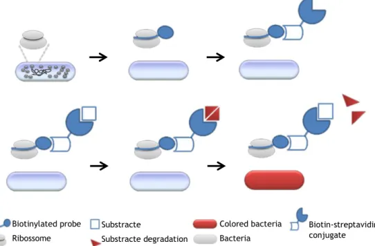
In order to overcome the major implementation barrier imposed to FISH procedures, which is the need for an epiflurescence microscope or a flow cytometer, a new experimental protocol that combines features of the PNA-FISH with characteristics of ELISA was designed. This alternative method is based on the replacement of the probe fluorochrome for a different reporter molecule, in this case biotin. The presence of this molecule allows the insertion of an enzymatic conjugate into the protocol. With the use of an appropriate substrate for this enzyme it became possible to determine the presence or absence of the specimen of study in a sample just by the substrate colour change. This colorimetric reaction can be analyzed using only a spectrophotometer, similarly to an ELISA test.
Procedure principles
In this study a PNA EUB338Bio probe (5’-TGCCTCCCGTAGGA-3’) that is complementary to a conserved region in the 16S ribosomal RNA of all Eubacteria was used [38]. The EUB probe can be conjugated, by a covalent bond, to biotin, a vitamin complex (B7) soluble in water that serves as coenzyme in the synthesis of fatty acids, isoleucine and valine, being also important in the gluconeogenese [39]. Biotin is widely used as a mean of conjugating
Chapter two
molecules, a process called biotinylation. In this case a covalent bond between the biotin and EUB probe, was formed [40]. Biotinylated PNA probes are already commercially available for customized probes. The small size of biotin ensures that the biological activity of the biotinylated protein/oligonucleotide is unaffected.
The use of biotin aims to allow the insertion of streptavidin into the protocol. Then streptavidin can be used in conjunction with an enzyme without losing its specificity or sensitivity, parameters that are crucial to the efficiency of a microbiological detection protocol. Conjugates of streptavidin are also commercially available, since they are commonly applied to EIA techniques.
Streptavidin is a crystalline structure with an extremely high affinity to biotin, actually the biotin-streptavidin interaction is one of the strongest non-covalent bonds known. Such affinity for biotin is due to several factors, such as: an excellent structural complementarity between these two compounds; the existence of a large network of hydrogen bonds involved in the interaction; and the stabilization of the binding of streptavidin by a loop, which links the L3 and L4 chains of biotin [41].
The biotin-streptavidin complex is very resistant to organic solvents, denaturants, detergents, proteolytic enzymes and extremes of temperature and pH. In addition, the streptavidin molecule has a neutral charge, which eliminates the possibility of electrostatic binding with the tissue, and does not contain carbohydrate groups which could bind to lectins [42].
Among the many existing methods for detecting the presence of biotin-streptavidin binding, the most popular method uses enzymes (which are conjugated with streptavidin) capable of generating a colour change when they are incubated in the presence of the respective substrate [43]. A typical option is the horseradish peroxidase (HRP), because, when compared with the most popular alternatives (such as alkaline phosphatase), the enzyme is smaller, more stable, less expensive and capable of generating strong signals in a relatively short time [44]. It was decided to use a biotinylated probe with the HRP-streptavidin conjugate because it allows to perform the protocol using always the same conjugate, and changing only the probe, moreover, it has been reported that best results are obtained when the probe biotinylation is previously performed and not during the experiment [45, 46].
It can be considered that streptavidin works like a strong link between biotin and HRP. The HRP produces a luminescent derivative when incubated with a specific substrate, such as tetramethylbenzidine (TMB), allowing the detection and quantification of the target molecule. TMB has a slightly yellow colour that, when incubated in the presence of HRP, produces a colour change to blue [47] (figure 2).
The TMB is a colorimetric substrate for the HPR enzyme. Since HRP is a peroxidase enzyme, in its presence the TMB oxidizes (Figure 3), with the hydrogen peroxide (originally contained in the TMB substrate) serving as a receiver of electrons. In its oxidised form, the
Conjugates of peptide nucleic acid-biotin for the detection of bacteria using in situ hybridization
13
TMB acquires a blue colour due to oxygen radicals produced during the hydrolysis of hydrogen peroxide made by HRP [48]. In order to increase the sensitivity of the reading and to stop the enzyme reaction, sulphuric acid is then added to the solution. The colour intensity can be measured by a spectrophotometer at a recommended wavelength of 450 nm, being the signal intensity directly proportional to the HRP activity [47, 49].
Figure 2 - Schematic representation of the molecular links and reactions that occur in the new protocol.
Figure 3 - Oxidation process of TMB. [49]
The methodology described here is still at an early stage of development and scientific validation is needed. In fact, similar strategies have been tested, but using DNA probes [50]. Different tests were performed to improve the protocol robustness and to determine its technical limits. For this, the following tests were performed: probe specificity; effect of the HRP concentration and its action time; effect of a blocking reagent in the signal to noise ratio; and finally the determination of the procedure detection limit.
Biotinylated probe Ribossome Substracte Substracte degradation Colored bacteria Bacteria Biotin-streptavidin conjugate
15
Chapter three
Material and methods
3.1 - Microorganisms selection
The probe selected for this study, EUB338Bio (Panagene, South Korea), targets all eubacteria. A general probe was chosen in order to allow the evaluation of the method performance in bacterial species with different properties. Nevertheless, to optimize the procedure, a model microorganism was selected. The bacterium Escherichia coli, was chosen. E. coli is a Gram-negative, facultative anaerobic and a rod-shaped bacterium. It is probably the most widely studied prokaryotic model organism, being one of the first organisms to have its genome sequenced. [51]
For further specificity tests other Eubacteria, with different membrane properties, have been selected. A set of gram-negative (E. coli CECT434, Pseudomonas fluorescens ATCC13525 and Delftia tsuruhatensis BM90) and gram-positive bacteria (Listeria innocua CECT910, Staphylococcus spp. and Bacillus cereus (strain isolated from a disinfectant solution and identified by 16S rRNA gene sequencing), was selected. In addition to these, as negative controls for specificity tests, the yeast Saccharomyces cerevisiae PYCC3507 and the archaea bacterium Methanobacterium formicicum DSM1535 were used.
3.2 - Culture maintenance
All eubacteria used were maintained on Tryptic Soy Agar medium (3% (wt/vol) of Tryptic Soy Broth (Merk, Germany); 1.5% (wt/vol) agar (Merk, Germany)) at 37ºC (FOC 225I, VELP® Scientifica), except for L. innocua and P. fluorescens that where incubated at 30ºC. For the S. cerevisiae culture, Yeast Extract Peptone Dextrose (YEPD) medium was used (1% (wt/vol) yeast extract (Merk, Germany), 2% (wt/vol) peptone (Liofilchem, Italy), 2% (wt/vol) glucose
Chapter three
(Merck, Germany) and 2% (wt/vol) agar) and incubation occurred at 30ºC. All these microorganisms were incubated overnight before use. The M. formicicum was provided by the Universidade do Minho, and it was conserved in a suspension of 4% (wt/vol) paraformaldehyde and stored at -20ºC. The cultured strains were stored at 4ºC and subcultured at least once a week.
3.3 - Probe specificity/sensitivity test
In order to test probe specificity/sensitivity without the influence of the changes that were implemented in the protocol proposed, a standard FISH protocol was performed as previously described in Guimarães et al. [24]. In this protocol the probe sequence was completely identical to the probe already described but instead of the biotin molecule it had a fluorochrome (Alexa fluor 488) as a reporter molecule. The universal PNA EUB338 probe (5’-TGCCTCCCGTAGGA-3’) was synthesized and labelled at the N terminus with AlexaFluor488 via a double 8-amino-3,6-dioxaoctanoic acid (AEEA) linker (Panagene, South Korea). In this test all the selected microorganisms referred in 3.1 were used.
After the preparation of the inoculum (with a loop of fresh biomass homogenized in sterile distilled water), 20 µl of this suspension were placed in each well of the glass slides (Thermo Scientific). The samples were left to dry and were subsequently covered with 40 µl of 4% (wt / vol) paraformaldehyde (ACROS, USA) for 10 minutes. Next, the paraformaldehyde was removed with absorbent paper and 40 µl of 50% (vol/vol) ethanol were added for 10 minutes, ending the permeabilization phase.
Subsequently, the sample was covered with 20 µl of hybridisation solution containing 10% (wt/vol) dextran sulphate (Sigma), 10 mM NaCl (Panreac, Spain), 30% (vol/vol) formamide (ACROS, USA), 0.1% (wt/vol) sodium pyrophosphate (ACROS, USA), 0.2% (wt/vol) polyvinylpyrrolidone (Sigma, China), 0.2% (wt/vol) Ficoll (Fisher Scientific, USA), 5 mM disodium EDTA (Panreac, Spain), 0.1% (vol/vol) Triton X-100 (Panreac, Spain), 50 mM Tris-HCl (Fisher Scientific, USA), 0,2% (wt/vol) polyvinylpirroline (Sigma, China) and 200 nM of probe. For the control samples the probe was not added to the hybridization solution. The samples were covered with coverslips and incubated at 57ºC (FD23, BINDER incubator, Germany) for 1 hour.
After this time of hybridization the coverslips were removed and the glass slides were immersed for 30 minutes in washing solution containing 5 mM Tris base containing (Fisher Scientific, USA), 15 mM NaCl and 1% (vol/vol) Triton X. Completed this time, the slides were removed and left to dry.
Finally the samples were mounted with a drop of non-fluorescent immersion oil (Merck, Germany) and covered with coverslips. After this it was possible to analyze the samples in a fluorescence microscope LEICA DM LB2, incorporating a CCD camera (LEICA DFC300 FX camera) and using the LAS V4.2 software (LEICA). The optical filter used consisted of a 450-490 nm excitation filter (LEICA I3).
Conjugates of peptide nucleic acid-biotin for the detection of bacteria using in situ hybridization
17
3.4. In situ hybridization (ISH) protocol
The proposed protocol comprises the standard steps of a PNA-FISH protocol; however, it presents a final binding phase of HRP-conjugated streptavidin and subsequent exposure to the HRP substrate.
Firstly an inoculum was prepared in distilled water (step 1), from this suspension thirty μl (approximately 108 cfu/ml) were pipetted into the wells of a 96 well plate (Scientific Orange) (step 2) and it was dried in an incubator at about 105ºC (Venticell incubater) (step 3).
Posteriorly thirty μl of 4% (wt/vol) paraformaldehyde were added into each well and let act for 10 min (step 4), after this period the paraformaldehyde was removed (step 5) and thirty μl of lysozyme (10 mg/ml diluted in phosphate buffered saline (PBS)) (Sigma-Aldrich) were added into each well (step 6). The plate was incubated at 37ºC for 1 hour (FOC 225I, VELP® Scientifica) (step 7).
Atfer the lysozyme be re removed (step 8), the wells were washed with 100 μl of PBS (4.8% (wt/vol) Sodium chloride (Panreac, Spain), 0.12% (wt/vol) potassium chloride (Panreac, Spain), 0.486% (wt/vol) di-sodium hydrogen phosphate dihydrate (ACROS, USA), 0.120% (wt/vol) monopotassium phosphate (ACROS, USA)) (step 9).
For the hibridization stage twenty μl of hybridization solution were pipetted into each well (with the EUB338Bio probe and in the negative controls without the probe) and incubated for 45 min at 57ºC (step 10). After that the hybridization solution was removed (step 11) and two hundred μl of wash solution were pipetted to each well and incubated again at 57ºC for 30 minutes (step 12).
When the washing stage has been ended the wash solution was removed (step 13) and forty μl of 0.2% (vol/vol) working solution HRP (diluted in 1% bloking reagent, from Invitrogen, USA) was added to each well and incubated at room temperature for 30 minutes (step 14). When ended the solution was removed and the wells were washed twice with 100 μl of PBS (step 15)
Two hundred μl of TMB revelation solution (Invitrogen, USA) were pipetted to each well and let to act for 30 minutes (step 16). Ended this stage 100 μl from the sample were transferred to a new plate and added to 100μl of sulfuric acid (0.5 M) (step 17).
Finally, the plate was read in a spectrophotometer (Molecular Devices, SpectraMax M2, USA) at wavelength of 450 nm (step 18).
3.5 - Effect of the blocking reagent
In an attempt to increase the signal to noise ratio of the test, the inclusion of a Blocking Reagent (BR), which is provided with the HRP conjugate, was tested. Blocking reagents are typically used to block the non-specific binding [52, 53].
Chapter three
For this, a step of 1% (wt/vol) BR solution was included in two different stages of the in situ hybridization protocol: prior to hybridization (before step 10 of the protocol described above in 3.4) and after the washing solution (before step 14 of the protocol in 3.4). These two points are the ones that contribute the most for the specificity of the test. At that step we will have the two specific bindings: probe - rRNA and conjugate (streptavidin-HRP) - biotin (reporter molecule of the probe). As such the inclusion of BR may prevent the occurrence of false positives by decreasing the noise of the reaction and increasing the reaction stringency. In both cases the BR was left to react by 10 minutes and after that the wells were washed with 100 μl of PBS.
At this stage it was also tried to further understand the origin of the noise value with the completion of one more test. On this test the procedure started only in the application of the conjugate (from step 14), i.e. without the cells and the others protocol components. This short test was conducted to evaluate the noise level that is obtained by the simple introduction of the conjugate in the experiment. More precisely, to verify if the conjugate can bind to undesired cells components, to other components of the protocol, or simply to the support material used (96-well tissue plate).
Six samples were analyzed for each test.
3.6 - Effect of HRP concentration and the incubation period of
TMB
Other parameters that may interfere with the signal-to-noise ratio are the HRP conjugate concentration and the incubation period for the revelation solution (TMB with H2O2). Both excessive concentrations of HRP and high incubation periods in the revelation solution may result in strong background signals. So, minimum concentrations and periods that do not compromise the signal intensity should be selected.
For this, a test was designed where different HRP conjugate concentrations and incubation period in the revelation solution were tested. For the HRP conjugate, three concentrations were selected (0.2, 0.5 and 1% wt/vol) and tested in the procedure, by changing step 14 (in section 3.4). For each of these concentrations, three incubation periods in the revelation solution (5, 15 and 30 minutes) were tested by changing step 16 (in section 3.4). Six samples were analyzed for each test.
3.7 - Specificity/sensitivity of the ISH procedure
A new specificity/sensitivity test was performed to access the performance of the ISH protocol in different bacterial species. The strains included in section 3.1 were used. This test had the main aim of verifying if the protocol had the same specificity of the probe, and especially if it had the ability to permeabilize the gram-positive bacteria. The procedure was
Conjugates of peptide nucleic acid-biotin for the detection of bacteria using in situ hybridization
19
performed as described above in section 3.4, but a TMB incubation period of 15 minutes (step 16), was used. Six samples were analyzed for each test.
3.8 - Detection limit of the ISH procedure
The detection limit is an important feature of any detection system. It measures the minimum concentration of the target microorganism needed to produce a clear positive result. The detection limit was carried out in E. coli cells. The test was performed in suspensions at different concentrations: 10; 100; 1.0x10³; 1.0x10⁴; 1.0x10⁵; 1.0x10⁶; 1.0x10⁷; 2.5x10⁷; 5.0x10⁷; 7.5x10⁷; 1.0x 10⁸; 5x10⁸; 109 cfu/ml. To prepare the different concentrations a stock suspension with OD of 1 was used, which is known to have a concentration of approximately 109 cfu/ml. From this inoculum successive dilutions were made to obtain the set of seven inocula mentioned. These different inoculums were introduced in step 1 – section 3.4. Six samples were analyzed for each test
3.9 - Statistical analysis
Results were compared using the t-Test (two-sample assuming unequal variances), using Microsoft Office Excel (Microsoft Corporation). All tests were performed with a confidence level of 95%. Differences between samples were considered statistically different when p-values were lower than 0.05 (p< 0.05).
21
Chapter four
Results and discussion
4.1 - Probe specificity/sensitivity test
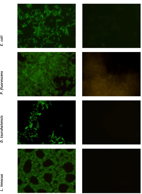
Before conducting the optimization of an alternative in situ hybridization protocol, the EUB338 probe specificity/sensitivity was evaluated in a standard FISH protocol, in order to be able to compare the effect of the new protocol on these parameters. The results obtained on PNA-FISH test with the EUB338 probe, showed that there were obvious differences between the samples with and without probe for all Eubacteria smears (E. coli, P. fluorescens, D. tsuruhatensis, L. innocua, B. cereus and Staphylococcus spp.) (Figure 4). This indicates a good sensitivity, since the selected bacteria are very diverse, but the probe was able to provide a good fluorescence signal for all the species. On the other hand, these differences were not apparent on S. cerevisiae and M. formicicum test, indicating that there was no hybridization between the probe and the ribosomal material of nonn-Eubacteria organisms. These results, as expected, confirm the probe specificity that was associated to this EUB338 probe.
From the images it is also possible to verify that Staphylococcus spp., compared with the other Eubacteria, showed lower fluorescence intensity and partial hybridization. This may be indicative of the greater difficulty in permeabilizing certain organisms, especially Gram-positive bacteria, as this is the case. Nonetheless, the other gram-Gram-positive bacteria tested provided strong signal. This behavioral difference between Staphylococcus spp. and the other gram-positive bacteria may be due to its tight cell wall, which consists essentially of peptidoglycan crosslinked by penta- or hexaglycine bridges and the high content of glycine [54].
The weak fluorescence signal detected in S. cerevisiae should also be noted. Since this behaviour was maintained on the test without probe, it may not be associated to probe hybridization but simply due the autofluorescence of this organism. Already reported by other authors [55].
Chapter four
With probe Without probe
E. coli P . fl uo re scen s D . t su ru ha ten si s L. inn ocua
Figure 4 - Fluorescence microscopy images resultants of PNA-FISH performed on different species
with and without the EUB338 probe. The samples tested without probes worked as controls. Images were obtained with equal exposure times, with an original magnification of x 1000. The optical filter used was a 450-490 nm excitation filter (LEICA I3).
Conjugates of peptide nucleic acid-biotin for the detection of bacteria using in situ hybridization
23
With probe Without probe
B. c ere us St aph yl ococc us spp . S. ce rev isi ae M. fo rm icicu m
Figure 4 (continuation) - Fluorescence microscopy images resultants of PNA-FISH performed on
different species with and without the EUB338 probe. The samples tested without probes worked as controls. Images were obtained with equal exposure times, with an original magnification of x 1000. The optical filter used was a 450-490 nm excitation filter (LEICA I3).
Chapter four
4.2 - Effect of the blocking reagent
On preliminaries tests the presence of some background noise in the obtained results was verified (data not shown). In an attempt to eliminate this effect, the use of a BR, usually used to avoid the non-specific biding of antibodies to non-target antigens, could be beneficial to the results by increasing stringency of the reaction. The procedure steps more susceptible to cross-reactivity are the hybridization (probe binding to the rRNA) and the addition of the conjugate solution, where the binding between strepatvidin and biotin takes place. As such, it was tested if the BR inclusion, before performing these steps, would be able to decrease the noise level of the overall procedure.
Results revealed no statistical difference (p>0.05) between the inclusion or not of the BR (Figure 5). Also, the test without cells allows to conclude that the noise value source is not caused by nonspecific binding of the probe or the conjugate. Instead, the background was due to the residual biding of the streptavidin-HRP conjugate to the plate material, which the inclusion of the BR solution does not avoid.
So, since the BR inclusion does not bring any advantage, it would only extend unnecessarily the protocol duration.
Figure 5 - Results for the inclusion of a BR in the procedure before probe hybridization or before HRP
conjugate biding. Results for standard protocol (without the BR) and a reduced protocol without cells (and also without fixation/permeabilization, hybridization and washing), are also present. Controls and samples refer to the test with and without probe respectively.
*
Statistically significant differences between samples and controls (p <0.05).0 0,2 0,4 0,6 0,8 1 1,2 1,4 1,6 1,8
control sample control sample control sample 1%BR before conjugate
addition
1%BR before hybridization Without 1%BR Without cells
OD ( 450nm )
*
*
*
Conjugates of peptide nucleic acid-biotin for the detection of bacteria using in situ hybridization
25
4.3 - Effect of HRP concentration and the incubation period on
TMB
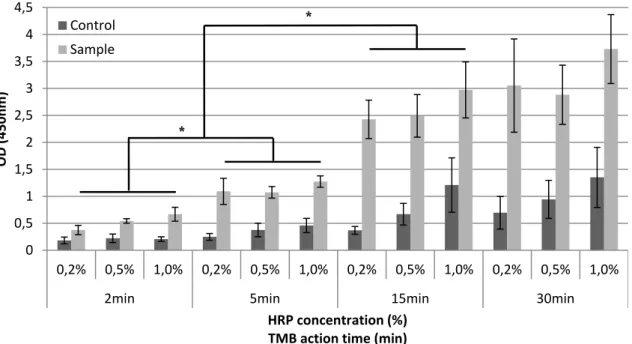
The study of the variation of HRP concentration and TMB incubation time allowed a better understanding of its influence in the noise level of the procedure. Both high concentrations of conjugate and extended incubation periods in the revelation solution (TMB), may increase the reaction background (noise) and consequently hamper the distinction between positive and negative results. An important observation from the results (Figure 6) is the lack of statistically significant differences (p<0.05) between positive samples exposed to different concentrations of conjugate but with the same period of incubation in TMB. On the other hand, the average noise obtained in controls increased with the conjugate concentration. This behavior is more accentuated for the incubation periods of 15 and 30 minutes, probably because higher concentrations of conjugate will result in higher residual amounts of non-bounded conjugate, even after a careful washing. Thus the controls noise will be further amplified when the TMB incubation time is increased.
Figure 6 - Results obtained by varying the conjugate concentration and the TMB incubation period.
Controls and samples refer to the test with and without probe, respectively. * Statistically significant differences between samples (p <0.05).
Since it was intended to adopt a combination of conjugate concentration and TMB incubation time that allow a good discrimination between controls and positive samples, the incubation periods of 2 and 5 minutes should not be considered as the best option. Nonetheless, it should be mentioned that statistically significant differences (p<0.05) were found between the majority of positive samples and controls for this shorter incubation periods (exception for the condition with 0.2% conjugate at 2 minutes in TMB).
0 0,5 1 1,5 2 2,5 3 3,5 4 4,5 0,2% 0,5% 1,0% 0,2% 0,5% 1,0% 0,2% 0,5% 1,0% 0,2% 0,5% 1,0%
2min 5min 15min 30min
OD
(
450nm
)
HRP concentration (%) TMB action time (min)
Control Sample
*
Chapter four
Regarding the longer incubation periods, there was no statistical difference (p>0.05) between samples with incubation periods of 15 and 30 minutes, which means there is no need to extend the incubation period after the 15 minutes. Taking into account these results, the combination that allowed a better discrimination between the positive and the control sample, with a reduced background level, using less reagent and in a shorter period was 0.2% of conjugate and 15 minutes in TMB. So this became the condition used in all subsequent tests.
4.4 - Specificity/sensitivity of the ISH procedure
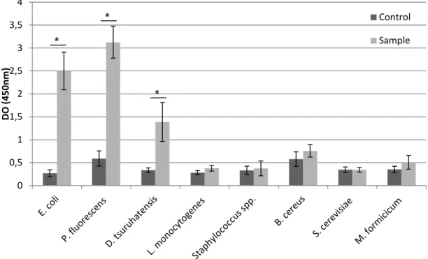
The specificity/sensitivity test of the ISH procedure allowed not only to verify the probe specificity and sensitivity to their targets using this new technology, but rather to ascertain whether the designed protocol would reveal the same behavior in bacteria with different membrane properties.
The results (Figure 7) showed that this protocol was effective for the Gram-negative eubacteria group, with statistically significant differences (p<0.05) between samples and controls on E. coli, P. fluorescens and D. tsuruhatensis. The same was not found in Gram-positive bacteria used in this study, L. innocua, Staphylococcus spp. and B. cereus.
Regarding non-Eubacteria microorganisms, the yeast S. cerevisiae and the archaea M. formicicum, as expected, did not achieve statistical differences (p>0.05) between the samples with or without (control) probe. As such, this alternative procedure revealed a similar pattern to that of standard PNA FISH probe specificity/sensitivity test (section 4.1); but it was unable to correctly identify Gram-positive bacteria
The main cause for the failure of the protocol sensitivity in gram-positive eubacteria is probably related to the permeabilization step. The major structural difference of gram-positive bacteria is the presence of a thicker peptidoglycan wall, 20-80 nm on gram-gram-positive in contrast to 2-3 nm on gram-negative [56]. It was precisely because of this fact that the lysozyme has been included into the permeabilizing step. Lysozyme hydrolyzes the β-1,4 linkages between the peptidoglycan disaccharide subunits; acetylglucosamine and N-acetylmuramic acid [57]. However, this failure does not mean that the lysozyme was not able to digest the peptidoglycan layer. Probably the time used (30 minutes) for the digestion to takes place, was not enough to allow the permeabilization of such thick layer. Some studies demonstrated that several gram-positive are resistant to lysozyme [58], and because lysozyme only partly digests the murein multilayers of fixed gram-positive cells [59], some Gram-positive bacteria cannot be permeabilized by lysozyme treatment alone.
Conjugates of peptide nucleic acid-biotin for the detection of bacteria using in situ hybridization
27
Figure 7 - Results obtained in the specificity/sensitivity test of the ISH procedure. Controls and samples
refer to the test with and without probe, respectively. Statistically significant differences between values indicated by a and b (p <0.05).
Streptavidin-HRP conjugate molecules are very large (5–6 nm and 40 kDa) comparing with a standard PNA/DNA probe (500 – 100Da) used [60]. The problem of the diffusion of large-molecular-weight molecules such as enzymes, antibodies, or (strept)avidin into whole fixed cells, was already described in some studies, e.g. using oligonucleotide probes directly labeled with horseradish peroxidase [61]. In permeabilization for large molecules the margin between the accessibility of target molecules and the loss of target molecules or complete cell lysis becomes very narrow [61]. This is an important point because the permeabilization problem could be overcome with an increasing of lysozyme incubation time, however, in a mixed sample could produce different effects depending on the species (permeabilize some and lyse others).
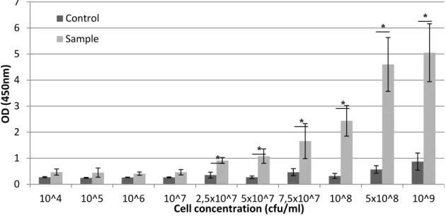
4.5 - Detection limit of the ISH procedure
The determination of the detection limit of a detection technique is a key step for its characterization. The limit of detection refers to the minimum amount of cells needed to generate a positive result easily distinguished from a negative result. A good detection limit will thus prevent the occurrence of false negative results.
The results of this test (figure 8) showed that the statistically significant differences (p<0.05) between controls and samples started on 2.5 x10⁷ cfu/ml concentration, at which concentrations it is also possible to find differences at the naked eye (Figure 9 - D). This
0 0,5 1 1,5 2 2,5 3 3,5 4 D O (450n m ) Control Sample
![Figure 1 - Basic steps of FISH: fixation/permeabilization, hybridization and washing. [20]](https://thumb-eu.123doks.com/thumbv2/123dok_br/15448761.1026759/23.892.158.780.104.497/figure-basic-steps-fish-fixation-permeabilization-hybridization-washing.webp)