UNIVERSIDADE DE LISBOA
FACULDADE DE CIÊNCIAS
DEPARTAMENTO DE BIOLOGIA ANIMAL
Retinoic acid:
a key regulator of vertebrate embryonic development
Mestrado em Biologia Evolutiva e do Desenvolvimento
André Dias
Dissertação orientada por:
Doutor Moisés Mallo
UNIVERSIDADE DE LISBOA
FACULDADE DE CIÊNCIAS
DEPARTAMENTO DE BIOLOGIA ANIMAL
Retinoic acid:
a key regulator of vertebrate embryonic development
Mestrado em Biologia Evolutiva e do Desenvolvimento
André Dias
Dissertação orientada por:
Doutor Moisés Mallo
Professora Doutora Sólveig Thorsteinsdóttir
2015
Resumo
O ácido retinóico é uma molécula sinalizadora, sintetizada a partir da vitamina A, necessária para o correto desenvolvimento embrionário dos vertebrados, uma vez que regula a transcrição de genes essenciais durante vários processos da embriogénese. A relação entre a sua produção e degradação, respetivamente através das enzimas Raldh2 e Cyp26a1, leva a que a sua atividade sinalizadora ocorra de uma forma bastante específica quer em termos de dose, localização da sua atuação (tecido) ou quer em termos temporais. De facto, a alteração dos níveis de ácido retinóico durante o desenvolvimento embrionário pode ser letal ou dar origem a doenças congénitas (como por exemplo, spina bífida)1–6.
Uma das etapas mais importantes na embriogénese dos vertebrados é a gastrulação. Durante esta, para que a formação dos três folhetos germinativos (que mais tarde irão dar origem a todos os tecidos e órgãos do organismo) possa ocorrer devidamente, é necessário um correto controlo da atividade sinalizadora mediada pelo ácido retinóico, que nesta fase do desenvolvimento é fundamentalmente fornecido por via materna (caso dos ratinhos), dado que alterações na sua degradação enzimática pelas Cyp26 podem resultar na morte do embrião. Tal ocorre uma vez que o ácido retinóico controla a expressão de Nodal (excesso de ácido retinóico provoca a indução ectópica de Nodal), proteína indispensável para que se dê o início da gastrulação através da formação da linha primitiva, decorrente da migração das células do epiblasto para a parte mais posterior do embrião7. Aparentemente a introdução de um transgene (T-streakCreERT) juntamente com o alelo repórter ROSA26R-β-gal em ratinhos
mutantes para o gene Gdf11, produziu letalidade embrionária durante a gastrulação8. Após verificarmos que o efeito não era devido ao transgene por si próprio, mas sim possivelmente ao local onde este foi inserido no genoma de uma linha particular (#47), procurámos caracterizar molecularmente os embriões Gdf11-/-::T-streak-CreERT#47+/0 de forma a tentar
encontrar a causa desta aparente letalidade. A alteração dos níveis de ácido retinóico observada nos embriões Gdf11-/- durante a transição entre a formação de tecidos do tronco e
da cauda, fez-nos considerar a possibilidade de durante a gastrulação ocorrer uma alteração semelhante desses níveis, que combinada com possíveis efeitos do transgene pudesse resultar na morte dos referidos embriões. No entanto, as experiências realizadas não só não permitiram a identificação de qualquer problema durante a gastrulação, decorrente da mutação no gene Gdf11 e/ou do transgene T-streak-CreERT#47+/0, como demonstraram a
correta formação da linha primitiva nos embriões Gdf11-/-::T-streak-CreERT#47+/0. Estudos
adicionais mostraram ainda viabilidade dos embriões Gdf11-/-::T-streak-CreERT#47+/0 a E10.5.
Uma possível explicação para este facto prende-se com a estratégia utilizada na genotipagem do alelo mutante de Gdf11, pois usava iniciadores desenhados para amplificar parte da cassete de neomicina (introduzida para criar a mutação nesse gene9) que também está presente no alelo repórter ROSA26-β-gal, que formava parte das experiências originais que levaram à nossa hipótese inicial. Assim sendo, é bastante provável que os erros cometidos na determinação do
que aparentemente essa alteração não se verifica durante a gastrulação, uma vez que a expressão tanto de Cyp26a1, como de Nodal permaneceram sem alterações.
Após a gastrulação, nos vertebrados, dá-se primeiro a formação da cabeça (na parte mais anterior do embrião) e depois por um processo de extensão axial, no sentido anterior para posterior, é formado o pescoço, a seguir o tronco e por último a cauda. Apesar do desenvolvimento destas estruturas ser progressivo e de depender dos progenitores axiais, parece que a forma como estas diferentes partes do corpo são criadas é diferente10. Mutações nos genes T, Cdx e Wnt3a11–17 suportam esta teoria, segundo a qual a posição dos membros superiores e inferiores delimita os referidos blocos estruturais do embrião (cabeça/pescoço, tronco e cauda). Como referido anteriormente, o mecanismo segundo o qual ocorre a transição tronco-cauda já foi demonstrado pelo nosso laboratório10, mas a transição entre a formação da cabeça e do tronco ainda permanece por esclarecer. Curiosamente a mutação da enzima Raldh2, que leva à inexistência de ácido retinóico nos tecidos neurais e na mesoderme, leva à morte do embrião após o desenvolvimento ser interrompido ao nível dos membros anteriores18,19. Mas, se for administrado ácido retinóico até essa altura do desenvolvimento (~E8.25) o embrião é capaz de ultrapassar esse bloqueio e formar as seguintes estruturas (tronco e cauda)20. Esse facto fez-nos considerar a hipótese de que o ácido retinóico pode estar a controlar o mecanismo de transição entre a formação da cabeça e do tronco. Para identificar essa necessária mudança nos progenitores axiais, dependente do ácido retinóico, efetuámos uma análise transcriptómica comparativa a partir de ARN isolado das caudas de embriões “tipo selvagem” a E8.75/E9.0 (ou seja, onde a transição já foi efetuada e estão a ser criados os tecidos do tronco) e de embriões Raldh2-/- da mesma idade (nos quais esta transição
encontra-se bloqueada). Após análiencontra-se dos resultados preencontra-sentes na RNA-encontra-seq, foi encontra-selecionado um grupo de genes candidatos com base na grandeza da expressão diferencial observada entre embriões tipo selvagem e Raldh2-/-, e tendo em conta a sua significância e função biológica. Vários genes
desse grupo (exemplo: Wnt3a, Dkk1 e Cav1) estão associados à sinalização Wnt, cuja atividade canónica (via -catenina) parece estar diminuída na cauda dos embriões mutantes. Esta observação é bastante interessante, tendo em conta a comparação dos fenótipos dos embriões mutantes para Wnt3 (que apresentam ausência total de mesoderme), com o fenótipo dos embriões Wnt3a-/- (em que o desenvolvimento apenas ocorre de forma normal
até ao nível do membro anterior)21, sugerindo que a atividade do ácido retinóico possa ser responsável por esta mudança na sinalização Wnt (de depender de Wnt3 e passar a depender de Wnt3a). Sendo que esta hipótese contrasta com a observação de que a expressão de
Wnt3a parece estar aumentada na cauda dos mutantes22, é possível que a existência de regulação diferencial dos vários componentes da sinalização Wnt nos embriões Raldh2-/- possa
resultar na incapacidade dos progenitores de responder apropriadamente a Wnt3a, ocorrendo dessa forma a inibição da sinalização canónica de Wnt. Por esse motivo, através de hibridação in situ procurámos observar a expressão de alguns desses genes envolvidos na sinalização Wnt e complementámos esses estudos com uma abordagem de sobre-expressão através da utilização de transgénicos (onde o gene avaliado foi associado ao promotor de Cdx2 que expressa nos progenitores do eixo). Estudos similares foram também feitos com outros genes não associados à sinalização Wnt (Mesp1 e Fgf4). No entanto as nossas experiências não permitiram determinar o mecanismo inerente à mudança nos progenitores, necessária para que o embrião termine de criar tecidos da cabeça e inicie a produção de tecidos do tronco.
Ainda sobre a forma como a atividade do ácido retinóico influência os progenitores axiais, através de hibridação in situ, confirmámos que na mesoderme pré-somítica, a área onde tanto
T como Sox2 são expressos é menor nos embriões Raldh2-/-. Em experiências preliminares foi
possível observar, através de imunofluorescência, a existência de células que expressam T e Sox2 (apesar de não ter sido possível contabilizá-las), indicando dessa forma a presença dos progenitores axiais nos embriões Raldh2-/-. Por último, surpreendentemente ao que está
publicado, verificámos que em alguns embriões Raldh2-/- é possível observar umas pequenas
protuberâncias onde Tbx5 (marcador da indução dos membros anteriores) encontra-se expresso mais tarde no desenvolvimento (do que aquilo que seria suposto) e em menor quantidade. Por isso, considerando o fenótipo decorrente da mutação de Cyp26a1 (onde ocorre uma transformação homeótica da vertebra cervical C5 para C6 e da C7 para uma vertebra torácica)23, é possível que o ácido retinóico seja crucial durante este período do desenvolvimento, definindo o tempo em que ocorre a transição entre a formação da cabeça (pescoço) e do tronco no embrião. Apesar desta hipótese necessitar de uma validação experimental, conectando a transição cabeça-tronco e a indução dos membros anteriores, todas as evidências apontam no sentido de que a atividade do ácido retinóico nos progenitores axiais é apenas necessária durante a transição cabeça-tronco (e não durante todo o processo de extensão axial) sendo depois restringida, pela Cyp26a1, a zonas mais anteriores do embrião24.
Palavras-chave
Abstract
Retinoic acid (RA) is a signalling molecule, derived from vitamin A, necessary for proper vertebrate embryonic development. It acts in a tissue, time and dose specific manner, shaping the embryo through the regulation of several master transcription factors. Alterations in RA levels during embryonic development are known to cause several problems, including embryonic lethality2.
During gastrulation, the formation of the three germ layers requires balanced interaction between RA and Cyp26 molecules, which is crucial for Nodal expression7. Interestingly, in Mallo’s lab, embryos carrying a transgene (T-streakCreERT), the cre reporter
allele ROSA26R-β-gal, and a total inactivation of Gdf11, seemed to die during this stage8. Considering the interaction between Gdf11 and Cyp26a125, we hypothesized that the transgene, together with Gdf11 might have affected RA signalling. However our experiments failed to find any indication of patterning problems that could justify the early lethality that was initially observed. Additional analyses indicate that the original phenotype might have resulted from incorrect genotyping of the parent lines. Also, we could conclude that despite what happens during the trunk to tail transition in Gdf11-/-, in these mutants RA signalling
seems not to be affected during gastrulation.
Embryos lacking RA (Raldh2-/-) become truncated at the forelimb level19. Since Raldh2 mutant embryos exposed to acute RA treatments at E8.25 acquire trunk and tail structures20 we concluded that RA signalling controls the mechanism regulating the head to trunk transition. To understand this process we performed a comparative transcriptomic analysis between tails of Raldh2-/- and wild type embryos. So far, we could not elucidate the
mechanism for this transition but our evidence suggests that it possibly involves Wnt signalling. Also our experiments, concomitantly with data regarding Cyp26a123, seem to indicate that RA activity in the axial progenitors is only necessary during this transition, thus setting the time at which it occurs.
Keywords
Table of contents
^ Resumo...2 Abstract...5 Chapter I – Introduction General Introduction...9 General Aims...17Chapter II – Retinoic acid and the mechanism involving the head to trunk transition in the axial progenitor cells Introduction……….19
Material and Methods……….22
Results……….31
Discussion……….42
Acknowledgements/Contributions……….45
Chapter III – A novel approach to the Gdf11-/-::T-streakCreERT#47+/0 problem: The combination effect Introduction……….………47
Material and Methods……….49
Results……….51
Discussion……….…56
Acknowledgements/Contributions……….58
Chapter IV – Final Considerations Final Considerations………..60
Supplementary Information
I – Cuffdiff RNA-seq analysis (EXCEL file)
II – Solutions (PDF file)
III – Manufacturer protocols (PDF file)
Chapter I
General Introduction
I - Retinoic Acid
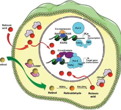
Retinoic acid (RA) is a signalling molecule, derived from vitamin A (retinol), necessary for proper embryonic development. Once retinol is inside the cell it is converted by retinol dehydrogenases (Rdh enzymes) into retinaldehyde, from where RA is synthesized through retinaldehyde dehydrogenases (Raldh enzymes). Endogenously produced or exogenous RA then binds to specific retinoic acid receptors, which interact with retinoic acid responsive elements (RAREs) in the genome, in order to activate the transcription of target genes (Fig.1). Regarding these target genes, RA is known to regulate several master transcription factors necessary for key processes during formation of the vertebrate body (e.g. gastrulation and axial elongation). RA is also degraded by cytochrome P450 enzymes (e.g. Cyp26a1), which limits RA activity in a tissue, time and dose specific manner1–3.
Alterations in RA signalling can produce a variety of problems during embryonic development, ranging from lethality to congenital spinal deformities in vertebrates (including spina bifida)4–6. Further below I will discuss several stages of vertebrate embryonic development to help better understanding how RA activity shapes the vertebrate embryo.
Fig. 1 – Summary of the retinoic acid signalling pathway, representing the activity of
exogenous or intracellularly-produced RA as a transcriptional activator of gene expression. (From Rhinn and Dollé, 2012)
II - Vertebrate embryonic development – the mouse case
Vertebrates display a large diversity of body shapes and sizes. They are formed through multiple tightly regulated and interdependent morphogenetic events during embryonic development. Despite the gross architectural differences observed early in development across vertebrate species, the fundamental principles of vertebrate embryonic development are maintained throughout vertebrate phylogeny (e.g. gastrulation, somitogenesis and axial extension). However, the environment where embryonic development occurs represents one of the main differences among species; for instance, birds develop inside independent eggs, whereas mammals develop inside the progenitor uterus. I will now focus on mammalian embryonic development, using the mouse embryo as example26,27.
II.a - From a fertilized egg to the gastrulating embryo
After fertilization, the egg undergoes cell divisions reaching the eight-cell stage, when apical-basal polarity is generated through a process known as compaction, forming a solid mass called morula27,28. Then the compacted embryo undergoes additional cleavages and cell fate decisions, eventually reaching the blastocyst stage27,28. At this stage it is possible to distinguish two different cell compartments, the trophectoderm (TE) and the inner cell mass (ICM). Cells from the TE will be necessary for implantation (which occurs around embryonic day (E) 4.5) and they will give rise to important extraembryonic tissues. The ICM contains pluripotent cells that undergo a second lineage separation, driven by Fgf signalling, to produce the visceral endoderm (positive for Gata4/6) and the epiblast (expressing Nanog and Oct3/4), which will give rise to the embryo proper27–29. Then, the epiblast changes morphologically to produce the so-called “egg cylinder” at around E5.5. At this stage the first signs of an AP axis are evident with the formation of the anterior visceral endoderm (AVE) (expressing Nodal and Wnt inhibitors). The AVE derives from the distal visceral endoderm (DVE) that migrates to the prospective anterior side of the embryo, shortly after being induced at the distal end of the embryo27–31. The molecular mechanism controlling the AP patterning of the embryo at this stage depends on interactions between β-catenin and Cripto. Other members of the Wnt and Nodal pathways, as well as Fgfs, are also involved in this important event27,28,32. Genetic experiments in the mouse and grafting experiments using other vertebrate model organisms showed that the AVE is involved in two main processes: the production of head structures later in the embryo and the induction/control of primitive streak (PS) formation in the opposite side of the egg cylinder, which will break the radial symmetry in the embryo and marks the onset of gastrulation27,30.
II.b – The gastrulating embryo: formation of the primitive streak
when RA activity is not correctly buffered by Cyp26 enzymes, gastrulation fails7. Nodal is a protein belonging to the transforming growth factor β (Tgfβ) family responsible for sorting epiblast cells towards the posterior part of the embryo to generate the PS 33,34. Wnt3-activated canonical Wnt pathway is also required for PS induction/maintenance and for the transcriptional activation of Brachyury (T) in the newly formed mesoderm27,35–39. PS induction requires expression of Wnt and Nodal inhibitors from the AVE to concentrate Nodal and Wnt signalling in the posterior epiblast. Accordingly, loss of these inhibitors (e.g. Cerl1 or Lefty1) resulted in the production of ectopic/enlarged PS27. Wnt/β-catenin signalling is also necessary for the maintenance of Nodal expression in the epiblast through a feedback loop involving
Nodal, Bmp4 and Wnt340. Epiblast cells in the PS will then undergo an epithelial to mesenchymal transition and ingress through the PS. These mesenchymal cells will give rise to the mesoderm and definitive endoderm27,40. This process also requires Fgf signalling (e.g. Fgf8) as its inactivation resulted in an accumulation of cells in the epiblast41. Epiblast cells located anterior to the newly formed PS are not affected by PS activity and therefore remain within the epiblast layer, eventually giving rise to the ectoderm27.
II.c – The node and the left-right asymmetry
Using transplantation experiments, Spemann and Mangold discovered a group of cells in the amphibian embryo that have the capacity to induce the formation of a new vertebrate body axis. They coined the term organizer to describe this tissue, which is thought to be conserved across vertebrates. In the mouse it is referred to as the node42. This organizer is now considered as a secondary or later organizer, because the new axis it can induce does not
Fig. 2 – (A) Development of a fertilized egg into a gastrulating embryo; (B) Gastrulae: the three germ layer embryo.(Adapted from Takaoka et al, 2011, and Arnold et al, 2009)
DVE AVE
ICM
Gastrulae
E7.5
include formation head structures (only trunk and tail, since the induction of head structures depend on the AVE). At the end of gastrulation, the node can be observed at the most anterior end of the PS, which at this stage is fully extended. Fate mapping experiments indicated that cells ingressing through the node are fated to produce the notochord43–45.
In addition to its role in gastrulation, the mouse node is also involved in the control of left-right asymmetry45. Disruption of the embryo’s bilateral symmetry is mainly controlled by cells located in the node, which have motile cilia that rotate in a clockwise direction to create a leftward flow of extracellular fluid43. Experiments using mutant mice with immotile cilia showed a total absence of flow in the node that later in development results in a randomization of the organ situs. Also, an artificial reversal in the flow’s direction was able to induce situs inversus. This flow seems then to direct some signalling activity from the node into the left side of the lateral plate mesoderm (LPM), once the reversal of the flow direction or its ablation results in changes of the organ situs43,44. The molecular components of this activity seem to vary among species (e.g. Shh in chicken46). However, Nodal signalling seems to be a key player in left-right asymmetry in most vertebrates. Nodal expression in the mouse can first be observed in the node during late PS and head-fold stages (~E7.75/E8.0). This expression is crucial for the creation of asymmetry in the embryo since blocking Nodal expression in the node inhibits later on Nodal expression on the left LPM, therefore creating left-right patterning defects43,47. A physical midline-barrier composed of the notochord and the floor plate, both derived from the node, has been proposed to maintain correct laterality of Nodal expression. The existence of a molecular midline-barrier, separating the expression of signals in the left-right LPM near the node has also been proposed based on studies of Lefty1-/- embryos. Lefty
genes are expressed on the left LPM and are thought to cooperate with Nodal to orchestrate a left fate in the embryo43,45. However, the processes that lead to left-right asymmetric morphogenesis in the embryo are not fully understood. RA signalling might play a role in this process since different RA levels seem to alter Nodal expression in the LPM48,49, thus creating several left-right patterning defects. Left-right alterations can also be observed in embryos lacking the RA synthesizing enzyme Raldh2, represented by asymmetric somite formation50.
A B C
II.d – From head to tail – axial elongation
After gastrulation, embryo growth is progressive. The first structure to form is the head at the anterior embryonic end. The axis then extends progressively in an anterior to posterior direction, first to produce the neck, then trunk structures and finally the tail10,51. Tightly associated with these processes is the continuous production of mesoderm, which seems to depend to a large extent on Wnt signalling. Loss of Wnt3 blocks mesoderm production, which can be evidenced by the absence of T expression35,37,38. This phenotype is similar to that of β-catenin mutants, suggesting that Wnt3 signals through the canonical pathway39,52. Later in development Wnt3 expression begins to sag and, concomitantly, Wnt3a expression becomes activated at the posterior embryonic end. This change in Wnt ligand expression coincides with a switch in the Wnt molecule driving mesodermal production53. Accordingly, a null mutation in Wnt3a leads to strong axial truncation caudal to the forelimb level derived from the failure to produce mesoderm17. At this point, T expression, which initially depended on Wnt3, now requires Wnt3a signalling to be maintained. Interestingly, however, observations in Lef1-/-::Tcf1-/- embryos suggest that this transcription complex
downstream of Wnt3a is required for maintenance but not initiation of T expression. This seems to indicate that Wnt3 and Wnt3a use a different set of effector complexes to regulate T expression17,21,54,55. Fgf signalling also plays a role in the regulation of T expression because
Fgf4 and Fgf8 double mutants display reduced T expression in the axial stem zone56.
Genetic experiments removing T and/or Cdx genes indicated that head structures are formed through a different process than trunk or tail. In particular, those experiments revealed that despite truncations in the main body axis these embryos were still able to produce head mesoderm and the first somites that originate cervical vertebra11–16. This important transition between head and trunk formation occurs at the level of the forelimb, which interestingly matches with the stage when Raldh2-/- stop developing (~E8.25)18.
The progressive production of new tissues at the caudal end of the embryo relies on a pool of cells, known as the long term axial progenitors, located at the caudal tip of the embryo. These include the bipotent neural-mesodermal progenitors (NMPs) that give rise to the neural tube and the paraxial mesoderm57,58. Early in development, around E8.25/E8.5, NMPs are located at the node-streak border (NSB) and in the epiblast, between the node and the anterior PS. Later in development, after the mouse embryo underwent axial turning (~E9.5), NMPs are reallocated into the tailbud, to a region known as the chordoneural hinge (CNH)58,59 – as roughly exemplified in Fig.4. Recent studies indicated that these cells co-express T and Sox2 and that they are able to self-renew within the embryo58. Also, they showed that Tbx6 is required to drive the NMPs into a mesodermal fate, through a down-regulation of Sox260. Accordingly, in the absence of Tbx6, embryos produce more neural tissue at the expense of paraxial mesoderm61. Other molecules, including Cdx proteins and signalling pathways activated by Wnt and Fgf ligands are also involved in the control of axial progenitors activity, although how is not fully understood62–66.
Again RA plays a key role at different stages of axial elongation. A variety of experimental evidence indicates that RA is required for the tight balance between maintenance and differentiation of NMPs. RA activity is somehow required for the transition
from head to trunk development as revealed by the truncated phenotype of Raldh2
-/-embryos19. Later in development, however RA signalling has to be kept on track (e.g. relocation of the NMPs into the CNH). This can be illustrated by genetic inactivation of Gdf11 signalling9,25 or of Cyp26a1 23. This last mutation, results in axial truncation at the level of the trunk to tail transition (TTT), due to excessive levels of RA, since ablation of Rarg rescued the caudal truncation in Cyp26a1-/- embryos67. Also, treatments with high RA doses during axial extension cause similar truncation phenotypes68.
II.e – From progenitors to body structures
During axial extension, the body forms all the primordiae of the different tissues and
h
ea
d
E8.25
E10.5
A
B
C
Fig. 4 – (A, B) Highlighted tailbud of an E8.25 (A) and E10.5 (B) embryo, showing the location of
NM progenitors in the NSB/epiblast (A) and later in development in the CNH (B). (C) Expression domain
of key molecular signals related with axial progenitors in an E8.5 embryo. (Adapted from Cambray et al, 2002, and Neijts et al, 2013)
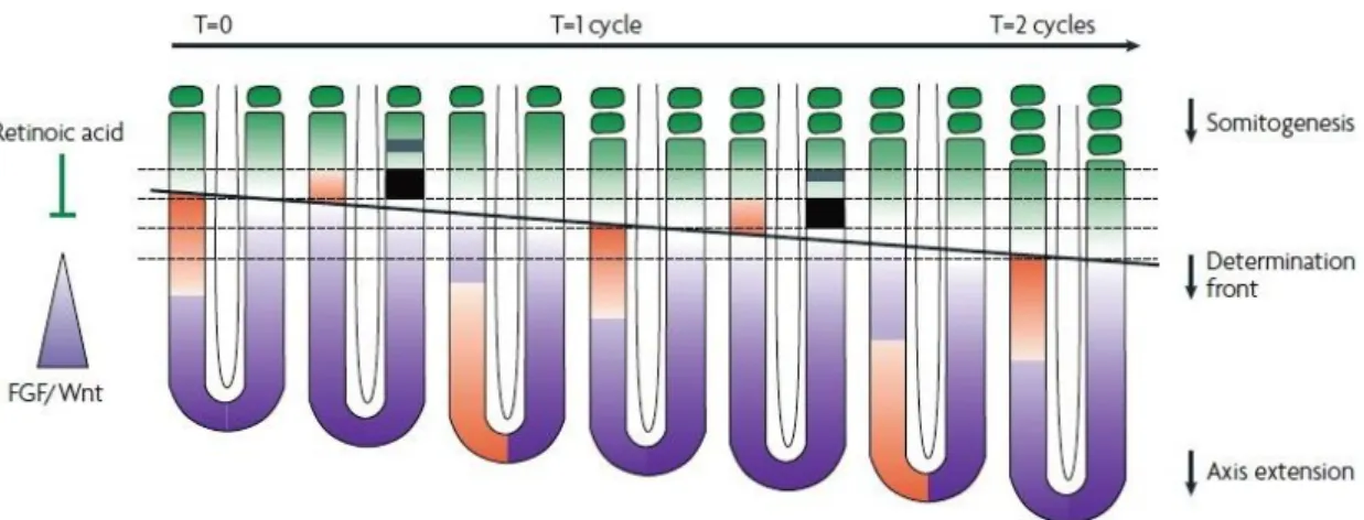
presomitic mesoderm51,69. All vertebrates undergo somitogenesis but the number of formed somites and the time necessary for the formation of new segments varies across species. The periodic formation of somites relies on the existence of a molecular oscillator coined as the “segmentation clock”69. These molecular oscillations can be observed by cyclic expression of several genes in the PSM. Although the specific identity of these genes seems to vary across vertebrate species (e.g. hairy1 in chicken70), most of them are members of the Notch signalling pathway or its target genes (e.g. Mesp2), whose requirement for the formation of inter-somitic boundaries seems to be conserved among vertebrates69,71. Apart from the “segmentation clock”, somitogenesis also relays on another system, the “wavefront”72(Fig.5). It consists of complex signalling gradients responsible for translation of signals from the molecular oscillator into patterning information, necessary for the formation of new somites51,69,72. This molecular control of segmentation is thought to derive from the convergence of two opposite functional gradients provided by Fgf and Wnt signalling (posterior to anterior inhibitory gradient) and RA signalling (anterior to posterior activating gradient), thus generating the determination front within the anterior PSM, where new intersomitic borders are created69,71.
As new segments are being produced, somites located at more anterior positions of the axis start differentiating. This differentiation incorporates positional information responsible for producing specific structures at the different axial levels in order to create a properly organized body51,69. In this process, AP patterning of the axial skeleton is mostly controlled by Hox genes69,73(Fig.6). Several Hox clusters/genes have retinoic acid responsive elements (RAREs) nearby18,74, which could explain the requirement of RA signalling for their proper expression and the many homeotic transformations resulting upon treatment with high doses of RA at different times of development68,75.
Fig. 5 – A somitogenesis model integrating the segmentation clock and the determination front.
Opposing gradients, Fgf-Wnt (purple) and RA (green) sets the determination front (black line), in the PSM, during axial elongation. The wave of cyclic gene expression (orange) is represented on the left side of the embryo, whereas on the right side is represented the acquisition of a future segmental domain by
In addition to the formation of the body structures described above, RA is also important in organ formation. Heart and kidneys are examples of organs where RA is crucial for morphogenesis3. RA signalling is also involved in the induction and/or AP patterning of non-axial structures, like the forelimbs, where it is necessary for induction and initiation of the pre-limb bud17,76,77(Fig.7).
In summary, RA is a key regulator of vertebrate embryonic development, required for a variety of process at different developmental stages. Importantly, its activity is often necessary during a specific time-window and in a dose and tissue dependent manner.
Fig.6 – Hox gene expression and genomic organization in the mouse embryo; the paralog
groups within the four clusters are color-coded according to their anterior-most expression domain in the mouse embryo.(Adapted from Pearson et al, 2005)
General Aims
The general aim of this MSc thesis was to contribute to a better understanding of how RA regulates vertebrate embryonic development. Two different projects were addressed during the period of this thesis and they are described here in Chapter II and III. Specific aims for each project of this thesis are specified in the referred chapters.
Chapter II
Retinoic acid and the mechanism involving the head to
trunk transition in the axial progenitor cells
Introduction
Retinoic acid activity during axial elongation
Studies using RA-responsive transgenic mice concluded that the Raldh2 enzyme is the main responsible for RA synthesis in neural and mesodermal tissues19. The genetic disruption of the Raldh2 enzyme leads to several abnormalities during development, after gastrulation, resulting in embryonic lethality at midgestation. This lethality could be rescued with RA treatments administered maternally at E8.25. Raldh2 mutant embryos (Fig 8) display a development block starting around E8.25, failing to form limb buds and they do not undergo axial rotation (normally occurring at E8.25-E8.5). These embryos have an open neural tube, small otocysts, only the one branchial arch and the heart has only a single and expanded cavity18–20. In the paraxial mesoderm these mutants form only 10 to 12 somites, which are smaller and more densely packed than in normal embryos, which results in shortened AP axis. Interestingly, however, analysis of rescued embryos using RA treatment indicated that RA is only essential for correct somite formation until the 6th somite18–20,78.
As referred in Chapter I, Raldh2-/- show asymmetric development in the paraxial
mesoderm around E8.25, during formation of the more anterior somites. This asymmetry is inverted in embryos with situs inversus. It is possible that this results from a requirement of RA to synchronize development in the paraxial mesoderm while maintaining asymmetric development in the lateral mesoderm50,79–81. Some of these patterning effects of RA have been shown to derive from interactions with Fgf8. In particular, it was found that RA antagonizes Fgf8 in the node ectoderm but not in the node mesoderm, where its expression is not uniform. In general RA sets the boundaries of Fgf8 expression domain in the epiblast and the heart 18,50,74,82. On the basis of these observations and considering the extended Fgf8 expression domain in the presomitic mesoderm (PSM) of chicken embryos lacking RA signalling, it has been postulated that the asymmetry in the somites of Raldh2 mutants derive from asymmetric
Fgf8 expression in the PSM2,83,84. The reciprocal interactions between RA and Fgf8 are also considered to be required for somitogenesis and axial extension by creating opposing gradients of activity in the PSM 6,22,63,69,84,85. However, the relevance of continuous RA signalling
Raldh2-
/-WT
Fig. 8 – E9.5 Raldh2 mutant versus wild type embryo (h- heart, b-branchial arch, s-somites,
for somitogenesis and axial extension is a question of debate because Raldh2-/- can be rescued
upon RA treatments at E8.25 and keep extending their axis and forming somites in the absence of RA. The fact is that all available evidence suggests that RA is fundamental at a specific time point during vertebrate embryonic axial extension, coincident with the stage when the forelimb buds are formed.
Raldh2 mutant embryos are unable to induce forelimb buds as estimated by both the
absence of a physical bud and of Tbx5 expression, possibly due to an extension of the Fgf8 domains and/or to a direct role of RA production in somites in the regulation of Tbx5 in the pre-bud18 . It has also been described that a strong and specific dose of RA (maternally administered) is necessary to induce forelimb outgrowth in these mutants76,78. Further analyses by Niederreither (2008) suggested that RA is essential for the induction and AP patterning of the pre-bud, but after this stage it must be removed for limb growth to continue normally. Interestingly, in rescued Raldh2-/- embryo hindlimb outgrowth was normal, indicating
different RA requirements for forelimb and hindlimb development76,78,86.
Fig. 9 – Model for Segment Determination (opposing RA and Fgf8 gradients during somitogenesis and
RA coordinates the Head to Trunk Transition?
As referred in Chapter I, vertebrate embryonic axial elongation relies on a pool of cells in the caudal tip of the embryo, characterized as long term axial progenitors58. In the last years, Mallo’s lab have been studying the way these cells control axial growth and are responsible for the existent diversity of body shapes among vertebrates. Arnon Jurberg showed that Gdf11 signalling plays an essential role in the TTT10. This activity is in part mediated by the activation of Isl1 in the NMPs, which will be responsible for the terminal differentiation of progenitors of the lateral mesoderm to produce the hindlimb10. More recently, Rita Aires during her PhD work (unpublished) observed that the tailbud of Gdf11-/- embryos contained some Oct4
expressing cells (much later than normal). This persistent Oct4 expression seems to be responsible for the increased body length of Gdf11-/- embryos since when Oct4 was
transgenically overexpressed in the tailbud, under the regulation of a Cdx2 promoter, embryos had longer trunks and severe trunk to tail transition defects87.
The phenotypic effects caused by inactivation of a variety of genes (e.g. T, Cdx1/2/4 and Wnt3a11–17) indicate that during vertebrate body formation, the mechanisms supporting head, and trunk development are also fundamentally different, indicating the existence of mechanisms specifically regulating the head to trunk transition (HTT). Interestingly the stage at which RA signalling seems to be fundamental during embryonic body axis formation, matches with the time when the production of head tissues stops and the formation of trunk tissues starts. So the specific aim of this project was to address the hypothesis of possible activated RA targets, in the node streak border, be responsible for a switch in the long term axial progenitors necessary for the HTT in the vertebrate embryo.
Material and methods
Mice/Embryos
All experiments and procedures conducted on mice followed the Portuguese (Portaria 1005/92) and European (Directive 2010/63/EU) legislations, concerning housing, husbandry, and welfare. These animals were kept in 12h dark/light cycle, some maintained in C57BL/6J background at the Rodent Facility and others (necessary for producing transgenic embryos) in FVB/N strain at the Pathogen-free Animal Facility, also at the IGC.
The transgenic embryos (Cdx2.Cav1T1 and Cdx2.Cav1T2) were generated using standard transgenic procedures (e.g. Nagy et al 2003), through pronuclear microinjection by the Transgenic Unit at the IGC.
The TOPGAL transgenic mice, Tg(TCF/Lef1-lacZ)34Efu/J, described in Gupta and Fuchs (1999) were purchased from Jackson Laboratory.
RA mutant mice were created by producing a Raldh2 allele unable to produce the protein product, by the introduction of stop codons in the second exon using the CRISPR-Cas9 technique. The mutation was confirmed by sequencing. Raldh2Ext designed oligos were used to
amplify part of the Raldh2 coding sequence from genomic DNA obtained from the tails of the transgenic progeny. The band containing the mutation was isolated in a 1% agarose gel in TAE using the QIAEX II Gel Extraction Kit*. Molecular cloning procedures were performed as described further below. In this case, the amplified fragment was digested with XhoI and XbaI, and inserted into the XhoI and SalI sites of the pKXM plasmid#. Plasmids containing the insert were sequenced to confirm the introduction of the mutation. The lethality of the mutation was also phenotypically confirmed since no Raldh2-/- pups were found upon crosses between Raldh2+/- mice, and the phenotype of Raldh2-/- embryos matched with what is described in the
bibliography.
Matings were done late in the afternoon and plugs were checked in the morning of the next day (corresponding to E0.5). Embryos were collected by caesarean section on cold PBS§ and fixed in 4%PFA§, at 4oC overnight. They were then washed in PBT§ and dehydrated in graded methanol series (25%, 50%, 75% made in PBT), washed in 100% methanol and stored
§
Solutions detailed information is present in Supplementary Information II *All manufacturer protocols are present in Supplementary Information III
#
PCR conditions and genotyping
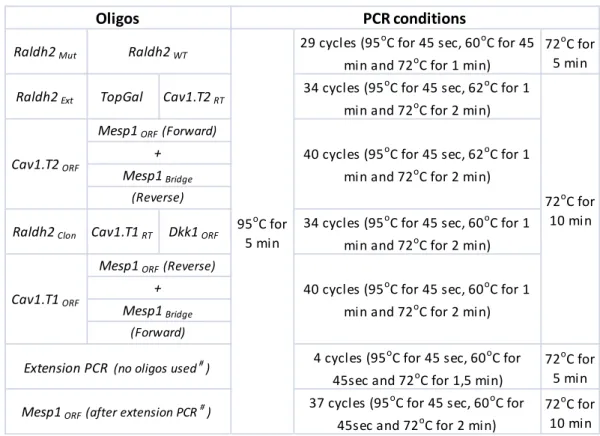
During the embryo harvesting process, the yolk sac was isolated and incubated overnight (ON) at 500C, in yolk sac lysis buffer§ containing proteinase K. Genomic DNA was isolated from mouse tail biopsies and incubated under the same conditions, but in PBND buffer§ containing proteinase K. After the ON incubation, proteinase K was inactivated at 950C for 15 min. Primers for several different PCR reactions and genotyping conditions are described in Tables 1 and 2. Exceptions are referred to in the corresponding sections.
Primers for Forward Reverse
Raldh2Mut TCTCCCACTGAATTCTATCAC
Raldh2WT AACACTCTCCCACTCTCTGAG
Raldh2Ext CATCTTCTAAGCAATACACAC
Raldh2Clon GACTCGAGTTTTCTGATCTCCCAGATCTC GATCTAGATCTTCTAAGCAATACACAC
Cav1.T1ORF CTGTCGACCTCCTCAGAGCCTGCAGCCAG GAGCGGCCGCGTCCCTCATATCTCTTTCTGCG
Cav1.T2ORF CTGTCGACTGTTCCCATCCGGGAACAG GAGCGGCCGCGTCCCTCATATCTCTTTCTGCG
Cav1.T1RT CTGTCGACCTCCTCAGAGCCTGCAGCCAG TTCTGGTTCTGCAATCACATC
Cav1.T2RT ACAGCCAGGCTGACTCTTGAC TTCTGGTTCTGCAATCACATC
Mesp1ORF CCGTCGACGGATAAAGCTACAGCGGACCC GAGCGGCCGCCAAAGGAAAAGTGTCTGTGC
Mesp1Bridge
CAGTCCCTCATCTCCGCTCTTCAGCAGCGACA TGCTG
GTCGCTGCTGAAGAGCGGAGATGAGGGACTG GGCTCC
Mesp1RT CGCAGAAACAGCATCCCAGG TGTCCCCTCCACTCTTCAGGC
Eno2RT CAAGCTGGCCCAGGAGAATGG CTGGTTGTACTTCGCCAGACG
Fgf4RT CCGGTGCAGCGAGGCGTGGTG GTACGCGTAGGATTCGTAGGCG
Dkk1ORF TGCGTCCTTCGGAGATGATGGTTG CTGTCGGTTTAGTGTCTCTGGCAG
ActinRT ATGAAGATCCTGACCGAGCG TACTTGCGCTCAGGAGGAGC
TOPGAL CGTGGCCTGATTCATTCC CGTGGCCTGATTCATTCC GTTTTCTGATCTCCCAGATCTC
Table 1 –Primers used for polymerase chain reaction
(normal PCR, RT-PCR and RT-qPCR).
PCR reaction contained Quantity
Template (DNA) ~ 1µL Primer Forward 0,25µL Primer Reverse 0,25µL dNTPs 25mM 0,2µL (0,2mM) MgCl2 25mM 2,5µL (2,5mM) Taq Buffer 10x 2,5µL (1x)
Taq polymerase (NZYTECH or Fermentas) 5und/µL 0,2µL (1und)
# used for Mesp1 cloning
Table 2 – PCR conditions
RT-PCR
To obtain the desired cDNAs from isolated RNA (described below), a Reverse Transcriptase reaction was performed using the NZYTECH RT Kit* according to manufacturer’s instructions. In these reactions, we used random hexamers for priming and the NZY Ribonuclease Inhibitor was substituted by nuclease-free water. At the end of the incubation, the cDNA was stored at -20oC.
PCR reactions were then performed using about 4µL of cDNA under the conditions specified in Table 3.
Raldh2Mut 29 cycles (95
oC for 45 sec, 60oC for 45
min and 72oC for 1 min)
72oC for 5 min
Raldh2Ext TopGal Cav1.T2RT 34 cycles (95
oC for 45 sec, 62oC for 1
min and 72oC for 2 min)
Raldh2Clon Cav1.T1RT Dkk1ORF 34 cycles (95
oC for 45 sec, 60oC for 1
min and 72oC for 2 min)
4 cycles (95oC for 45 sec, 60oC for 45sec and 72oC for 1,5 min)
72oC for 5 min 37 cycles (95oC for 45 sec, 60oC for
45sec and 72oC for 2 min)
72oC for 10 min Extension PCR (no oligos used#)
Mesp1ORF (after extension PCR#)
40 cycles (95oC for 45 sec, 62oC for 1 min and 72oC for 2 min)
Cav1.T1ORF
Mesp1ORF (Reverse)
+ Mesp1Bridge
(Forward)
40 cycles (95oC for 45 sec, 60oC for 1 min and 72oC for 2 min)
Oligos PCR conditions Raldh2WT 95oC for 5 min 72oC for 10 min Cav1.T2ORF
Mesp1ORF (Forward)
+ Mesp1Bridge
(Reverse)
Cav1.T1RT Cav1.T2RT Eno2RT Mesp1RT ActinRT
40 cycles (95oC for 45 sec, 60oC for 1 min and 72oC for 2 min) 40 cycles (95oC for 45
sec, 62oC for 1 min and 72oC for 2 min)
Oligos RT-PCR conditions 95oC for 5 min 72oC for 10 min Fgf4RT
qPCR
SYBR Green quantitative PCR analysis* was performed using cDNA obtained from RNA isolated from tails of Raldh2 embryos, according to the manufacturer’s protocol and under the conditions described in Table 4. The results were analysed by Bio-Rad CFX Manager software
Agarose gel electrophoresis
Agarose was dissolved in 1X TAE§, usually at the concentration of 0.8%, 1% or 2%. Ethidium bromide or GelRed (1:39 in H2O) was added ~1:4 in order to visualize the DNA when UV light was applied. 6x Gel loading dye was added to each sample (1x final concentration). An electric current of about 120V was applied to the gel immersed in 1X TAE. The QIAEX II Gel Extraction Kit* was normally used to extract the DNA from the agarose gels (eluted in TE§).
Phenol-Chloroform extraction and standard digestions
In several situations described below, DNA was purified by phenol-chloroform extraction. For this, TE buffer was used to make a final volume of 100μL and an equal volume of phenol-chloroform was added. The sample was mixed and centrifuged for 3 min at 14000 rpm. The DNA was recovered from the aqueous phase and precipitated with a 1:10 volume of 3M NaOAc pH 5.3 and 2.5 volumes of 100% ethanol for 30min on dry ice. The precipitated DNA was recovered by centrifugation at 14000rpm for 30min at 4ºC. The retrieved (air-dried) DNA pellet was resuspended in an appropriate volume of water or TE for further experiments (DNA concentrations were determined with a Nanodrop). Standard digestions using restriction enzymes were some of those applications. For that, to 5µL of DNA we normally added to 13 µL of H2O, about 0,5µL of Enzyme and 2µL of the 10X concentrated buffer. The resulting mixture was incubated at least during 1h 30 min at 370C.
RNA extraction from the tails of Raldh2 embryos
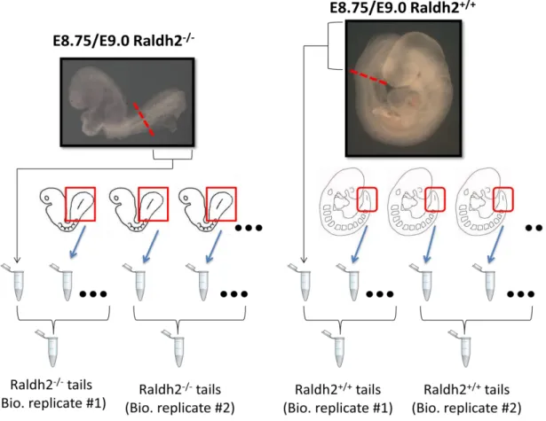
To analyze the transcriptome of axial progenitor cells, we isolated tails from E8.75/E9.0
Raldh2 embryos (resulting from Ralhd2+/- mouse intercrosses), which were stored immediately
at -800C. Upon genotyping, tails were grouped according to their category in groups of about 8 tails, as shown in Fig. 10. RNA was then isolated from these selected tissues, including two replicate groups for each category (WT and MUT) using TRI Reagent (Trizol), under the
950C 950C 10 sec 600C 30 sec 720C 30 sec 650C 950C Plate Read 5 min 39 cycles Melting Curve 5 sec increment 0,50C Table 4 – qPCR conditions
conditions specified in the TRI REAGENT SIGMA protocol*. 11µL of nuclease-free water was added to the RNA pellet, which was dissolved at 650C during 10 minutes and then put on ice; finally samples were stored at -800C.
Fig. 10 – Process for Raldh2 isolation and storage in biological groups and category (Raldh2+/+ or (Raldh2-/-)
RNA-seq
After the RNA was isolated from the selected group of tail tissues, a small amount was sent to the Gene Expression Unit (IGC), where the sample quality was assessed. Once the RNA integrity and concentration had the necessary levels, the four samples (RNAmut1, RNAmut2, RNAwt1 and RNAwt2) were sent to the EMBL in Germany where the RNA-seq was conducted. The RNA-seq results were analysed by the Bioinformatics Unit (IGC) using the Cuffdiff algorithm, which estimates expression at transcript-level resolution and controls for variability across replicates.
Molecular cloning
sample concentration was measured using a Nanodrop and the ligation reactions were performed with T4 DNA ligase using vector and insert in a 1-5 proportion. After 1h at room temperature the transformation was performed using DH5- competent cells in a 1/10 reaction on ice followed by a heat shock (42oC) during 45 seconds and chilled on ice before being grown in LB medium at 37oC for 1 hour. Finally the bacteria were plated on solid LB medium with ampicillin (50 µg/mL) and incubated ON at 37oC. Single colonies were then picked and grown on LB medium with ampicillin at 37°C, during 3 hours with shaking. These cultures were used for screening purposes (see next section) and stored at 40C.
Molecular screenings and Plasmid DNA, Mini- and Midi-scale preparations
To perform a fast screen for positive colonies, 1µL of the above cultures was used for a PCR reaction, using the appropriate primers for each case. For colonies giving a positive PCR signal, 20µL of the culture was retrieved and added to 5mL of LB+ampicillin. Then, 1,5mL of that culture was spun down and the pellet resuspended in 100μl of TE with RNase (10μg/mL). Then, 300μl of TENS§ and 150μl of 3M KOAc (pH5,2) were added to the sample. After mixing, the mixture was centrifuged (4min at 14000rpm) and the supernatant was transferred into a fresh tube containing 900μl of 100% EtOH. Then a spin was performed to pellet DNA and RNA, which were dissolved in 50µL TE in order to be used in screening digestion reactions. When higher purity DNA was required, plasmids were purified using commercial plasmid preparation kits: “NZYTECH MINIPREP” kit* was used when small amounts of plasmid were needed (for sequencing reactions) and “MN Plasmid DNA purification (NucleoBond Xtra Midi)” kit* was used when larger amounts of plasmid DNA were necessary (e.g. for later in vitro transcription experiments).
Sequencing reactions
To confirm the sequences of the cloned DNA products, cycle sequencing reactions were performed as described below (Table 5).
Table 5 – Standard cycle reaction conditions
Reagent Quantity
Template ( plasmid DNA mini prep) ~3µL (normally about 350ng)
Primer (T7 or T3) 1 µL (~5pmol) Buffer 2µL Terminator 2µL H20 Mili-Q Up to 10µL 960C 960C 10 sec 500C 5 sec 600C 4 min 40C 1 min 25 cycles
-The amplified DNA was then precipitated with 2µL of 3M NaOAc and 50µL of 95% EtOH (plus 10µL H20) for 30 min at RT, centrifuged during 30min at 4ºC, the pellet was washed with 250μL of 70% ethanol and finally centrifuged at 14000 rpm for 15min at 4ºC. The supernatant was again removed and the pellet air-dried. The samples were then sent to the Genomics Unit at the IGC. The output sequences were analysed by the combined use of Finch TV software and BLAST (NCBI).
Microinjection construct
20 µg of the DNA construct was digested with the appropriate restriction enzymes to remove plasmid sequences and gel purified using the QIAquick Gel Extration Kit*. The DNA was eluted with 40µL of buffer EB and stored at -20ºC.
Probe generation by in vitro transcription
RNA probes for in situ hybridization were produced by transcription in vitro. First, a specific restriction enzyme was used to linearize 10g of plasmid containing the relevant cDNA, purified by a phenol-chloroform extraction and resuspended in 20µL of water. Then, about 1g of cDNA was used for RNA transcription (Table 6) for 3h at 370C. The RNA was recovered by NaOAc precipitation and its length confirmed in an agarose gel (normally 2%).
Table 6 – RNA transcription reaction
Cav1 cloning, probe and transgenic construct
Cav1 has two transcript variants, Cav1.T1 representing the longer one and Cav1.T2
representing the shorter one. Cav1.T1 was amplified by PCR using cDNA produced by reverse transcription of RNA isolated from ES cells. The PCR reaction used Pfu polymerase, 5mM MgCl2
and Cav1.T1ORF oligos. Cav1.T2 was amplified (also with Pfu and with Cav1.T2ORFoligos) using the
Cav1.T1 amplified sequence as template. They were inserted into the SalI and NotI sites of pBluescript II KS# using standard cloning procedures.
To create the Cav1.T1 and Cav1.T2 transgenic constructs under the Cdx2 promotor, the
DNA ~1ug
T7/T3 polymerase 1µL
Buffer 10x 2µL
DIG label 1µL
For in vitro transcription, the pKS.Cav1.T1 plasmid was linearized with SalI and the probe was synthesized with T7 RNA polymerase.
Dkk1 probe
The Dkk1 coding sequence was amplified using cDNA obtained from RNA extracted from Raldh2-/- embryo tails. For this PCR, we used HotStart Taq* (QIAGEN) under the
manufacturer conditions. The cloning was made with a PCRII-TOPO plasmid# using the TOPO TA cloning Kit*. Standard molecular cloning procedures were adopted and the plasmids transformed into DH5α competent cells. For in vitro transcription, the plasmid was linearized with SpeI and the probe was synthesized with T7 RNA polymerase.
Mesp1 probe
Once the entire coding sequence of Mesp1 is split by just one intron we designed oligos to amplify the cDNA from genomic DNA, linking the two exons in vitro. For this we performed two separated PCRs: “A” using Mesp1ORF (Forward) + Mesp1Bridge(Reverse) oligosand in
“B” Mesp1ORF (Reverse) + Mesp1Bridge,both using Pfu polymerase. Then, after the amplified DNA
was gel purified with QIAEX Gel Extraction Kit, equimolar amounts were mixed and used as a template for PCR cycles without primers, also using Pfu polymerase. Finally, we added the
Mesp1ORF (forward and reverse) oligos to the PCR reaction. After the required band was isolated
from a 1% agarose gel, we followed the standard cloning techniques to introduce the Mesp1 coding sequence into the SalI and NotI sites of pKS bluescript (BLAST was performed against
“Mus musculus mRNA for MespI, complete cds”, NCBI accession: D83674). For in vitro transcription,
the plasmid was linearized with SalI and the probe was synthesized with T7 RNA polymerase.
Other probes
All the other probes used in this work were available in the lab synthesized by in vitro transcription.
In situ hybridization
All the whole mount in situ hybridizations were performed using DIG-labeled antisense RNA probes as described in Kanzler et al 1998. On the first day, the embryos were rehydrated, and washed in PBT. They were then bleached in 6% H2O2 at RT for 1 h and washed in PBT. They were then treated with proteinase K for time lengths that depended on their developmental stage. After inactivating proteinase K with glycine§ and several washes in PBT, the embryos were post-fixed with PFA/glutaraldehyde§. Pre-hybridization solution§ was then added and incubated for 1 hour at 65oC, after which it was changed for hybridization solution containing the probe (3 to 6μL of probe per mL of pre-hybridization solution), and incubated overnight
between 65oC and 70oC. On day 2, after several washes using a post-hybridization solution§ and TBST§ the embryos were incubated in blocking solution (MABT/block/10% sheep serum§), first without antibody during 2,5h and then with the antibody against DIG (1:2000), overnight at 4oC. On day three several washes were performed with MABT and on day four the embryos were incubated with a developing solution (NTMT§ plus NBT/BCIP), at RT, protected from the light. The reaction was stopped with PBT, the embryos fixed in 4% PFA and finally stored in PBT.
Whole mount embryo immunofluorescent staining
After rehydration from methanol to PBS, the embryos were washed 4x30 min in PBST§, incubated with 1M glycine in 0,1% PBST for 30 min to reduce unspecific binding and washed 3x in PBST to remove glycine residues. The embryos were then blocked in donkey serum blocking buffer§ overnight at 40C. On the next day, the embryos were incubated ON at 40C with new blocking serum containing (~1:250) anti-T (Goat AF2085 from R&D) and anti-Sox2 (Rabbit ab92494
monoclonal from abcam) primary antibodies. On the third day, after several washes in PBST, the
embryos were incubated in new blocking serum containing the secondary antibodies (donkey anti goat rabbit and donkey anti rabbit, ~1:1000). The last day, after several washes of PBST, the embryos were incubated with DAPI in PBST (~1:5000) during 2,5 hours and finally through a process involving graded washes in methanol to methyl salicylate to clear the embryos, which were prepared in a blade for confocal microscopy.
Wnt reporter activity
To observe β-catenin signalling activity in Raldh2 mutant embryos, we introduced the TOPGAL Wnt reporter mice into the Raldh2+/- background. Raldh2+/-::TOPGAL+/0 males were
crossed with Raldh2+/-::TOPGAL+/0 females to obtain Raldh2-/-::TOPGAL+/0 embryos, which were
fixed in Mirky’s ON at 4ºC. After 3x10 min washes with 0,02%Tween-20/NP40 in PBS, the embryos were stained (protected from the light) at 370C with X-gal staining solution§ and finally post fixed ON, at 4ºC, with Mirky’s§.
Results
Looking for a change in the axial progenitors
Previous studies indicated that RA signalling is essential for the vertebrate embryo to undergo HTT. We thus decided to use Raldh2 mutant mouse embryos, which lack neural and mesodermal RA activity, to search for the mechanisms controlling this transition. In the mouse embryo this transition occurs around E8.25/E8.5, roughly corresponding to the stage when the forelimb bud is induced. We therefore isolated tails from wild type E8.75/E9.0 embryos, which already started trunk development, and tails from Raldh2-/- littermates, which display a strong
developmental delay, possibly resulting from a failure to undergo the HTT. We then analysed gene expression in these tissues by RNA-seq and compared their mRNA profiles (Fig. 10, 11 and Supplementary Information I). 0 2 4 6 -15 -10 -5 0 5 10 15
-l
o
g
10(p
-v
al
ue
)
log
2(fold change)
Fig. 10 – Volcano plot (t-test) using RNA-seq data, with some highlighted genes that had a high fold
change in the analysis.
Cav1 Fgf4
Dkk1
Comparison of the data obtained with the biological replicates indicated high robustness of the RNA-seq assay. Similarly, in this study, it was possible to observe in this study variations in gene expression fitting previously described gene expression experiments using other strains of Raldh2 mutants. In particular, Fgf8 and Wnt3a expression values were consistent with in situ hybridization (ISH) studies from Duester et al (2006 and 2009) 50,88;
Fgf17, Fgf18, Wnt8a, Axin2, Cdx1, Cdx2 and Cdx4 showed modifications similar to those
described in Duester et al (2006)88; Meis1 and Meis2 were down-regulated, similarly to what was observed by Cunningham (2014)89; T, Sox2, Sprouty2 and Mkp3 showed expression differences similar to those in Ribes et al (2009)90; Hand1 and Fgf3 values were consistent with ISH experiments reported in Dollé et al (1999)19; and Pax6, Bhlhe40 as well as Crabp2 expression profiles were congruent with those obtained by Niederreither (2013)91. We also performed a few control tests, through ISH (Fig.11), to further assess the quality of the transcriptomic data and our Raldh2 mutation.
Fig. 11 – Transcriptome analysis data reflecting fold change (log2 (fold change)) differences between
selected mRNA gene expression in wild type and Raldh2-/- isolated tails. Regarding Hox expression: no
major changes were noticed in anterior Hox genes, the differences in posterior Hox genes can be due to developmental stage of wild type embryos and the high fold change observed in the Hox12 cluster
derives from very low absolute values.
Sh
h
W
n
t2
w
n
t3
a
Bm
p
4
Ral
d
h
2
WT
Raldh2
-/-Raldh2
-/-A B C D E F G H I JFig. 12 – ISH analysis was congruent with data from RNA-seq. Shh, Bmp4 and Wnt3a were up-regulated
in the mutant tails (A, E, I respectively ) when compared with their expression in wild type littermates (B, F, J respectively. Wnt2 and Raldh2 expression was downregulated in the mutants (C, G) compared to
Considering the previous experiments and preliminary RT-qPCR analysis (Fig.13) on selected genes that had a high-fold change in the RNA-seq, which seem to confirm the analysed data, we concluded that the mRNA profiles obtained provide a faithful representation of gene expression in the analyzed tissues.
Mesp1 was asymmetrically expressed in the Raldh2
-/-presomitic mesoderm
Mesp1 is one of the genes whose expression was severely decreased in the Raldh2
mutant tails according to our transcriptome analysis. Considering that the phenotype derived from the mutation of this gene is very similar to that of Raldh2-/- embryos (e.g. growth
retardation, failure to overcome axial turning and accumulation of cells in the PS)92,93, and because lineage tracing experiments showed that Mesp1-expressing cells seem to contribute to all head structures up to (and including) the forelimb buds94, we performed ISH experiments to observe Mesp1 gene expression in Raldh2-/- embryos. Shortly after gastrulation and during
formation of the first somites, Mesp1 expression was unchanged in the absence of embryonic RA (Fig.14).
Fig.13 – RT-qPCR analysis on selected genes from the RNA-seq: Cav1 (Cav1.T1 and Cav1.T2) was found
overexpressed, while Mesp1 was downregulated in Raldh2-/-.
At later stages, when the embryo is undergoing HTT, Mesp1 expression in Raldh2 mutant embryos followed an asymmetric pattern similar to that observed by Vermot et al (2005)80 for Mesp2. We could also observe equivalent patterns for other genes involved in segmentation69 (Fig.15).
Considering the expression patterns of these genes in the PSM, it is possible that the apparent downregulation observed in the RNA-seq data resulted from the left-right somite asymmetry present in Raldh2 mutant embryos80, which could have led to recovery of different amounts of expressing tissue in wild type and Raldh2 mutant embryos. Altogether, we can conclude that Mesp1 is not responsible for the failure of Raldh2 mutant embryos to undergo HTT.
Possible Fgf4 overexpression in Raldh2 mutants is not the cause for the observed
embryo truncation
As referred in Chapter I, Fgf signalling is involved in a variety of important process during embryonic development (e.g. axial extension, somitogenesis and limb morphogenesis). Together with Fgf8, Fgf4 is the main Fgf signal responsible for controlling those processes. According to Duester et al (2009), the absence of RA does not seem to affect Fgf4 expression in the tails of E8.25 embryos. However, according to our RNA-seq data (which was performed
C
D
E
F
A
B
Fig. 15 – Gene expression analysis for Mesp1 (A, B), Paraxis (C, D) and Ripply2 (E, F) in E8.75/E9.0 embryos. An asymmetric expression of these genes can be observed in the Raldh2-/- embryos (A, C and
in a slightly different developmental stage) Fgf4 expression in the Raldh2 mutant embryos was higher than in wild type embryos. Therefore to understand if a deviation from the normal timing of posterior Fgf4 down-regulation could be the cause for the mutants truncated phenotype, we tested Fgf4 expression by ISH in Raldh2 embryos (Fig.16). The patterns that we obtained for Fgf4 expression, and most particularly in the Raldh2-/- embryos, were not
completely consistent. At E8.5, several Raldh2-/- embryos had expression in the axial stem
zone, whereas from E8.75 onwards Fgf4 expression in the posterior part of the embryo was observed only in a subset of the embryos. When this expression was present, it was restricted to a small number of cells. After 8,75 we could not detect Fgf4 expression in the tailbud of any
Raldh2-/-. So we concluded that even if posterior Fgf4 expression in some Raldh2 mutant
embryos was maintained longer than in wild type embryos, the low number of Fgf4 expressing cells and the timing of their expression in Raldh2-/- embryos cannot explain the truncation
observed in these mutants and therefore Fgf4 does not seem to play a major role in the HTT.
Canonical Wnt signalling activity was reduced in the tails of Raldh2 mutant embryos
In the RNA-seq data sets we could observe differential expression of several genes involved in the Wnt signalling in the Raldh2 mutant tails (e.g. Wnt3a, Cav1 and Wif1). Considering the role of Wnt signalling during axial extension, particularly the interactions and molecular functions of Wnt3 and Wnt3a (fully described in Chapter I) we decided to explore a possible RA-mediated change in Wnt signalling at the time of the HTT. For that we used the TopGal Wnt reporter transgenic mice in order to observe the activity of the canonical WntA B B’
C C’ D
B’
C’
Fig. 16 – Fgf4 expression was observed in the PSM of only some Raldh2 mutants: A – Example of E8.75
Raldh2-/- were no expression was detected; B and C – mutants embryos (E8.75 and E9.0, respectively) were Fgf4 was detected in the PSM. D – Wild type E9.0 were Fgf4expression was absent in the PSM.