UNIVERSIDADE DE LISBOA
FACULDADE DE MEDICINA VETERINÁRIA
VIRULENCE CHARACTERIZATION AND ANTIMICROBIAL RESISTANCE OF MAJOR BACTERIAL GENERA FROM DIABETIC FOOT INFECTIONS
CARLA MOTTOLA
Orientadores: Professora Doutora Maria Manuela Castilho Monteiro de Oliveira Professora Doutora Patrícia Maria Cavaco Silva de Sá Montez
Tese especialmente elaborada para obtenção do grau de Doutor em Ciências Veterinárias na Especialidade de Ciências Biológicas e Biomédicas
Júri:
Presidente: Professor Doutor Rui Manuel de Vasconcelos e Horta Caldeira Vogais:
- Professor Doutor Miguel Viveiros Bettencourt - Professor Doutor Luís Manuel Morgado Tavares
- Professor Doutor Paulo Manuel Rodrigues Martins da Costa - Professora Doutora Maria Manuela Castilho Monteiro de Oliveira - Professor Doutor João João Duarte Alves Mendes
“Look up at the stars and not down at your feet.
Try to make sense of what you see, and wonder about what makes the universe exist. Be curious.”
Stephen Hawking
- In the memory of my uncle Roberto, You left us too early, we miss you everyday
- In the memory of Professor Cristina Vilela, Who taught me to never give up
i
AKNOWLEDGEMENTS
This thesis is the harvest of a very long journey, full of will, sacrifices, good and less good moments, with some stones in the way. Nevertheless, it was hugely worth it. None of this would have been possible without the help and support of my family, my friends and the people who contributed to the ideas and knowledge on this work. I will always be grateful to all of you. I will bring with me a great baggage full of many professional and life teachings, many moments to remember and many people for life and that I will keep forever.
To my parents, who always gave me so much love and let me follow my dreams. Mamma e papà, grazie per aver sempre creduto in me e per avermi sempre permesso di seguire i miei sogni. Siete il mio porto sicuro, vi amo di bene.
To my sisters, that are my light and without whom I cannot imagine my life. Laura e Lory, siete la più grande certezza della mia vita, le mie migliori amiche e la mia vita senza di voi non avrebbe alcun senso. Vi amo all’infinito.
Un grazie al resto della mia famiglia, per avermi sempre supportato durante questi anni e per avermi insegnato ad essere forte e non desistere mai. Sono fortunata ad avere una famiglia così.
À Professora Cristina Vilela, que nos deixou demasiado cedo, o meu maior obrigada por ter acreditado em mim e não me ter deixado desistir quando achei que não tinha forças para continuar. A sua sabedoria, a sua garra, o seu carinho, o seu ser especial ficará sempre comigo.
À Professora Manuela Oliveira, minha orientadora, o meu muito obrigada por ter aceite este inesperado desafio, pelos ensinamentos, pelos conhecimentos partilhados, pela paciência constante, por acreditar em mim e pela amizade.
À Professora Patrícia Cavaco-Silva, minha orientadora, obrigada pelo apoio, pelas sugestões valiosas, pelo suporte e palavras amigas. Foi um prazer trabalhar consigo.
Ao Professor Luís Tavares, um muito obrigada por me ter acolhido na sua equipa, pelo apoio e pelo carinho demonstrado ao longo deste percurso.
Ao Dr João Mendes, a sua energia é contagiante. Obrigada por ter contribuído para que este trabalho tenha sido possível, pelas sugestões valiosas e pela confiança. Gostei muito de trabalhar consigo e espero voltar a fazê-lo no futuro.
À Professora Teresa Semedo, pelos ensinamentos, pela ajuda e pelo apoio constante. À Carla Carneiro, por me ter orientado quando me iniciei no mundo da Bacteriologia, pela paciência, pelo apoio em todos os momentos, pelas longas conversas e pela amizade. Ao Dr Alexandre Leitão, por me ter apoiado sobretudo nos momentos mais difíceis, pelas palavras amigas e por me ter mostrado o que é ser uma pessoa justa e coerente.
À Clara Leandro, obrigada por me ter feito conhecer o mundo dos fagos e me ter ajudado no trabalho.
ii
Ao Professor Carlos Martins, obrigada por me ter acolhido na sua equipa quando me iniciei na Investigação e pelos conhecimentos partilhados.
À Solange, obrigada pelos ensinamentos nos exórdios desta viagem e pelo apoio. Ao Professor José Melo-Cristino, pelos conhecimentos partilhados e pela confiança.
Ao Rui C., sem o qual a minha vida não teria sido o que é hoje e sem o qual não teria seguido este caminho. Obrigada por teres sempre acreditado em mim, pelo apoio incondicional e pelo amor ao longo destes anos. Infelizmente decidiste abandonar o barco a breve distância da chegada, mas foste essencial para que eu atingisse a meta.
Rui Seixas, esta viagem sem ti nunca teria sido a mesma coisa. Muito obrigada por tudo, pelo apoio profissional e emocional, pela troca de ideias e conhecimentos, por teres vivido isto comigo a cem por cento, por me teres aturado nos maus momentos, pelas experiências lindas que fomos partilhando nestes anos e por saber que estiveste e que estás lá sempre. Não podia ter desejado um melhor companheiro de viagem. És um amigo para a vida. Marga, desde o momento em que nos conhecemos nos corredores da FMV percebi que ias ser minha amiga para sempre. Até hoje muito caminho temos percorrido juntas, muitas coisas partilhadas, muitos momentos para recordar e ainda mais há de vir! Obrigada pela amizade, pelo apoio constante, por me dares sempre força, pelo teu optimismo contagioso. Adoro-te.
Joana, ter vivido isto tudo contigo foi uma sorte, obrigada por seres uma ótima amiga. A tua sinceridade, a tua garra, as tuas palavras de conforto, o teu carinho e as nossas infinitas conversas foram preciosas ao longo deste percurso. Não tenho dúvidas de que irei poder contar contigo para sempre. Gosto mesmo muito de ti.
A voi amici di una vita: Annalisa, Enrico M., Enrico A., Federica, Irma, Isidoro, Luca, Marco Rosalucia, Rossana. Sono davvero fortunata ad avervi nella mia vita, anche se spesso lontani vi porto sempre con me e so che potrò sempre contare su di voi. Mille parole non basterebbero per ringraziarvi di tutto, siete una costanza nella mia vita, un bene prezioso che terrò per sempre stretto a me.
A Raffaele, esempio di forza e costanza. Sei e sarai sempre un grande amico, grazie di tutto. Cátia, és a minha minina. Um obrigada é pouco por agradecer o apoio constante, a força, a confiança, e o facto de me chamares geniazinha quando mais preciso. És uma amiga excecional, sei que estás lá sempre e isso não tem preço.
Sara, és a minha Saretta. É como se te conhecesse há uma vida, e sabes que sou a irmã que nunca tiveste. Obrigada por me apoiares sempre, pelas palavras amigas, pelo teu riso contagioso e por acreditares em mim.
Manuel, é nas alturas mais difíceis que nos apercebemos com quem podemos realmente contar. Não foi uma surpresa saber que és uma destas pessoas mas foi uma confirmação. Obrigada por todo o apoio, pelo carinho e por saber que não estou sozinha. Nunca hei de esquecer.
iii
Miguel, muito obrigada pela amizade ao longo destes anos, pelo apoio, pelos conselhos de vida e por me aturares quando faço perguntas sobre o português.
Vocês são a minha família portuguesa e sempre serão.
André, foste umas das melhores surpresas ao longo desta caminhada. Obrigada pela tua amizade, tive muita sorte em cruzar-me contigo e sei que fiz um amigo para a vida.
Ao João C. e à Silvia, pelos ensinamentos preciosos, pela ajuda, por me terem apoiado nos momentos difíceis e pela amizade.
Ao João e ao Hernâni, obrigada pelos bons momentos e por me apoiar sempre. Tenho a certeza de que muitos outros bons momentos iremos passar juntos; João obrigada por começares esta nova etapa comigo, a nossa amizade só poderá crescer ainda mais. Gosto muito de vocês.
À Carina, pela vontade de aprender e boa energia. Obrigada por teres trabalhado comigo. À Clara, pela ternura e conselhos maternos, por todo o apoio ao longo destes anos.
À Ju, ao Ferdinando, ao Gonçalo, ao Pedro, por todos os momentos partilhados, as brincadeiras e o suporte neste percurso.
À FCT, pelo financiamento através da minha bolsa de Doutoramento e do projecto do pé diabético.
À FMV e ao CIISA, por ter sido a minha segunda casa durante estes últimos sete anos, por me terem dado a oportunidade de me iniciar no mundo da Investigação e pela confiança. At last but not least, I would like to thank some people that tested my resilience in this path. I really learned a lot from this, made me stronger and ready to face what’s coming next.
iv
FUNDING
The present work was funded by the PhD fellowship SFRH/BD/72872/2010, by project grant PTDC/SAU-MIC/122816/2010 - “Biofilms in diabetic foot: microbial virulence characterization and cross-talk of major isolates”, from the Fundação para a Ciência e Tecnologia – FCT Portugal, and by project UID/CVT/00276/2013 from “Centro de Investigação Interdisciplinar em Sanidade Animal” (CIISA) of Faculty of Veterinary Medicine, University of Lisbon, Portugal.
v
Abstract
Virulence characterization and antimicrobial resistance of major bacterial genera from diabetic foot infections
Diabetes mellitus is a major chronic disease that continues to increase significantly. One of the most important and costly complications of diabetes is the development of foot ulcers, colonized by pathogenic and antimicrobial resistant bacteria, which may be responsible for impairing its successful treatment. Diabetic foot ulcer (DFU) bacterial communities can be organized in polymicrobial biofilms, which may be responsible for its chronicity. The ability of these communities to produce biofilm was evaluated and was higher when compared to biofilm formation by individual species.
Staphylococcus aureus is one of the most prevalent species in diabetic foot infections (DFI). Staphylococci isolated from DFU in patients from the Lisbon area were identified, genotyped and screened for virulence and antimicrobial resistance traits. The isolates showed high genomic diversity, were resistant to important clinically antibiotics and expressed relevant virulence determinants.
As biofilm formation is one of the most important virulence traits of S. aureus, the antimicrobial susceptibility patterns of biofilm-producing S. aureus strains were also analysed. The minimum biofilm inhibitory and eradication concentrations were determined for ten antimicrobial compounds. Staphylococci biofilms were resistant to antibiotic concentrations ten to thousand times higher than those effective for planktonic cells. Furthermore, the enterococci frequently isolated from DFI, were also identified and characterized, showing high antimicrobial resistance and important virulence traits.
Since DFI are often caused by resistant bacteria, it is necessary to find alternatives to antibiotic therapy, such as phage therapy. The inhibitory potential of five bacteriophages, previously characterized, was evaluated against established biofilms formed by S. aureus, P. aeruginosa and A. baumannii. A significant cell reduction after phage exposure was observed, mainly after multiple treatments.
DFI are very complex and studies on this topic are scarce. It is necessary to intensify research in order to develop more adequate therapeutic protocols for this type of infection.
Keywords: Diabetic foot infections, biofilm, staphylococci, virulence determinants,
vii
Resumo
Caracterização da virulência e resistência a antimicrobianos dos principais géneros bacterianos envolvidos em infeções de pé diabético
Diabetes mellitus é uma doença crónica com grande impacto em saúde pública e cuja incidência continua a aumentar significativamente em todo o mundo, atingindo atualmente mais de 400 milhões de pessoas. Uma das complicações mais importantes da diabetes e associada a gastos económicos significativos são as úlceras de pé diabético. Uma vez que a camada protetora de pele é danificada, os tecidos profundos ficam expostos à infeção bacteriana, a qual pode evoluir rapidamente. As infeções das úlceras de pé diabético são a causa mais comum de internamento hospitalar de pacientes diabéticos e uma importante causa de morbilidade, levando frequentemente à amputação dos membros inferiores. Estas infeções podem ser promovidas por bactérias potencialmente patogénicas e resistentes aos compostos antimicrobianos, prejudicando assim o sucesso do tratamento. As comunidades bacterianas presentes nas úlceras podem estar organizadas em biofilmes polimicrobianos, que contribuem para que as infeções se tornem crónicas e muito difíceis de resolver.
Foi avaliada a capacidade de produção de biofilme por comunidades polimicrobianas de isolados bacterianos de pé diabético, utilizando um ensaio de microtitulação em placa com “Alamar Blue” (AB) e uma técnica de Hibridação In Situ Fluorescente Múltipla (MFISH). Esta avaliação foi realizada em três períodos de incubação distintos (24, 48 e 72 horas), depois da determinação da capacidade de formação de biofilme por 95 isolados de úlceras de pé diabético pertencentes a vários géneros bacterianos (Staphylococcus, Corynebacterium, Enterococcus, Pseudomonas e Acinetobacter). Todos os isolados apresentaram a capacidade de produzir biofilme às 24 horas, sendo que a quantidade de biofilme produzido aumentou com o tempo de incubação. Pseudomonas apresentou a capacidade mais elevada de produção de biofilme, seguida de Corynebacterium, Acinetobacter, Staphylococcus e por fim, Enterococcus. Foram encontradas diferenças estatisticamente significativas na capacidade de formação de biofilme entre os três períodos de incubação. As comunidades polimicrobianas produziram mais biofilme do que as espécies individualmente. As comunidades formadas por Pseudomonas + Enterococcus, Staphylococcus + Acinetobacter e Corynebacterium + Staphylococcus formaram mais biofilme do que as comunidades formadas por Enterococcus + Staphylococcus e por Enterococcus + Corynebacterium. O comportamento biológico das diferentes espécies bacterianas nos biofilmes polimicrobianos tem implicações clínicas muito importantes para o sucesso do tratamento deste tipo de infeções. A sinergia entre as bactérias presentes em biofilmes multiespécies foi descrita previamente, sendo que este trabalho representa o primeiro estudo sobre a evolução temporal da formação de biofilme por parte de comunidades polimicrobianas isoladas de úlceras de pé diabético, incluindo várias espécies.
viii
Staphylococcus é um dos géneros bacterianos mais prevalentes nas infeções de pé diabético. Neste estudo, isolados de Staphylococcus (n = 53) obtidos a partir de úlceras de pé diabético de doentes da região de Lisboa foram identificados, caracterizados genotipicamente, e rastreados para genes de virulência e de resistência a antimicrobianos. A relação genética entre os isolados foi avaliada através da técnica de eletroforese em gel de campo pulsado (PFGE) e da tipagem de sequências “multilocus” (MLST) de representantes dos pulsotipos identificados. A reação em cadeia de polimerase (PCR) foi aplicada para deteção de doze genes de virulência e a técnica E-teste foi realizada para determinar a concentração mínima inibitória (MIC) em relação a dez antibióticos. Verificou-se que Staphylococcus isolados de úlceras de pé diabético são genotipicamente muito variados, apresentam resistência a antibióticos importantes do ponto de vista clínico, nomeadamente ciprofloxacina e eritromicina, e expressam diversos determinantes de virulência. Essas propriedades sugerem que os estafilococos podem contribuir para a persistência e gravidade deste tipo de infeções, levando ao insucesso da terapêutica. Além disso, existe a possibilidade de eles poderem transmitir estas caraterísticas a outros microrganismos que partilham o mesmo nicho ecológico. Neste contexto, os pacientes diabéticos podem tornar-se um veículo de transmissão de clones bacterianos entre o ambiente hospitalar e a comunidade.
S. aureus resistentes à meticilina (MRSA) têm emergido como um dos principais problemas clínicos e epidemiológicos a nível hospitalar. As estirpes MRSA têm a capacidade de resistir à ação da maioria dos antibióticos β-lactâmicos, mas também a uma vasta gama de outros agentes antimicrobianos pertencentes a diferentes classes, tornando o tratamento destas infeções muito difícil e dispendioso. Até à data, existem disponíveis duas cefalosporinas de quinta geração eficazes contra MRSA, a ceftarolina e o ceftobiprole, com espetro de ação semelhante. Tendo em conta que a formação de biofilme é uma das mais importantes caraterísticas de virulência de S. aureus e que o seu desenvolvimento desempenha um papel importante na patogénese da infeção, uma vez que representa um mecanismo de defesa bacteriano, os padrões de suscetibilidade antimicrobiana dos isolados de S. aureus produtores de biofilme foram analisados, através da determinação da concentração mínima inibitória de biofilme (MBIC) e da concentração mínima de erradicação de biofilme (MBEC), para dez antibióticos incluindo a ceftarolina. Foi igualmente avaliada a presença de genes relacionados com antibiótico-resistência pela técnica de PCR. Foi observado que relativamente aos antibióticos mais utilizados no tratamento de infeções de pé diabético, são necessárias concentrações muito mais elevadas para inibir a formação de biofilme por isolados de S. aureus in vitro, o que pode explicar o facto da monoterapia com estes agentes não ser frequentemente eficaz em erradicar a infeção. De facto, os biofilmes analisados foram resistentes a concentrações de antibióticos dez até mil vezes mais elevadas do que as necessárias para matar as células planctónicas correspondentes. Os
ix
únicos antibióticos capazes de erradicar os biofilmes produzidos por 50% dos isolados de S. aureus foram a ceftarolina e a gentamicina. Os resultados sugerem que os padrões de sensibilidade aos antibióticos não podem ser extrapolados para infeções com biofilme estabelecido.
Além de estafilococos, existem outras espécies bacterianas frequentemente identificadas em úlceras de pé diabético. Os enterococos são considerados bactérias oportunistas, mas nos últimos anos foram relacionados com infeções clínicas muito graves. Enterococcus sp. isolados a partir de infeções de pé diabético foram identificados por multiplex PCR, caracterizados pela análise de macro-restrição através de PFGE, e rastreados para características de virulência e resistência antimicrobiana. A maioria dos Enterococcus foi identificada como E. faecalis, espécie considerada mais patogénica dentro deste género bacteriano; genotipicamente os isolados mostraram elevada similaridade, revelando uma relação clonal. Todos os isolados foram considerados multirresistentes, produtores de citolisina e gelatinase, e a maioria mostrou ter capacidade de produzir biofilme, demonstrando a importância de Enterococcus no desenvolvimento das infeções de pé diabético e na sua persistência, especialmente em relação à sua capacidade de formação de biofilme e resistência a antibióticos clinicamente relevantes.
Uma vez que estas infeções são frequentemente promovidas por bactérias resistentes, torna-se necessário encontrar alternativas terapêuticas, tais como a terapêutica fágica. Para além das bactérias Gram-positivas já mencionadas, estas complexas infeções de pé diabético incluem muitas vezes bactérias Gram-negativas altamente patogénicas, como Pseudomonas aeruginosa e Acinetobacter baumannii. Um dos objetivos do estudo foi verificar o efeito de cinco bacteriófagos, produzidos pela empresa TecnoPhage, e caracterizados anteriormente, em biofilmes formados pelas espécies bacterianas S. aureus, P. aeruginosa e A. baumannii. A aplicação dos bacteriófagos a células planctónicas revelou-se eficaz às 4 horas após inoculação, mas às 24 horas pós-aplicação obrevelou-servou-revelou-se um ressurgimento do crescimento bacteriano. Em relação aos biofilmes e utilizando a atividade metabólica como medida da viabilidade celular determinada através da aplicação de AB, verificou-se uma redução celular significativa após a exposição aos fagos, quer às 4 quer às 24 horas pós-aplicação, mas mais significativa às 4 horas. Um tratamento múltiplo, com aplicação de fagos a cada 4 horas, promoveu uma diminuição ainda mais significativa da atividade celular. Os efeitos inibitórios mais elevados para células planctónicas e biofilme ocorreu num índice de multiplicidade bacteriófago:bactéria de 10. Estes resultados reforçam o potencial clínico da terapêutica fágica para o tratamento de infeções de pé diabético. As infeções de pé diabético são uma realidade muito complexa e a epidemiologia dos agentes bacterianos envolvidos encontra-se em evolução. Os estudos disponíveis sobre este tema são escassos e é necessário intensificar a investigação quer na vertente
x
microbiológica quer na vertente clínica no sentido do desenvolvimento de protocolos terapêuticos adequados para este tipo de infeções.
Palavras-Chave: Infeções de pé diabético, biofilme, estafilococos, determinantes de
xi TABLE OF CONTENTS AKNOWLEDGEMENTS ... i FUNDING ... iii Abstract ... v Resumo ... vii
LIST OF TABLES ... xii
LIST OF FIGURES ... xiv
LIST OF ABBREVIATIONS ... xv
CHAPTER I ... 1
1.1. Introduction on Diabetic Foot Ulcer ... 2
1.1.1. Definitions and epidemiology ... 2
1.1.2. Pathophysiology ... 3
1.1.3. Diagnosis and classification ... 5
1.2. Diabetic Foot Infections... 7
1.2.1. Definition and pathophysiology ... 7
1.2.2. DFI diagnosis ... 8
1.3. Microbiology of DFI ... 9
1.3.1. Bacteria involved in DFI ... 9
1.3.2. Staphylococci ... 11
1.3.2.1. Generalities and classifications ... 11
1.3.2.2. Staphylococci virulence traits ... 13
1.3.2.2.1. Cell wall-anchored (CWA) surface proteins ... 13
1.3.2.2.2. Secreted factors (exotoxins) ... 15
1.3.2.2.3. Virulence regulatory systems ... 17
1.3.2.2.4. Phenotypic switching ... 18
1.3.2.3. Staphylococci antimicrobial resistance ability ... 20
1.3.3. Enterococci ... 22
1.3.3.1. Enterococci virulence traits ... 23
1.3.3.2. Enterococci antimicrobial resistance ability ... 24
1.3.4. Other bacteria frequently involved in DFI ... 25
1.3.4.1. Corynebacterium ... 25 1.3.4.2. Pseudomonas aeruginosa ... 26 1.3.4.3. Acinetobacter baumannii ... 27 1.4. Biofilms in DFI ... 29 1.4.1. Introduction ... 29 1.4.2. Biofilm formation ... 29
1.4.3. Biofilm and Quorum Sensing ... 30
1.4.4. Multi-drug resistance in biofilm ... 32
xii
1.5. DFU Treatment ... 35
1.5.1. Therapeutic protocols ... 35
1.5.1.1. Debridement, wound healing agents and surgery ... 35
1.5.1.2. Antibiotic therapy ... 37
1.5.1.3. Advanced therapeutics ... 38
1.5.2. Bacteriophage therapy ... 39
1.5.2.1. Bacteriophages: general features and life cycle ... 39
1.5.2.2. Pros and cons of bacteriophage therapy ... 41
1.5.2.3. Bacteriophage therapy over time... 43
1.5.2.4. Bacteriophage therapy and biofilm ... 45
1.6. Thesis objectives and scope ... 46
CHAPTER II ... 47
2.1. Polymicrobial biofilms by diabetic foot clinical isolates ... 48
CHAPTER III ... 63
3.1. Molecular typing, virulence traits and antimicrobial resistance of diabetic ... 64
foot staphylococci ... 64
CHAPTER IV ... 79
4.1. Susceptibility patterns of Staphylococcus aureus biofilms in diabetic ... 80
foot infections ... 80
CHAPTER V ... 95
5.1. Characterization of multidrug-resistant diabetic foot ulcer enterococci ... 96
CHAPTER VI ... 103
6.1. In vitro design of a novel lytic bacteriophage cocktail with therapeutic potential against organisms causing diabetic foot infections ... 104
CHAPTER VII ... 121
7.1. General Discussion ... 122
CHAPTER VIII ... 133
xiii
LIST OF TABLES Chapter 1
Table 1. PEDIS classification system by the International Working Group of the Diabetic Foot
that classifies all ulcers in five main categories………6
Chapter 2
Table 1. Composition of polymicrobial communities tested by a microtiter biofilm assay and
by MFISH………..52
Table 2. Probes used in the Multiplex Fluorescent In Situ Hybridization (MFISH) protocol...54 Table 3. Classification of biofilm production by the DFU clinical isolates, as determined by a
microtiter biofilm assay………...56
Table 4. Comparison of biofilm production between the polymicrobial community and the
individual species, using Friedman’s test and Wilcoxon post hoc with Bonferroni
correction………..58
Chapter 4
Table 1. PCR target genes and primers used in this work………...87
Table 2. In vitro MIC, MBIC and MBEC values for the antibiotics tested against S. aureus
DFU isolates (*CLSI range susceptibility)………88
Table 3. Antibiotic resistance phenotypes and genotypes of S. aureus DFU isolates……….89 Chapter 5
Table 1. Data regarding enterococcal isolates and DFI patients………99 Chapter 6
xiv
LIST OF FIGURES Chapter 1
Figure 1. Diabetic foot infection pathophysiology………5
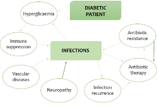
Figure 2. Interplay among factors associated with diabetes, infections and antibiotic resistance in causative agents in diabetic patients………..9
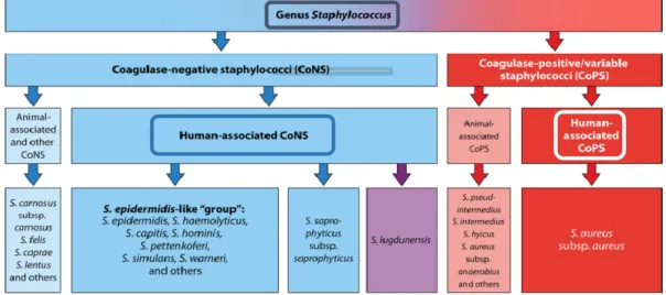
Figure 3. Clinical and epidemiological classification of staphylococcal species………..12
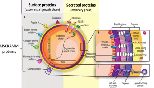
Figure 4. Structural and secreted pathogenic factors of S.aureus……….16
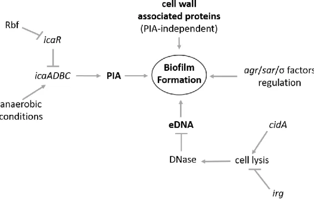
Figure 5. Regulatory factors involved in S. aureus biofilm formation……….19
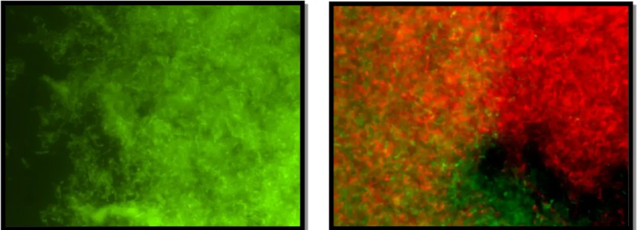
Figure 6. E. faecalis biofilm-producer by DFU strain (left picture) after FISH (×1000; original); polymicrobial biofilm formed by Corynebacterium (green) and E. faecalis (red) DFU strains after FISH (right picture) (×1000; original)………...25
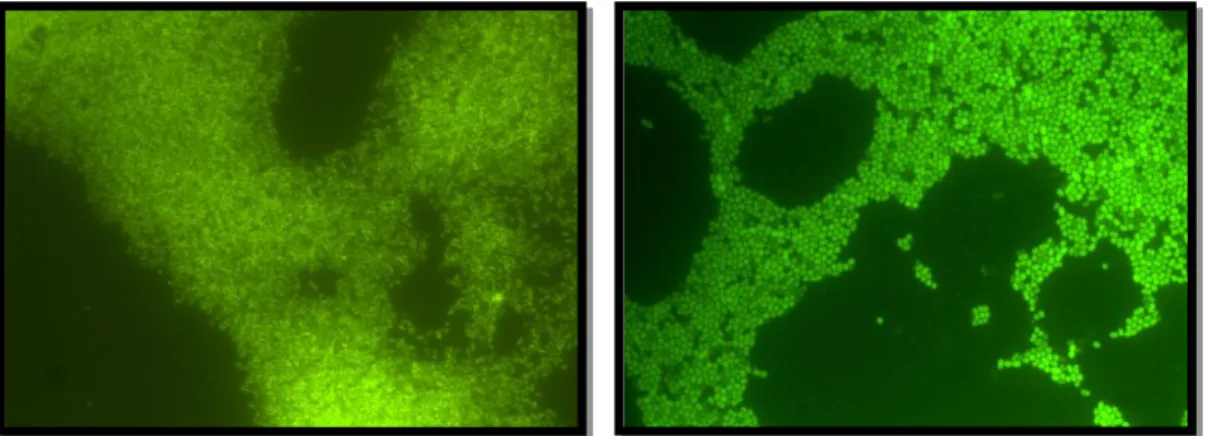
Figure 7. P. aeruginosa (left picture) and A. baumannii (right picture) DFU strains after FISH (×1000; original)………...28
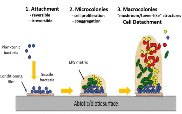
Figure 8. Schematic representation of the biofilm formation steps: initial attachment to surface, cell proliferation with formation of a monolayer and biofilm maturation and cell dispersal………30
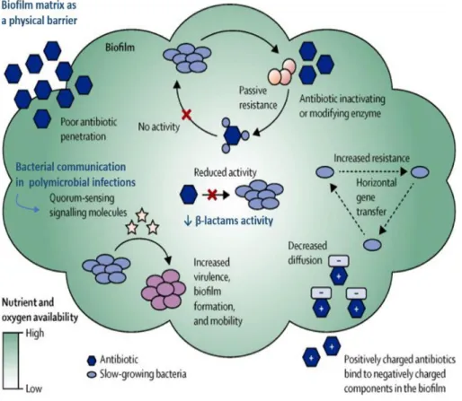
Figure 9. Antibiotic resistance in bacterial biofilm……….34
Figure 10. The phage replication cycles: schematic of lytic, lysogenic and pseudolysogenic cycles……….40
Chapter 2 Figure 1. Time course of one-species biofilm formation (24, 48, and 72 h)………..55
Figure 2. Time course of polymicrobial communities’ biofilm formation (24, 48, and 72h)….56 Figure 3. Polymicrobial biofilm formed by Corynebacterium (green) and Staphylococcus (red) after a 24h incubation (×1000; original)……….……….59
Figure 4. Polymicrobial biofilm formed by Acinetobacter (green) and Staphylococcus (red) after a 72h incubation (×1000; original)…….………..60
Chapter 3 Figure 1. Dendrogram based on SmaI-PFGE patterns of the S. aureus diabetic foot isolates………..71
Figure 2. Minimum spanning tree of 23 S. aureus representing the 23 different pulsotypes detected amongst the diabetic foot isolates………73
Chapter 4 Figure 1. Minimum inhibitory concentration (MIC), minimum biofilm inhibitory concentration (MBIC) and minimum biofilm eradication concentration (MBEC) of S. aureus DFU isolates as determined by a modified version of the Calgary Biofilm Pin Lid Device………92
Chapter 5 Figure 1. Dendrogram based on SmaI-PFGE patterns...101
Chapter 6 Figure 1. Morphological and genomic characteristics of the bacteriophages used for BT...111
Figure 2. Time-kill curves of the target bacteria during planktonic growth when challenged with their specific bacteriophages (alone or in combination)……….114
xv
LIST OF ABBREVIATIONS
A absorbance
Aae S. epidermidis autolysin
Aap accumulation-associated protein
Aas S. saprophyticus autolysin
AB alamar blue
Abi abortive infection
agr acessory gene regulator
AHL acyl homoserine lactones AIP-I autoinducing peptide I Atl autolysin
AtlC S. caprae autolysin AtlE S. epidermidis autolysin AtlL S. lugdunensis autolysin AtlWM S. warneri M autolysin
BES bio-engineered skin
blaZ penicillin resistance gene
BT bacteriophage therapy
CA-MRSA community-acquired MRSA
CC clonal complex
ccr cassette chromosome recombinase
CHIPS chemotaxis inhibitory protein of S. aureus
CIP ciprofloxacin
ClfA clumping factor A
ClfB clumping factor B
Cna colagen binding protein
CoNS coagulase-negative staphylococci CoPS coagulase-positive staphylococci
CRISPR Clustered Regularly Interspaced Short Palindromic Repeats
CWA cell wall-anchored
cyl cytolysin
DFI diabetic foot infection DFU diabetic foot ulcer
DM diabetes mellitus
Eap Extracellular adherence protein
eDNA extracellular DNA
Efb extracellular fibrinogen binding protein Embp extracellular matrix-binding protein
EMRSA epidemic MRSA
EPS extracellular polymeric substances erm erythromycin resistance gene
xvi Esp Enterococcal surface protein
ET exfoliative toxins
Fbe fibrinogen-binding protein
FDA U.S. Food and Drug Administration FEP functional equivalent pathogroups FISH Fluorescent In Situ Hybridization FLIPr formyl peptide receptor-like-1 inhibitory FnbpA fibronectin binding proteins A
FnbpB fibronectin binding proteins B
fsr E. faecalis regulator
GelE enterococcal gelatinase
gyrA gyrase subunit A
G-CSF Granulocyte-Colony Stimulating Factor HA-MRSA hospital-associated MRSA
HBOT hyperbaric oxygen therapy ica intercellular adhesion
icaA intercellular adhesion A
icaADBC intercellular adhesion operon ABCD
icaD intercellular adhesion D
icaR intercellular adhesion regulator
ICTV International Committee for Taxonomy of Viruses IDSA Infectious Disease Society of America
IgG immunoglobin G
Isd iron-regulated surface protein
IM input multiplicity
IWGDF International Working Group of the Diabetic Foot LED light-emitting diodes
LLLT low-level light therapy
Luk leukocidin
MBEC minimum biofilm eradication concentration MBIC minimum biofilm inhibitory concentration
MDR multidrug-resistant
MDT maggot debridement therapy
MFISH multiplex fluorescent in situ hybridization MHC-II major histocompatibility complex class II MIC minimum inhibitory concentration
MLSb macrolide-lincosamide-streptograminB MLST multilocus sequence typing
MRS methicillin-resistant Staphylococcus MRSA methicillin-resistant S. aureus MRSE methicillin-resistant S. epidermidis
xvii MSSA methicillin-susceptible S. aureus NEAT Near-iron transporter
NICE National Institute for Health and Care Excellence norA ciprofloxacin resistance gene
NPWT Negative pressure wound therapy PBP Penicillin binding protein
PBP2a penicillin binding protein 2a
PIA polysaccharide intercellular adhesin Pls plasma-sensitive surface protein PNAG poly-N-acetylglucosamine PVL panton-valentine leucocidin
PSM phenol-soluble modulin
Rbf Regulator of biofilm formation
rhPDGF recombinant human platelet derived growth factor RM Restriction-Modification
σA primary sigma factor
σB alternative sigma factor
sae staphylococcal accessory element sarA staphylococcal accessory regulator A sarS staphylococcal accessory regulator S sarT staphylococcal accessory regulator T sasG S. aureus surface protein G
SCCmec staphylococcal cassette chromosome mec SCIN staphylococcal complement inhibitor SCV small colonial variant
Sdr serine-aspartate repeat protein SE staphylococcal enterotoxins Sie superinfection exclusion system spa staphylococcal protein A
SSF staphylococcal scarlet fever
SSSS staphylococcal scalded skin syndrome TCRS Two-Component Regulatory System tet tetracycline resistance gene
TSS toxic shock syndrome
TSST-1 toxic shock syndrome toxin-1
VAN vancomycin
van vancomycin resistance gene
VISA vancomycin-intermediate MRSA VRE vancomycin-resistant enterococci VREF vancomycin-resistant E. faecium VRSA vancomycin-resistant MRSA WHO World Health Organization
1
CHAPTER I
Literature Review and Objectives
2
1.1. Introduction on Diabetic Foot Ulcer
1.1.1. Definitions and epidemiology
Diabetes mellitus (DM) is one of the most epidemic chronic diseases worldwide. Its prevalence is increasing mainly due to population growth, but also aging, urbanization and changing lifestyles that lead to reduced physical activity and increased obesity. The last World Health Organization (WHO) report states that in 2014 globally 422 million adults aged over 18 years were living with diabetes (WHO, 2016), an important rise comparing with the 108 million adults affected in 1980. In fact, the global prevalence has increased from 4.7% in 1980 to 8.5% in 2014. Diabetes prevalence is expected to double by the year 2030, also as a result of better health care conditions, which will increase the longevity of people with diabetes (Wild, Roglic, Green, Sicree & King, 2004).
Diabetes distribution varies substantially according to countries’ economic status. The majority of diabetic patients in developed countries are aged over 60 years, whereas in developing countries most people with diabetes are of working age, between 40 and 60 years (Shaw, Sicree & Zimmet, 2010). In Portugal, there were over 1 million cases of diabetes registered in 2015. The cost associated to each diabetic patient was estimated to be about 1.880 euros (http://www.idf.org/membership/eur/portugal), proving that diabetes represents a significant health burden in Portugal.
A major concern with diabetes is the high risk of patients in developing one of the many major complications associated with the disease, such as cardiovascular, including myocardial infarction, stroke, angina and heart failure, blindness or nephropathy (Hopkins, Burke, Harlock, Jegathisawaran & Goeree, 2015). A common and most devastating complication of diabetes is the development of diabetic foot ulcers (DFU). Historically, foot ulcers have been estimated to affect 1 to 4% of patients with diabetes annually, but a recent study in the United States indicated that the annual incidence may be as high as 6% (Rice et al., 2014). In Canada and United States, it was observed that diabetic patients have a 25% risk of developing a DFU in their lifetime (Hobizal & Wukich, 2012; Rice et al., 2014).
A DFU can be defined as any full-thickness wound with skin necrosis or gangrene, below the ankle, induced by peripheral neuropathy or peripheral arterial disease in a diabetic patient, independently of its duration (Chuan, Tang, Jiang, Zhou & He, 2015). Most DFU are chronic wounds, defined in standard surgical textbooks as those that have not healed in 3 months. The most common forms are related to diabetes mellitus, venous stasis, peripheral vascular diseases and pressure ulcerations (Siddiqui & Bernstein, 2010).
Acute wounds are caused by external damage to intact skin, including more severe traumatic wounds and are expected to heal within a predictable time frame, although the treatment required to facilitate healing will vary according to the type, location and wound depth. In contrast, chronic wounds are most frequently caused by endogenous mechanisms that
3
compromise the integrity of dermal and epidermal tissues, such as pathophysiological abnormalities including leg ulcers, foot ulcers and pressure sores that include compromised tissue perfusion as a consequence of impaired arterial supply (peripheral vascular disease) or impaired venous drainage (venous hypertension), and metabolic diseases such as diabetes mellitus (Eron et al., 2003). Wound healing is characterized by three phases of inflammation, fibroplasia and maturation, resulting in a fine scar with little fibrosis and a return to an almost normal tissue architecture and organ function; if a wound does not heal in an orderly sequence or timely, or if the healing process does not result in structural integrity, then the wound is considered chronic and the healing occurs with the formation of abundant granulation tissue and often with excessive fibrosis leading to scar contraction and loss of function (Stadelmann, Digenis & Tobin, 1998).
DFU require minor or major amputations of lower limbs in 15% to 27% of cases. Infection is the preponderant factor for amputation in 50% of ulcers (Mendes & Neves, 2012), representing a major cause of morbidity and mortality and the most common cause of diabetes-related admission to hospitals, with huge financial, social and psychological consequences (Richard, Sotto & Lavigne, 2011; Mendes & Neves, 2012). A recent report estimated that the risk of hospitalization and lower-extremity amputation was approximately 56 and 155 times greater for diabetic people who had a foot infection than for those without (Richard et al., 2011). In the longer term, DFU have recurrence rates of up to 70%, resulting in repeated interventions and progressive disability that increase sanitary costs (Mendes & Neves, 2012). In fact, nearly one in six patients die within 1 year after their first infection (Hobizal & Wukich, 2012).
1.1.2. Pathophysiology
DFU have a multifactorial nature, but it is well established that absolute or relative insulin deficiency is the primary biochemical abnormality that leads to the organic complications of diabetes mellitus. It has also been established that a persistent glycaemic control, with either insulin or oral antidiabetic drugs, is able to stop and probably regress DFU associated microvascular and macrovascular complications (Turner, 1998; Mendes & Neves, 2012). The two major underlying causes of diabetic foot complications are peripheral neuropathy and peripheral vascular disease, by several mechanisms (Figure 1). One of the most frequently described mechanisms is the polyol pathway, in which the hyperglycaemic state leads to an increase in the action of the enzymes aldose reductase and sorbitol dehydrogenase, resulting in the conversion of intracellular glucose to sorbitol and fructose. The accumulation of these sugar products causes a decrease in the synthesis of myoinositol, required by nerve cells for normal progression of the neural impulse (Clayton, 2009). Additionally, the chemical conversion of glucose results in a depletion of nicotinamide adenine dinucleotide phosphate, leading to accumulation of reactive oxygen species and diminished synthesis of the
4
vasodilator nitric oxide (Yagihashi, Mizukami & Sugimoto, 2011). These factors result in oxidative stress in the nerve cell and also in an increased vasoconstriction leading to ischemia, which will promote nerve cell injury and death (Clayton W., 2009).
Several studies have also referred to a potential link between diabetic neuropathy and the mitochondria of sensory neurons located in dorsal root ganglia. These mitochondria are particularly vulnerable, because in the hyperglycaemic state they produce reactive oxygen species, which can damage mitochondrial DNA and membranes, impairing cell functions and leading to degeneration (Leinninger, Edwards, Lipshaw & Feldman, 2006; Said, 2007).
It appears that the size of neurons is also important in diabetes because longer nerve fibres show an earlier decrease in the velocity of nervous impulse transmission. This is why the loss of sensation and reflexes are often observed in the feet first, then progress to other areas, in particular the hands, causing the “glove and stocking” syndrome, which symptoms include numbness, dysesthesia, sensory loss and nocturnal pain (Forbes & Cooper, 2013). The damage to the nerves of the intrinsic foot muscles leads to an imbalance between flexion and extension capacities of the affected foot, producing anatomic deformities that create pressure points, which gradually cause skin breakdown and ulceration (Bowering, 2001) (Figure 1). Moreover, the autonomic neuropathy leads to a decrease in sweat and oil gland function, so the foot becomes dry and keratinized prompting the development of cracks and fissures that constitute a portal for infection development in wounds (Clayton W., 2009). Advanced neuropathy is characterized by altered sensitivities to vibrations and thermal thresholds, which progress to loss of sensory perception. For this reason, many wounds go unnoticed and progressively worsening as the affected area is continuously subjected to repetitive pressure and forces from walk and weight (Forbes & Cooper, 2013). Hyperalgesia, paraesthesia and allodynia can also occur in a proportion of patients, with pain evident in 40 to 50% of those with diabetic neuropathy, decreasing life quality (Obrosova, 2009).
Peripheral vascular disease plays a secondary role in DFU pathophysiology, as a consequence of the persistent hyperglycaemic state. In diabetes there is a decrease in endothelium-derived vasodilators leading to constriction, and an increase in thromboxane A2, a vasoconstrictor and platelet aggregation agonist, which leads to an enhanced risk for plasma hypercoagulability (Clayton W., 2009). Macroangiopathy is also observed, due to atherosclerosis, an obstructive disease of large vessels typically involving the tibial and peroneal arteries, resulting in capillary basement membrane thickening, altered nutrient exchange, tissue hypoxia and microcirculation ischemia (Hobizal & Wukich, 2012) (Figure 1). Cumulatively, these alterations can lead to occlusive arterial disease that result in ischemia in the lower extremity and an increased risk of ulceration in diabetic patients. Moreover, smoking, hypertension and hyperlipidaemia are other factors that contribute to the development of peripheral arterial disease in diabetic patients (Mulder, Tenenhaus & D’Souza, 2014).
5
1.1.3. Diagnosis and classification
The establishment of the most adequate therapeutic protocol for DFU must take into account all the risk factors involved and a consistent DFU classification. This evaluation requires the careful assessment of the global medical, foot and wound history, a systemized and detailed physical examination and the execution of complementary diagnostic procedures (Mendes & Neves, 2012).
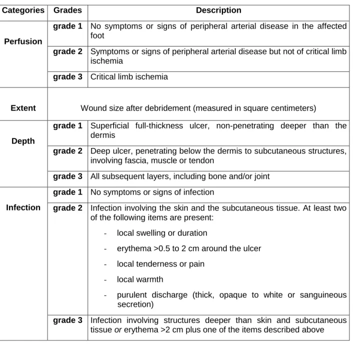
Many DFU classification systems have been proposed to predict clinical outcome, but many of them have limitations. The International Working Group of the Diabetic Foot (IWGDF) developed the PEDIS classification system to categorize and define DFU objectively and facilitate communication between health-care providers, also allowing the prediction of associated health-care costs. In this system, all DFU are classified according to five categories that consider the most relevant signs and symptoms: perfusion, extent/size, depth/tissue loss, infection and sensation. Moreover, each subcategory is defined according to strict criteria based upon objective techniques, being applicable worldwide (Table 1) (Chuan et al., 2015). Comparing with previous systems, such as the Wagner and SINBAD systems (Site, Ischemia, Neuropathy, Bacterial, Infection and Depth), PEDIS predicts the clinical outcome more
Figure 1. Diabetic foot infection pathophysiology. Diabetic foot ulcer
results from a complex interaction of risk factors, in which neuropathy plays the central role and causes ulcerations due to trauma or excessive pressure in a deformed foot without protective sensibility. Once the protective layer of skin is broken, deep tissues are exposed to bacterial colonization and an infection can settle (original).
6
effectively, as its definition of DFU is based on more strict criteria based upon objective techniques and more comprehensive ulcer healing parameters. PEDIS DFU categorization is based on: 1) perfusion examination, essential for diagnosing peripheral arterial disease, resulting from the physical observation performed by a specialized health-care worker; 2) wound size measurement, which should be determined after debridement, if possible; 3) depth establishment, which is difficult to perform, allowing ulcers to be divided into lesions confined to the skin and those deeper than the skin; 4) infection diagnostic, mainly based on expert opinion; and finally, 5) sensation evaluation, by determining the presence or absence of protective sensation in the affected foot (Schaper, 2004).
Table 1. PEDIS classification system by the International Working Group of the Diabetic Foot
(IWGDF) that classifies all ulcers in five main categories. Adapted from Schaper et al. (2004).
Categories Grades Description
Perfusion
grade 1 No symptoms or signs of peripheral arterial disease in the affected
foot
grade 2 Symptoms or signs of peripheral arterial disease but not of critical limb
ischemia
grade 3 Critical limb ischemia
Extent Wound size after debridement (measured in square centimeters)
Depth
grade 1 Superficial full-thickness ulcer, non-penetrating deeper than the
dermis
grade 2 Deep ulcer, penetrating below the dermis to subcutaneous structures,
involving fascia, muscle or tendon
grade 3 All subsequent layers, including bone and/or joint
Infection
grade 1 No symptoms or signs of infection
grade 2 Infection involving the skin and the subcutaneous tissue. At least two
of the following items are present: - local swelling or duration
- erythema >0.5 to 2 cm around the ulcer - local tenderness or pain
- local warmth
- purulent discharge (thick, opaque to white or sanguineous secretion)
grade 3 Infection involving structures deeper than skin and subcutaneous
7
grade 4 Any foot infection with two or more signs of a systemic inflammatory
response syndrome:
- temperature >38 or <36 °C - heart rate >90 beats/minute
- respiratory rate >20 breaths/minute (or PaCO2 <32 mmHg)
- white blood cell count >12.000 or <4.000 cells/mm3
(or 10% band forms)
Sensation
grade 1 No loss of protective sensation grade 2 Loss of protective sensation
PaCO2: partial pressure of carbon dioxide in the arterial blood.
1.2. Diabetic Foot Infections
1.2.1. Definition and pathophysiology
Infection is a frequent (40-80%) and costly complication of DFU and represents a major cause of morbidity and mortality (Boyanova & Mitov, 2013). The healing impairment of DFU is caused by several factors that favour the overgrowth of bacteria, and has an essential role in the rapid spread of infection in diabetic ulcers (Jeffcoate & Harding, 2003) (Figure 2). These include intrinsic factors, such as neuropathy and vascular problems, and extrinsic factors, including callus formation and excessive local pressure. Traditionally, this set of predisposing abnormalities in diabetes has been referred to as “the pathogenic triad of neuropathy, ischaemia, and trauma” (Falanga, 2005).
The significance of bacteria in wounds presents a continuum, from contamination through colonization, to critical colonization and, finally, to infection (Siddiqui & Bernstein, 2010). Some wound specialists believe that the presence of a high concentration of microorganisms, usually superior to >105 colony-forming units [CFU] per gram of host tissue, represents ‘increased
bioburden’ or ‘critical colonization’ even in the absence of clinical evidence of infection. There is, however, no universal techniques to define critical colonization, no routine laboratory tests available for quantitative bacteriology and no convincing evidence of its association with adverse clinical outcomes, for example, failure of healing or development of overt infection (Spichler, Hurwitz, Armstrong & Lipsky, 2015). This threshold may be altered by the status of the immune system and number and type of bacteria involved. Signs of critical colonization include atrophy or deterioration of granulation tissue, discoloration of granulation tissue to deep red or grey, increased wound friability and increased drainage (Siddiqui & Bernstein, 2010).
8
A diabetic foot infection (DFI) is defined as any infra-malleolar infection in a person with diabetes mellitus. These infections may include paronychia, cellulitis, myositis, abscesses, necrotizing fasciitis, septic arthritis, tendonitis and osteomyelitis. However, the most common and classical lesion is the infected diabetic “mal perforans” foot ulcer, in which, after the breach of the protective layer of skin, underlying tissues are exposed to bacterial colonization; this wound may progress to become actively infected, involving deeper tissues. This sequence of events can be rapid, occurring over days or even hours especially in ischemic limbs (Lipsky, 2004a). Although most infections, such as cellulitis, remain superficial, in around 25% of the cases will spread from the epidermal layer to deeper regions, reaching subcutaneous tissues and bones, as observed in necrotic fasciitis, septic arthritis and osteomyelitis (Noor, Zubair & Ahmad, 2015).
Wound contaminants are likely to originate from three main sources: the environment, including exogenous microorganisms in the air or those introduced by traumatic injury; the surrounding skin; and endogenous sources, involving mucous membranes, such as the gastrointestinal, oropharyngeal and genitourinary mucosae (Bowler, Duerden & Armstrong, 2001). After colonization, transition to infection occurs when bacterial proliferation overcomes the host's immune response (Siddiqui & Bernstein, 2010). This transition involves a multitude of microbial and host factors, including type, location, size and depth of the wound, the level of blood perfusion of the wound, the health and immune status of the host, the microbial load and the virulence potential expressed by the microorganisms involved. The immunocompromised state of diabetic patients, namely the neutrophil dysfunction, constitutes a relevant aspect because facilitates infections. The hyperglycaemia state seems to be the main factor altering the neutrophil chemotaxis, phagocytosis and intracellular killing of bacteria (Boyanova & Mitov, 2013).
Due to frequent infections, diabetic patients are more exposed to antibacterial agents, which can lead to increased antibiotic resistance rates; also, the presence of peripheral vascular disease can leads to poor antibiotic penetration in the infected tissue, favouring the development of antibiotic resistance (Lipsky et al., 2012a). There is a vicious cycle in which the infections can worsen the glycaemic control of the diabetic patient and, vice-versa, the poor glycaemic control and other factors associated with DM can facilitate or aggravate the development of infections (Figure 2).
1.2.2. DFI diagnosis
DFI diagnosis begins with a clinical suspicion accompanied by a comprehensive history and physical exam, validated with a complete laboratory evaluation, microbiology assessment and diagnosis imaging. Since all chronic wounds are colonized by microorganisms, the DFI diagnosis is based on clinical findings. According to the Infectious Disease Society of America (IDSA) guidelines, infection is present if there is obvious purulent drainage and/or the
9
presence of two or more signs of inflammation (erythema, pain, tenderness, warmth or induration), being divided in four grades of severity according to the extension of tissue involved and the presence of systemic toxicity or metabolic derangement (Hobizal & Wukich, 2012). In some patients, especially those with peripheral neuropathy or vasculopathy, symptoms and signs may be diminished, leading some clinicians to evaluate the presence of “secondary” findings before diagnosing infection, such as foul odour and the presence of friable or discoloured granulation tissue (Spichler et al., 2015).
1.3. Microbiology of DFI
1.3.1. Bacteria involved in DFI
To properly identify the microorganisms involved in DFI it is important to choose the correct sampling procedure. Tissue biopsy following initial debridement and cleansing of superficial debris is known as the most advantageous and standard sampling method. However, due to fear of infections spreading and loss of adjacent structure in limb and ischemia tissue, sometimes biopsy is not advisable, being replaced by swab sampling. This method renders sample collection easier and can be performed in any kind of wound. This method is widely used for the identification of causative microorganisms and antimicrobial susceptibility testing, despite the fact that isolated bacteria also include colonizers (Noor et al., 2015).
Figure 2. Interplay among factors associated with diabetes, infections and
10
In the presence of a large volume of wound fluid, sampling by needle aspiration can be performed, being well established as the most useful procedure for sampling purulent fluid from intact cutaneous abscesses (Bowler et al., 2001). Cultures of specimens obtained from patients with mixed infections generally yield 3 to 5 isolates, including Gram-positive and Gram-negative aerobes and anaerobes (Lipsky, 2004a). Many studies from western countries report that prevailing microorganisms cultured from DFI are Gram-positive aerobes (Dang, Prasad, Boulton & Jude, 2003; Citron, Goldstein, Merriam, Lipsky & Abramson, 2007; Mendes et al., 2012), whereas studies from eastern countries have described the presence of Gram-negative aerobes as predominant organisms (Gadepalli et al., 2006; Zubair, Malik, Ahmad & Rizvi, 2011; Noor et al., 2015).
Generally, aerobic Gram-positive cocci are the predominant microorganisms that colonize and cause acute DFI, with Staphylococcus aureus being the most common isolated pathogen, followed by Staphylococcus epidermidis and Streptococcus spp., especially the ones belonging to β-haemolytic groups (Shankar, Mohan, Premalatha, Srinivasan & Usha, 2005; James et al., 2007; Galkowska et al., 2009; Tascini et al., 2011). Staphylococci are perhaps the most virulent pathogens in DFI, showing a correlation between specific virulence genotypic markers from DFU isolates and ulcer outcome (Sotto, Lina & Richard, 2008). Chronic wounds develop a more complex colonizing microbiota and infections are more frequently polymicrobial, often including aerobic or facultative Gram-positive bacteria such as Enterococcus and Corynebacterium, Gram-negative bacilli such as Pseudomonas aeruginosa and Acinetobacter, and obligate anaerobic bacteria (Citron et al., 2007; Dowd et al., 2008a; Zubair et al., 2011; Małecki, Rosiński & Adamiec, 2013; Sekhar, Vyas, Unnikrishnan, Rodrigues & Mukhopadhyay, 2014). In fact, foot ulcers with ischemia and deep tissue necrosis have been reported to present anaerobic growth (Hobizal & Wukich, 2012; Lipsky, Richard & Lavigne, 2013; Noor et al., 2015). It is also important to remember that fungi have also been reported in DFI and can be a major contributor to the bioburden or biofilm formation in wounds (Dowd et al., 2011).
Acute infections in patients who have not recently received antimicrobials are often monomicrobial, being the majority promoted by an aerobic Gram-positive coccus, whereas chronic infections are often polymicrobial (Noor et al., 2015). The pathogenic role of each isolate in a polymicrobial infection is often unclear, but some studies suggest that the interactions of organisms within these polymicrobial communities lead to the production of virulence factors, such as collagenases, proteases and haemolysins, and short-chain fatty acids that cause inflammation, obstructed wound healing, increasing the chronicity of infection (Noor et al., 2015). The impaired host defences around necrotic soft tissue or bone may allow low-virulence colonizers, such as coagulase-negative staphylococci and Corynebacterium spp. (“diphtheroids”), to assume a pathogenic role (Lipsky, 2004a).
11
Generally, the presence of one microorganism generates a niche for the colonisation by other pathogenic microorganisms; on the other hand, two or more non-pathogenic microorganisms together can cause disease. The major polymicrobial interactions that have been studied are synergism and antagonism. In synergy there is a cooperative interaction between two or more species of microbes, allowing increased growth, virulence, antimicrobial tolerance and enhanced production of exopolysaccharide (EPS) (Brogden, Guthmiller & Taylor, 2005). Antagonism, also called antibiosis, is characterized by the suppression of one microbial species by another; it can include production of chemical signs and factors inhibiting the growth of neighbours and/or storage of nutrients that promote their starvation (Gabrilska & Rumbaugh, 2015).
Hospitalization, surgical procedures and, especially, prolonged or broad-spectrum antibiotic therapeutics may predispose patients to colonization and/or infection with antibiotic-resistant microorganisms. Among hospitalized patients, the prevalence of methicillin-resistant Staphylococcus aureus (MRSA) in DFI can range from 15 to 30%, depending on the geographical location of affected patients (Hobizal & Wukich, 2012). Although MRSA strains have been previously isolated mainly from hospitalized patients, community-associated strains are now becoming common, being already associated with severe outcomes in patients with DFI (Dang et al., 2003). For example, the first reported cases of vancomycin-resistant S. aureus in the United States and in Europe have been isolated from diabetic patients with foot infections (Lipsky, 2004a; Melo-Cristino, Resina, Manuel, Lito & Ramirez, 2013).
In summary, bacterial genera frequently associated with DFI include Staphylococcus, Enterococcus, Corynebacterium, Pseudomonas and Acinetobacter.
1.3.2. Staphylococci
1.3.2.1. Generalities and classifications
Staphylococcus, named after the Greek words staphyle (bunch of grapes) and kokkos (berry), is a genus of Gram-positive bacteria frequently associated with surgical and skin infections, respiratory disease and food poisoning (Licitra, 2013). They were observed for the first time in 1880, by the Scottish surgeon Sir Alexander Ogston that observed staphylococci in pus samples from a surgical abscess in a knee joint, describing them as “the masses looked like bunches of grapes.” In 1884, German physician Friedrich Julius Rosenbach differentiated the bacteria species by the colour of their colonies growing in agar medium: S. aureus (from the Latin aurum, gold) due to the presence of carotenoids, and S. albus (Latin for white); S. albus was later renamed as S. epidermidis due to its ubiquitous presence on human skin (Orenstein, 2008). Staphylococci are characterized by being non-motile, non-spore-forming, spherical cells with 0.5 to 1.5 µm in diameter, usually forming clusters; they are facultative anaerobes with
12
complex nutritional requirements including several amino acids and vitamins B; they tolerate high concentrations of sodium chloride and are resistant to heat (Becker & Eiff, 2011).
Staphylococci are catalase-positive and their cell wall peptidoglycan structure contains multiple glycine residues in the cross-bridge, responsible for their susceptibility to lysostaphin action (Plata, Rosato & Wegrzyn, 2009). The Staphylococcus genus is traditionally divided into two groups based on the bacteria’s ability to produce coagulase, an enzyme that causes blood clotting by converting fibrinogen to fibrin: coagulase-positive staphylococci (CoPS), the group that includes S. aureus, a frequent ethological agent of diseases that exhibits resistance to a growing number of therapeutic agents; and the coagulase-negative staphylococci (CoNS), a group composed by common skin commensals (Figure 3). CoNS include S. epidermidis and S. haemolyticus as the most prevalent species, along with S. capitis, S. hominis, S. simulans, S. warneri and S. saprophyticus (Becker, Heilmann & Peters, 2014) (Figure 3).
S. aureus is the most pathogenic species of the genus Staphylococcus, implicated in both community-acquired and nosocomial infections and associated with considerable morbidity and mortality rates (Carbon, 2000). It has been estimated that about 20 to 30% of the population are permanently colonized by this bacterium, constituting healthy carriers, while other 30% are transient carriers. The anterior nose nares are the most frequent location for S. aureus colonization, followed by skin, perineum, pharynx, and less frequently, the gastrointestinal tract, axillae and vagina (Wertheim et al., 2005). Colonization represents an increased risk of infection particularly for the immunocompromised host. In such cases, S. aureus can be responsible for the development of bacteraemia, infective endocarditis as well as osteoarticular, skin and soft tissue, pleuropulmonary and device-related infections (Tong, Davis, Eichenberger, Holland & Fowler, 2015).
Figure 3. Clinical and epidemiological classification of staphylococcal
13
Staphylococcus, especially S. aureus, is by far the most common and virulent pathogen in DFI (Jones, Edwards, Finch & Jeffcoate, 1985; Sotto et al., 2008; Lipsky et al., 2013). Many studies have described a high S. aureus prevalence in these infections, ranging from about 50% (Dang et al., 2003; Citron et al., 2007; Galkowska et al., 2009; Mendes et al., 2012) to 30% (Richard et al., 2011; Tascini et al., 2011; De Vries, Ekkelenkamp & Peters, 2014; Zenelaj, Bouvet, Lipsky & Uckay, 2014). These studies also describe CoNS as the second most frequently encountered aerobic Gram-positive isolates. MRSA have emerged as a serious and common problem inpatients with diabetic foot ulcers, and infections promoted by such strains may result in prolonged stay hospital and increased economic costs (Tentolouris et al., 2006). High rates of MRSA were already reported in several studies on DFI, normally representing 20 to 40% of all S. aureus isolates (Bowling, Salgami & Boulton, 2007; Tascini et al., 2011; Boyanova & Mitov, 2013); also one study described a 68% MRSA rate in DFI (Malik, Mohammad & Ahmad, 2013). In case of diabetic foot osteomyelitis, the majority of infections are polymicrobial, with S. aureus being the most common etiologic agent, isolated in about 40% of cases, followed by S. epidermidis, isolated in about 25% (van Asten et al., 2015).
1.3.2.2. Staphylococci virulence traits
S. aureus virulence determinants can be divided into cell wall-anchored (CWA) surface proteins (adhesins) and secreted factors (exotoxins). These factors allow this bacterial species to adhere to surfaces/tissues and avoid or invade the host immune system causing toxic effects.
1.3.2.2.1. Cell wall-anchored (CWA) surface proteins
The first step of staphylococcal invasion is bacterial adherence that can occur via direct interaction with host cells or via interaction with host-factor binding proteins (Schroeder et al., 2009). These include autolysins, adhesins, binding proteins, clumping factors, iron transporters and surface proteins.
Direct S. aureus adherence to a surface may be mediated by the major autolysin Atl that has both adhesive and enzymatic functions. It is involved in the initial attachment of the cells to a polymeric surface and in biofilm formation. It also binds to vitronectin, suggesting a role in not only colonizing polymer surfaces, coated materials and host tissue (Heilmann, Hartleib, Hussain & Peters, 2005). This enzyme also participates in the hydrolysis of cell wall peptidoglycan, leading to autolysis and release of extracellular DNA (eDNA), which has been shown to be an important component of staphylococcal biofilms (Becker et al., 2014). S. aureus Atl is highly homologous to the S. epidermidis autolysin/adhesin AtlE, which is involved in the attachment to polymer surfaces. Recently, homologous Atl proteins with similar functions were also reported for other CoNS, such as S. caprae (AtlC), S. saprophyticus (Aas),