Cardiac Angiosarcoma - A Review
[37]
ANTÓNIOMURINELLO, PAULAMENDONÇA, ANAABREU, A. LARANJEIRASANTOS, JOSÉROQUETE, EUGÉNIAPINTO, JOÃOALPENDRA, JÚLIOSEMEDO, ANARODRIGUES, DANIELACUNHA, J. FIGUEIRACOELHO, SOFIALOURENÇO, SIMÃOMIRANDA
Serviços de Medicina I e de Dermatologia do Hospital Curry Cabral, Lisboa, Portugal
Serviços de Cardiologia, de Cirurgia Torácica e de Anatomia Patológica do Hospital Stª Marta, Lisboa, Portugal Serviços de Radiologia e de Pneumologia do Hospital Pulido Valente, Lisboa, Portugal
Rev Port Cardiol 2007; 28 (5): 577-584
577
ABSTRACT
Based on a case of a patient with angiosarcoma (AS) of the right atrium with superior vena cava syndrome associated with urticaria and polyarthralgias, who died soon after surgery, the authors present a brief review of the subject of cardiac AS, an extremely rare pathology, usually diagnosed late due to its non-specific symptomatology. Several topics are discussed, including mechanisms of clinical manifestations caused by blood flow obstruction and valve dysfunction, local invasion with arrhythmias and pericardial effusion, embolic phenomena and constitutional symptoms. Imaging and histopathologic methods of diagnosis are considered, as well as references to cytogenetic analysis. Surgery is the first treatment choice, but heart AS are frequently not completely resectable and concomitant metastases at the time of surgery are common, both usually leading to a dismal prognosis. Chemotherapy, radiotherapy and even heart transplantation do not substantially improve the survival of these patients. Urticaria is not generally assumed by most authors to be associated with malignancy, but there are rare reports of its association with some malignant tumors.
Key words
Heart sarcoma; Heart angiosarcoma; Superior vena cava syndrome; Urticaria
RESUMO
Angiosarcoma Cardíaco - uma Revisão
Baseados no caso dum doente com
angiosarcoma da aurícula direita manifestando quadro de síndrome da veia cava superior associado a urticária e poliartralgias, com óbito poucos dias após cirurgia paliativa, os autores fazem revisão concisa dos angiosarcomas cardíacos, tumores extremamente raros e geralmente diagnosticados tardiamente em virtude da não especificidade da
sintomatologia. São discutidos os mecanismos de expressão clínica, resultantes de obstrução ao fluxo sanguíneo, de disfunção valvular, de invasão local condicionando arritmias e derrame pericárdico, fenómenos embólicos e sintomas constitucionais. O diagnóstico é considerado em termos de imagiologia, histopatologia, imunocitoquímica e análise citogenética. O tratamento de primeira linha é cirúrgico, mas geralmente os tumores não são totalmente ressecáveis, para além da frequente presença de metastização concomitante, ambos os factores condicionando prognóstico
geralmente muito desfavorável. A
quimioterapia e a radioterapia não oferecem geralmente grande benefício e, mesmo a transplantação cardíaca não tem obtido resultados favoráveis. A maioria dos autores não encontra dados convincentes da possível associação entre urticária e tumores malignos, mas existem algumas referências de casos clínicos de doentes em que as duas situações ocorreram concomitantemente.
Palavras-Chave
Sarcoma cardíaco; Angiosarcoma cardíaco; Síndrome da veia cava superior; Urticária
Recebido para publicação: Outubro de 2006 • Aceite para publicação: Janeiro de 2007 Received for publication: October 2006 • Accepted for publication: January 2007
INTRODUCTION
Primary tumors of the heart occur with an incidence of approximately 0.02% in autopsy series, being far less common than metastatic tumors of the heart, which occur 20-40 times more frequently(1). About 25% of primary heart
tumors are malignant and most of these are sarcomas(2).
Cardiac sarcomas are often asymptomatic until the advanced stages and, even then, cause a variety of non-specific symptoms and may mimic other pathologies, delaying diagnosis. The dismal prognosis results from extensive local invasion and/or distant metastization at presentation(3). A
high index of suspicion remains the key for early diagnosis of a malignant cardiac tumor.
According to Shanmugan(4), cardiac sarcomas
manifest via several mechanisms: (1) obstruction to blood flow and interference with valve function; (2) local invasion causing arrhythmias or pericardial effusion with tamponade; (3) embolic phenomena from tumor fragments or peritumoral thrombi; (4) systemic or constitu-tional symptoms including dyspnea, syncope, chest pain, fever, malaise and weight loss. Left chamber tumors can cause cerebral, coronary, and retinal emboli. Right-sided tumors may be the source of pulmonary emboli that can cause pulmonary hypertension when extensive.
Advances in noninvasive cardiovascular imaging techniques - especially transesophageal echocardiography (TEE)(5), computed tomography
(CT) and magnetic resonance imaging (MRI)(6)
-have greatly facilitated diagnostic evaluation, enabling rapid identification of intracardiac masses.
Histopathologic classification of cardiac sarcomas is generally obtained by combining both morphologic features and immunohisto-chemical staining(7, 8), which can identify several
tumor types: angiosarcoma, undifferentiated sarcoma, osteosarcoma, fibrosarcoma, malignant fibrous histiocytoma, leiomyosarcoma, myxosar-coma, synovial sarmyxosar-coma, neurofibrosarcoma and rhabdomyosarcoma. AS is the most common type in almost all series(3, 9, 10, 11). Cardiac AS seem to
arise in a younger population (42-43 years) than in other soft tissue locations (60 years). They are generally large, grossly hemorrhagic, multilobular right atrial masses that spread along the epicardial surface and replace the right atrial
wall, and may protrude into or fill the adjacent cardiac chamber(s).
Urticaria is due to local increase in permeability of capillaries and venules. These changes are dependent on activation of cutaneous mast cells and basophils, which contain a range of mediators, predominantly histamine (12).
Urticaria is one of the least common markers of internal disease, and two of the major textbooks in dermatology do not consider its association with malignant diseases proved(12,13). However,
there are occasional reports of chronic urticaria occurring in association with malignant neoplasia in Braverman's textbook(14). The authors describe
a case of a patient with heart AS located in the right atrium, who was previously admitted twice to our hospital due to unexplained recurrent urticaria and polyarthralgias, and whose diagnosis of heart AS was only made during his third admission, with a clinical picture of superior vena cava syndrome associated with recurrence of urticaria and polyarthralgias. To our knowledge this association has not previously been described.
CASE REPORT
On May 9 2006, a 47-year-old white man was transferred from the Dermatology Unit of our hospital to our Internal Medicine Unit due to a clinical diagnosis of acute superior vena cava syndrome. He had previously been admitted to the Dermatology Unit from August 29 to September 8 2005 for 15 days with foot edema and prurigo, with pruriginous erythematous papulomatous exanthema, appearing first on the legs, but quickly extending to all skin surfaces, including the face and palmar and plantar surfaces. Five days before that admission the evolution was complicated by fever (38-39 ºC), prostration and malaise, odinalgia and symmetric arthralgias of several joints: ankle, tarsal, knees, wrists, hip, proximal interphalangeal joints of the hands, and shoulder. The patient reported a Billroth type II duodenal ulcer operation 18 years before and chronic atrial fibrillation for several years, and had been treated for 3 months with nonsteroidal anti-inflammatory and analgesic drugs for frequent back pain caused by chronic osteoarthropathy of the lumbar spine. He denied taking any other drugs. No personal or family
history of atopic allergic diseases was obtained. He was a heavy smoker but reported no alcohol consumption. On physical examination the patient was febrile (38 ºC), with blood pressure of 89/60 mmHg and complete arrhythmia with an irregular pulse of approximately 74 bpm. The oropharynx was normal, a slight hepatomegaly was detected, and there were widespread erythematous cutaneous plaques and macular exanthema, confluent in certain areas (shoulders and knees). There was some limitation to movement due to joint pains although without joint swelling, but no other abnormal physical signs were detected. Blood tests showed no anemia, ESR 80 mm (1st hour), leukocytosis 14.5x10⁹/L (N 4.0-10.0) with 83% neutrophils,
platelets 499x109/L (150-400), GOT 101 U/L
(17-59), GPT 93 U/L (21-72), gamma-GT 272 U/L (15-73), normal values of glycemia, serum urea, creatinine, and alkaline phosphatase, normal INR and APTT, negative serology for viral hepatitis A, B, C, CMV, herpes, EBV, HIV, syphilis, and Borrelia, slightly elevated IgA 570 mg/dL (88-440) and IgE 347 UI/L (N <100). Bacteriologic cultural examination of ocular exudate was positive for colonies of methicillin-sensitive Staphylococcus aureus. A complete immunologic study for the diagnosis of autoimmune diseases was performed, including collagen/vasculitic diseases and autoimmune thyroiditis, which was completely negative. C1 inhibitor protein and C1 inhibitor function were normal. Although most of the skin lesions disappeared within 24 hours with hydroxyzine therapy, with a behavior resembling urticaria, a skin biopsy in one of the areas with confluent lesions to exclude a diagnosis of Sweet syndrome showed a slight perivascular diffuse mixed inflammatory infiltrate of neutrophils, lymphocytes and histiocytes of the reticular dermis, findings that were in favor of a diagnosis of urticaria. No vasculitis was found. The patient was advised to stop taking the drugs used before admission. After discharge the patient was admitted again 15 days later to the Internal Medicine Unit for one week, with the same symptoms and also rapidly improving with the same therapy. Investigation was also negative for a different diagnosis at that time. The patient was subsequently followed in the outpatient Dermatology Unit, due to intermittent episodes of unexplained recurrent urticaria and
polyarthralgias. He stated that he had not continued with the previous therapy, except on one occasion. He was admitted again to the Dermatology Unit on April 16 2006 because of rapidly developing and progressive rest and effort dyspnea, face, neck and upper thorax edema, and external jugular vein dilatation. No abnormal venous circulation was detected. Blood pressure was 160/40 mm Hg, there was atrial fibrillation with ventricular rate of approximately 100 bpm and superficial respiratory rate of 24 bpm. Pleural effusion, clinically suspected, was confirmed on thorax X-ray, which also showed cardiomegaly. Abdominal examination revealed slightly painful hepatomegaly with hepatojugular reflux and abdominal wall edema. A moderate degree of dependent edema was also seen on the legs. No lymphadenopathies were detected. He had come to the Emergency Unit several days before with the same symptoms, for which he had been treated with corticosteroids, but without amelioration. Blood tests revealed slight normocytic normochromic anemia (hemoglobin 12.2 g/dL), leukocytes 18,600 (62% N), ESR 18 mm, platelets 98x109/L falling in two days to
59x109/L, INR 1.65, APTT 28.9 s, normal values
of chromogenic protein C, protein S, antithrombin III, factor VII, VIII, IX, and X; factor V 47% (N 50.0-150.0), negative test for activated protein C resistance; normal values of glycemia, serum urea and creatinine, uric acid, proteinogram, cholesterol, triglycerides, calcium, phosphate, magnesium, and alkaline phosphatase; GOT 86 U/L, GPT 99 U/L, gamma-GT 257 U/L, DHL 1094 U/L (313-618). Thyroid function tests were normal and antithyroid antibody tests negative. Arterial blood gases: PaO2 65.6 mmHg (83.0-108.0), PaCO2 34.6 mmHg (32.0-48.0), O2 sat 93.3%, pH 7.442, base excess -0.3, HCO3- 24.5 mmHg, lactic acid 1.9 mmol/L (N 0.6-1.6). Several serum tumor markers were normal (CEA,
α-FP, CA 19.9, PSA) except CA 125 = 278.0 U/mL (N <35.0) No clinical, laboratory or radiological signs in favor of infection were found. Due to technical difficulties and to the patient's hemodynamic and respiratory instability, two-dimensional transthoracic echocardiography (TTE) was not conclusive and showed no abnormalities. Cytopathologic examination of serohematic pleural fluid was negative for neoplastic cells and for mycobacterial culture. Abdominal ultrasonography and CT scan of the 579
thorax without contrast revealed no sign of neoplastic lesions. A pleural catheter was inserted to permit symptomatic respiratory relief. Meanwhile an urgent helical angio CT of the thorax showed a voluminous tumor mass measuring 7x4 cm with heterogeneous contrast enhancement, involving the affluence of the superior vena cava in the right atrium, extending to the atrial septum and also to the initial part of the ascending aorta (Figs. 1 a, b, c). The lumen of the superior vena cava was compressed, although permeable. There was no compromise of the jugular veins or of the initial part of either innominate vein. Apart from the right pleural effusion there were no other abnormalities. TEE confirmed the existence of a heterogeneous echogenic mass partially occupying the right atrial cavity and adjacent to the superior vena cava orifice (Figs. 1 d, e, f). The superior vena cava was dilated but no other abnormalities were detected. Urgent thoracic surgery was decided on May 12 2006 to obtain a correct diagnosis and to permit palliative symptomatic treatment. Pronounced adhesive constrictive pericarditis
was found that made dissection between the epicardium and pericardium almost impossible. The right atrium was hard and distended and its external surface was infiltrated. The right atrial cavity of the was partially occupied by a rough tumor merging with its free wall, obstructing the superior vena cava, and extending to the atrial septum. The entry to the superior vena cava was free and with partial removal of the tumor, it was unobstructed. In the postoperative period, multiorgan failure developed rapidly, with death on the fourth day. On necropsy we found global cardiomegaly, with involvement of the entire heart by a hemorrhagic infiltrative tumor extending to the first part of the great vessels of the heart. Microscopically the tumor diffusely infiltrated the myocardial tissue (Figs. 2 a, b) and showed several anastomosing vascular channels lined by atypical endothelial cells (Fig. 2 c). Immunohistochemical stain for CD34 was positive in the endothelial cells (Fig. 2 d). Atypical endothelial cells were also seen infiltrating the pericardium (Fig. 2 e). No lung metastases were found, but one hemorrhagic
580
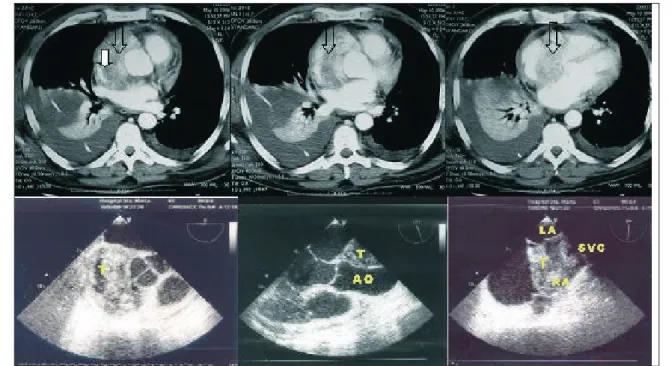
Figure 1 a, b, c: Thoracic angio-CT: three consecutive thoracic CT images after intravenous contrast, showing extensive solid
lesion (open arrow), with heterogeneous contrast enhancement, diffusely infiltrating the right atrial wall, reducing its lumen. The lesion is compressing the superior vena cava (closed arrow), although the latter remains permeable, and involves the ascending aorta. All CT characteristics suggested malignant disease. There was moderate right pleural effusion with inserted pleural catheter and passive collapse of the inferior lobe, with slight left pleural and pericardial effusion.
Figure 1 d, e, f: TEE: d) TEE (0 degrees): tumor (T) in right atrium; e) TEE (113 degrees): tumor (T) in right atrium next to
aortic root (AO); f) TEE (84 degrees): heteroechogenic tumor (T) in right atrium (RA) extending to superior vena cava (SVC). LA: left atrium.
nodule was also found on lymph node, left suprarenal gland and liver. Microscopically all these nodules were metastases of the angiosarcoma of the right atrium (Figs. 2 f, g, h).
DISCUSSION
Clinical presentation of primary malignant cardiac neoplasms is determined by many factors, including tumor location, size, growth rate, tendency for embolization (friability), and degree of invasiveness. Intracavitary tumors tend to obstruct cardiac valves or major vessels or to produce emboli. Myocardial lesions may affect the conduction system of the heart resulting in arrhythmias. Pericardial lesions lead to pericardial tamponade(15).
Heart AS mostly arise in the right atrium and
have a tendency to occur in the third to fifth decade and more commonly in males(16). They
frequently extend to the pericardium, vena cava, or tricuspid valve, causing tamponade and/or heart inflow obstruction with superior vena cava syndrome. The exact primary site of an extensive pericardial tumor in continuity with the right atrial or ventricular component may be unclear, but most originate in the right atrial chamber (17).
Metastases in the lungs are common and often widespread at diagnosis, in 66 to 89% of patients
(8, 18). Other sites of metastization are lymph nodes,
bone, liver, brain, bowel, spleen, adrenal glands, pleura, diaphragm, kidneys, thyroid, and skin(19).
Besides the symptoms described, heart AS may present with hemoptysis secondary to diffuse pulmonary hemorrhage(20) and other clinical
features related to the sites of metastases. Sometimes pulmonary artery or right ventricular outflow obstruction can cause cardiac arrest(4).
Most patients die within a few months after symptom onset(17), despite surgical resection and
even in cases treated by heart transplantation(21).
Chest X-ray is of little help for the detection of cardiac AS, as it only reveals changes secondary to hemodynamic consequences of the tumor growth: cardiomegaly, heart failure, pleural effusion, focal cardiac mass, pulmonary consolidation, or pericardial effusion.
Echocardiography is a reliable, noninvasive, and widely available tool for detecting cardiac tumors, the site of tumor attachment, the pattern of tumor movement and tumor size. Due to technical reasons TTE may have limited diagnostic accuracy(22, 23). Moreover, TEE has
a much higher resolution for differentiating between benign and malignant tumors, which typically disrupt, infiltrate, and obscure the tissue planes of adjacent cardiac anatomy(24). The
presence of a tumor in the pulmonary veins or extension into the vena cava has been described as an useful aid in the differentiation of a malignant neoplasm from myxoma(5, 24).
Despite these advantageous qualities of TEE, its performance in soft tissue characterization is not as good as that achieved with helical CT and MRI, and myocardial disease such as tumor infiltration is not clearly depicted. Most importantly, both TTE and TEE provide limited views of the mediastinum and cannot be used to evaluate extracardiac manifestations of the disease. However, both CT and MRI are not as 581 Figure 2 a, b, c, d, e, f, g, h: Histopathology: a) right atrial
AS; b) AS of the right atrium infiltrating myocardial cells; c) AS: cellular area with atypical endothelial cells; d) AS: immunohistochemical stain for CD34 revealing positive endothelial cells; e) AS: atypical endothelial cells infiltrating the pericardium; f) AS: mediastinal lymph node metastasis; g) AS: atypical papillary fronds lined by hyperchromatic endothelial cells. Adrenal metastasis; h) AS: liver metastasis.
useful in the characterization of moving structures such as the cardiac valves(6).
According to Janigan(17), there are two main
morphological types of cardiac AS, characterized by: (1) a well-defined mass protruding into a cardiac chamber, often sparing the atrial septum. Macroscopically the tumors are hemorrhagic, necrotic and often adherent to the pericardium. CT often shows a low-attenuation right atrial mass, irregular or nodular, usually arising from the right atrial free wall. Tumor infiltration of the myocardium, compression of the cardiac chambers, direct extension into the pericardium, and involvement of the mediastinal great vessels are generally well demonstrated in CT and MRI; (2) a diffusely infiltrative mass extending along the pericardium. The pericardial space may be obliterated with hemorrhagic and/or necrotic tumor debris, which may appear on CT as pericardial effusion or thickening.
Because of the propensity of heart AS for hemorrhage and necrosis, it typically has heterogeneous signal intensity on MRI. Areas of increased signal intensity on T1-weighted images may be focal or peripheral and are thought to represent blood products(6). Local
nodular areas of increased signal intensity interspersed within areas of intermediate signal intensity on T1 and T2-weighted images have a cauliflower-like appearance(25). Linear contrast
material enhancement along vascular lakes gives a “sunray” appearance to cases with diffuse pericardial infiltration(26).
AS are considered malignant neoplasms of endothelial differentiation. They vary from well-differentiated tumors that are composed of anastomosing vascular channels lined by elongated, mononucleated and fusiform malignant cells occasionally protruding into the vascular lumen, to undifferentiated tumors arranged as solid sheets of anaplastic and spindle-shaped tumor cells. Immunohisto-chemical stains for CD31, CD34 and factor VIII-related protein are useful markers to confirm the endothelial origin of these tumors (20).
Zu et al. (27) recently reported 55,XY,+der
(1:A)(q10:q10),+2,+7,+8,+19,+20,+21,+22 chromosomal abnormalities in cytogenetic analysis. Multicolor fluorescent in situ hybridization on paraffin-embedded tissue sections illustrated polysomy of chromosome 8. Immunohistochemical analysis showed high
expression of mutated p53 gene products in tumor cell nuclei(28, 29).
Surgery is the first-line treatment for cardiac AS. However, most patients present with marginally resectable or technically unresectable disease at diagnosis, generally resulting in poor survival rates(30). Given the likely inadequacy of
surgical margins and the mortality risk of distant metastases, both adjuvant radiotherapy and systemic chemotherapy have been tried, but unfortunately the results of these multimodality therapies are disappointing(3, 10). For
Lombard-Cussac et al.(3)heart AS had the worst prognosis
of all cardiac sarcomas, in a series including adjuvant chemotherapy with doxorubicin. The usual presence of metastases at the time of diagnosis only aggravates the problem. Orthotopic heart transplantation has been proposed, but the results also are generally not good(21, 32). Survival after multimodality therapy is
generally short, and only slightly better if the tumor is completely resectable in selected patients(33).
Urticaria is due to degranulation of mast and basophil cells with release of histamine and other mediators, and is caused by various etiopathogenic mechanisms. The causes of urticaria can be divided into four categories(34):
(1) IgE-mediated (allergic), (2) complement-mediated (immunologic), (3) direct degranu-lating effect of drugs, neurohormones, opioid peptides, or other chemicals, (4) direct or indirect effects of physical stimuli.
Chronic urticaria (CU) is defined as any pattern of urticariform lesions that occurs at least twice a week during a period of more than six weeks(35). Urticaria is generally not considered a
manifestation of neoplasm(36, 37), but it has
occasionally been described together with choriocarcinoma and rectal cancer (38). In both
instances, successful removal of the tumor prompted disappearance of the urticaria. To accept a causal relationship between neoplasm and concomitant CU it would be necessary to see complete or partial amelioration of the urticaria with medical or successful treatment of the neoplasm. CU may be a manifestation of hematologic malignancies. In a review by Karakelides et al. (39) of 1639 patients
presenting with CU between 1994 and 2001, 47 had monoclonal gammopathy of unknown significance (MGUS), 142 had a malignancy, and
24 had both. Fifteen of the CU patients with MGUS had a hematologic malignancy compared with 0.9% of those without MGUS (p<0.001). Patients presenting with a new diagnosis of CU at an older age (>56 years) were more likely to have associated underlying MGUS. The occurrence of MGUS in this group was higher than its reported incidence in the general population. There are also references to the association of CU with malignancies of the colon, rectum and lung, Hodgkin disease and non-Hodgkin lymphoma (40),
and hairy-cell leukemia(41).
The case of heart AS in our patient is in certain respects atypical, because of the previous concomitant unexplained occurrence of urticaria and polyarthralgias, which began several months before the final admission to the hospital with a clinical picture of superior vena cava syndrome. A complete study for an etiologic diagnosis to explain the occurrence of urticaria had been
performed during the previous episodes but was unsuccessful. Although we cannot establish a cause and effect relationship between the tumor and the occurrence of urticaria, defining a paraneoplastic syndrome, we think such an association is possible. The proof would be the resolution of urticaria after treatment of the tumor, which was not possible due to the patient's death.
Pedidos de separatas para: Address for reprints: ANTÓNIO MURINELLO Av. Eng. António Azevedo Coutinho, lt 8, r/c Dtº 2750-644 Cascais PORTUGAL Telemóvel: 918 626 874
583
BIBLIOGRAFIA / REFERENCES
1 - Sabatine MS, Colucci WS, Schoen FJ. Primary Tumors of the Heart. In: Braunwald's Heart Disease. Zipes DP, Libby P, Bonow RO, Braunwald E (eds.). Philadelphia: Elsevier Saunders, 7th ed. 2005: 1741-55
2 - Silverman NA. Primary cardiac tumors. Ann Surg 1980; 191: 127-38
3 - Burke AP, Cowan D, Virmani R. Primary sarcomas of the heart. Cancer 1992; 69: 387- 95
4 - Shanmugan G. Primary cardiac tumors. Europ J Cardio-Thor Surg 2006; 29: 925-32
5 - Hsieh P-L, Lee D, Chiou K-R, Kung M-H, Lin S-L, Liu C-P, et al. Echocardiographic features of primary cardiac sarcomas. Echocardiography 2002; 19: 215-20
6 - Araoz PA, Eklund HE, Wech TJ, Breen JF. CT and MR imaging of primary cardiac malignancies. Radiographics 1999; 19: 1421-34
7 - Donsbeck AV, Ranchere D, Coindre JM, Le Gall F, Cordier J-F, Loire E. Primary cardiac sarcomas: an immunohistochemical and grading study with long-term follow-up of 24 cases. Histopathology 1999; 34: 295-304
8 - Tazelaar HD, Locke TJ, McGregor LG. Pathology of surgically excised primary cardiac tumors. Mayo Clin Proc 1992; 67: 957-65
9 - Burke AP, Virmani R. Tumors and tumor-like conditions of the heart. In: Cardiovascular Pathology. Silver MD, Gottlieb AI, Schoen FJ (eds.). Philadelphia: Churchill Livingstone, 1st ed. 2001: 583-65
10 - Putnam J, Sweeny M, Colon R, et al. Primary cardiac sarcomas. Ann Thor Surg 1991; 51: 906-1
11 - Piazza N, Chughtai T, Toledano K, et al. Primary cardiac
tumors. Eighteen years of surgical experience on 21 patients. Can J Cardiol 2004; 20: 1443-8
12 - Black AK, Champion RH. Urticaria. In: Rook, Wilkinson, Ebling's Textbook of Dermatology. Champion RH, Burton JL, Burns DA, Breathnach SM (eds.). London: Blackwell Science, 6th ed. 1998: 2113-39
13 - Soter NA. Urticaria and Angioedema. In: Fitzpatrick's Dermatology in General Medicine. Freedberg IM, Eisen AZ, Wolff K, Austen KF, Goldsmith LA, Katz SI, Fitzpatrick TB (eds.). New York: McGraw-Hill, 5th ed. International edition. 1999: 1409-25
14 - Braverman IM. Hypersensitivity syndromes - Urticaria. In: Braverman IM's Skin Signs of Systemic Disease. Braverman IM (ed.). Philadelphia: Saunders, 1st ed. 1981: 44-6; 453-76 15 - Grebenc ML, Rosado de Christensen ML, Burke AP, Green CE, Galfin KR. Primary cardiac and pericardial neoplasms: radiologic-pathologic correlation. Radiographics 2000; 20: 1073-1103
16 - Glancy DL, Morales JB, Roberts WC. Angiosarcoma of the heart. Am J Cardiol 1968; 2: 413-9
17 - Janigan DT, Husain A, Robinson NA. Cardiac angiosarcoma - a review and a case report. Cancer 1986; 57: 852-9
18 - Amonkar GP, Desanpande JR. Images in Cardiovascular Pathology - cardiac angiosarcoma. Cardiovascular Pathology 2006; 15: 57-58
19 - Burke A, Virmani R. Tumors of the heart and great vessels. In: Atlas of Tumor Pathology. 3rd series. Fasc 16. Washington DC: Armed Forces Institute of Pathology. 1996: 136-40 20 - Adem C, Ambry MC, Tazeler HD, Meyers JL. Metastatic angiosarcoma masquerading as diffuse pulmonary hemorrhage.
Arch Pathol Lab Med 2001; 125: 1562-5
21 - Crespo MG, Pulpon LA, Pradas G, Serrano S, Segovia J, Vegazo I, et al. Heart transplantation for cardiac angiosarcoma: should its indication be questioned? J Heart Lung Transplant 1993; 12: 52-30
22 - Meng Q, Lai H, Lima J, Tong W, Qian Y, Lai S. Echocardiographic and pathologic characteristics of primary cardiac tumors: a study of 149 patients. Int J Cardiol 2002; 84: 69-75
23 - Kurian KC, Weisshaar D, Parekh H, Berry GJ, Reitz B. Primary cardiac angiosarcoma: case report and review of the literature. Cardiovascular Pathology 2006; 15: 110-2 24 - Freeman WK, Reeder GS. Cardiac neoplasias and thrombi. In: Transesophageal Echocardiography. Freeman WK, Seward JB, et al. (eds.). New York: Little, Brown, 1st ed. 1994: 339-84 25 - Kim EE, Wallace S, Abello R, et al. Malignant cardiac fibrous histiocytoma and angiosarcoma: MR features. J Comput Assist Tomogr 1989; 13: 627-32
26 - Yahata S, Endo T, Houma H, et al. Sunray appearance on enhanced magnetic resonance image of cardiac angiosarcoma with pericardial obliteration. Am Heart J 1994; 127: 468-71 27 - Zu Y, Perle MA, Yan Z, Liu J, Kumar A, Waisman J. Chromosomal abnormalities and p53 gene mutation in a cardiac angiosarcoma. Appl Immunohistochem Mol Morphol 2001; 9: 24-8
28 - Naka M, Tomita Y, Nakanishi H, Araki N, Hongyo T, Ochi T, et al. Mutations of p53 tumor-suppressor gene in angiosarcoma. Int J Cancer 1997; 71: 952-5
29 - Garcia JM, Gonzalez R, Silva JM, Dominguez G, Vegazo IS, Gamallo C, et al. Mutational status of K-ras and TP53 genes in primary sarcomas of the heart. Brit J Cancer 2000; 82: 1183-5 30 - Herman MA, Shankerman RA, Edwards WD, Shub C, Schaff HV. Primary cardiac angiosarcoma: a clinicopathologic study of six cases. J Thorac Cardiovascul Surg 1992; 103: 655-64 31 - Llombart-Cussac A, Pivot X, Confesso G, Rhor-Alvarado R, Delord JP, Sjielmaun M, et al. Adjuvant chemotherapy for primary cardiac sarcoma: the IGR experience. Brit J Cancer 1998; 78: 1624-8
32 - Armitage JM, Kormos RL, Griffith BP, Fricker FJ, Hardesty RL. Heart transplantation in patients with malignant disease. J Heart Transplant 1990; 9: 627-9
33 - Kakizaki S, Takagi H, Hosaka Y. Cardiac angiosarcoma responding to multidisciplinary treatment. Int J Cardiol 1997; 62: 273-5
34 - Adelsberg BR. Chronic urticaria. In: Difficult Diagnoses in Dermatology. Lebwohl M (ed.). New York: Churchill Livingstone, 1st ed. 1988: 317-29
35 - Grattan CE, Sabroe RA, Greaves MW. Chronic urticaria. J Am Acad Dermatol 2002; 46: 645-57
36 - Lindelof B, Sigurgeirsson B, Wahlgren CF, et al. Chronic urticaria and cancer: an epidemiologic study of 1155 patients. Br J Dermat 1990; 123: 453-6
37 - Greaves MW. Chronic urticaria. New England J Medicine 1995; 332: 1767
38 - Urbach E. Endogenous allergy. Arch Dermatol 1942; 45: 697
39 - Karakelides M, Monson K L, Volcheck GW, Weiler CR. Monoclonal gammopathies and malignancies in patients with chronic urticaria. Int J Dermatol 2006; 45 (9), 1032-8. doi: 10.1111/ j.1365-4632.2006.02982.x
40 - Kaplan P. Urticaria and angioedema. In: Allergy: Principles and Practice. Middleton E Jr et al. (eds.). St Louis: Mosby Yearbook, 1998: 1104-22
41 - Clore L, Stafford C. Chronic urticaria as a presenting sign of hairy-cell leukemia. Allergy Asthma Proc 1999; 20: 51-5